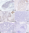
Reduction of phosphorylated signal transducer and activator of transcription-5 expression in feline mammary carcinoma
- PMID: 38777776
- PMCID: PMC11251807
- DOI: 10.1292/jvms.23-0470
Reduction of phosphorylated signal transducer and activator of transcription-5 expression in feline mammary carcinoma
Abstract
Signal transducers and activators of transcription (STATs) are a family of transcription factors involved in various normal physiological cellular processes. Moreover, STATs have been recently identified as novel therapeutic targets for various human tumors. STAT3, STAT5a, and STAT6 have been suggested to be involved in tumorigenesis in human breast cancer. Owing to the similarity between feline mammary carcinomas (FMCs) and human breast cancers, these factors may play an important role in FMCs. However, studies on the expression of STATs in animal tumors are limited. Therefore, in this study, we aimed to characterize the expression of total STAT5 (tSTAT5) and phosphorylated STAT5 (pSTAT5) in FMCs, feline mammary adenomas, non-neoplastic proliferative mammary gland lesions, and normal feline mammary glands using immunohistochemistry. High expression of tSTAT5 was observed in the cytoplasm of all the samples assessed in this study. Moreover, high expression of tSTAT5 was observed in the nucleus; however, its levels varied depending on the lesion. The percentage of pSTAT5-nuclear positive cells varied among normal feline mammary glands (40.1 ± 25.1%), and non-neoplastic lesions, including mammary hyperplasia (43.2 ± 28.6%) and fibroadenomatous changes (18.0 ± 13.6%). Moreover, the percentage of pSTAT5-nuclear-positive cells in feline mammary adenomas was 24.5 ± 19.2%, which was significantly reduced in feline mammary carcinomas (2.4 ± 5.6%), regardless of histopathological subtype. This study suggests that decreased STAT5 activity may be involved in the development and malignant progression of feline mammary carcinomas.
Keywords: feline mammary carcinoma; immunohistochemistry; signal transducers and activator of transcription-5.
Conflict of interest statement
The authors have no conflict of interest to declare with respect to the research, authorship, and/or publication of this article.
Figures


Similar articles
-
CXCR4 expression in feline mammary carcinoma cells: evidence of a proliferative role for the SDF-1/CXCR4 axis.BMC Vet Res. 2012 Mar 14;8:27. doi: 10.1186/1746-6148-8-27. BMC Vet Res. 2012. PMID: 22417013 Free PMC article.
-
Immunohistochemical evaluation of STAT3-p-tyr705 expression in feline mammary gland tumours and correlation with histologic grade.Res Vet Sci. 2007 Apr;82(2):218-24. doi: 10.1016/j.rvsc.2006.06.010. Epub 2006 Aug 24. Res Vet Sci. 2007. PMID: 16934302
-
Expression of Stat3 in feline mammary gland tumours and its relation to histological grade.Vet Res Commun. 2006 Aug;30(6):599-611. doi: 10.1007/s11259-006-3335-z. Vet Res Commun. 2006. PMID: 16838202
-
Prognostic histopathological and molecular markers in feline mammary neoplasia.Vet J. 2012 Oct;194(1):19-26. doi: 10.1016/j.tvjl.2012.05.008. Epub 2012 Jul 28. Vet J. 2012. PMID: 22841451 Review.
-
Signal transducer and activator of transcription 5 as a key signaling pathway in normal mammary gland developmental biology and breast cancer.Breast Cancer Res. 2011 Oct 12;13(5):220. doi: 10.1186/bcr2921. Breast Cancer Res. 2011. PMID: 22018398 Free PMC article. Review.
Cited by
-
Prognostic Insights in Feline Mammary Carcinomas: Clinicopathological Factors and the Proposal of a New Staging System.Animals (Basel). 2025 Mar 10;15(6):779. doi: 10.3390/ani15060779. Animals (Basel). 2025. PMID: 40150308 Free PMC article.
References
MeSH terms
Substances
LinkOut - more resources
Full Text Sources
Research Materials
Miscellaneous

