Review
. 2024 May 22;7(1):616.
doi: 10.1038/s42003-024-06263-w.

Replication stress as a driver of cellular senescence and aging

Affiliations
Review

Replication stress as a driver of cellular senescence and aging

Lauren M Herr et al. Commun Biol. .

Abstract

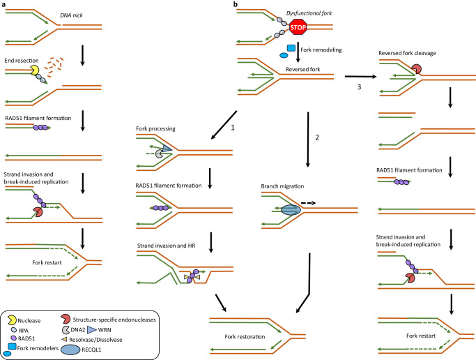
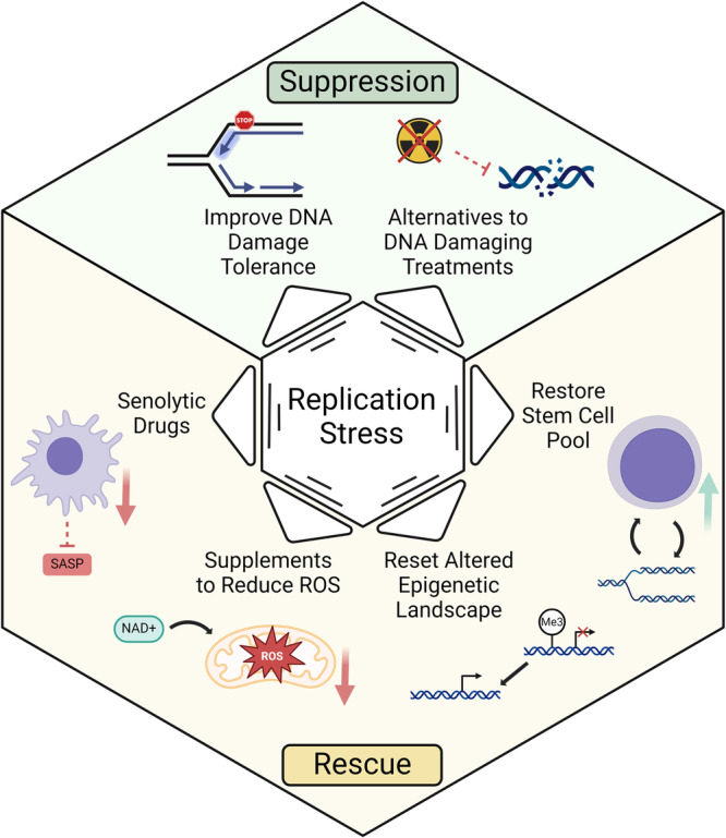
Replication stress refers to slowing or stalling of replication fork progression during DNA synthesis that disrupts faithful copying of the genome. While long considered a nexus for DNA damage, the role of replication stress in aging is under-appreciated. The consequential role of replication stress in promotion of organismal aging phenotypes is evidenced by an extensive list of hereditary accelerated aging disorders marked by molecular defects in factors that promote replication fork progression and operate uniquely in the replication stress response. Additionally, recent studies have revealed cellular pathways and phenotypes elicited by replication stress that align with designated hallmarks of aging. Here we review recent advances demonstrating the role of replication stress as an ultimate driver of cellular senescence and aging. We discuss clinical implications of the intriguing links between cellular senescence and aging including application of senotherapeutic approaches in the context of replication stress.


Conflict of interest statement

The authors declare no competing interests.

Figures

Fig. 1
Fig. 1. Mechanisms of stalled fork recovery under conditions of replication stress.
Replication stalling or fork collapse upon exposure to various exogeneous and endogenous stressors activates DNA damage response pathways critical for stalled fork stabilization or restoration and replication restart. a A replication fork encountering an ssDNA break/gap can be recovered by generating a DSB, thereby allowing HR-dependent fork restoration. Nucleases involved in DNA end resection such as MRE11 or EXO1 process the break and generate a 3’ ssDNA overhang necessary to initiate HR at the broken fork. ssDNA-bound RPA is replaced by RAD51 recombinase which in turn allows for strand invasion into the sister chromatid, thereby forming a displacement (D)-loop structure to initiate break-induced replication using the sister chromatid as template. The D-loop can be cleaved by structure-specific endonucleases such as MUS81 to restore the replication fork and continue DNA synthesis. b A dysfunctional fork such as one stalled due to nucleotide depletion is acted upon by the coordinated actions of fork remodeling enzymes and RAD51 to generate a reversed fork intermediate. Fork protecting factors such as BRCA1, BRCA2, RAD51, WRN, and RECQL1 protect reversed forks from unscheduled pathological degradation by nucleases. However, helicase-assisted nucleolytic processing (e.g., WRN-DNA2) of stalled forks (1) in a controlled manner promotes replication restart through HR. Alternatively, a reversed fork can be restored via the branch-migration activity of RECQL1 helicase (2). The reversed fork can also be cleaved by structure-specific endonucleases (e.g., MUS81) to generate a single-ended double strand break (seDSB) at the fork that undergoes homology-directed fork restoration (3), as described in a. Cells genetically deficient in fork-stabilizing proteins (e.g., WRN helicase mutated in WS) accumulate DSBs at forks and experience gross chromosomal instability which may lead to replicative senescence and accelerated aging phenotypes.
Fig. 2
Fig. 2. Relationships of replication stress to hallmarks of aging.
Evidence from the literature suggests that many of the previously described hallmarks of aging are driven by replication stress. Representative examples are cited below as well as in the text. Chronic Inflammation. Innate immune response activation by LINE-1 element derepression and cytosolic ssDNA incurred by replication stress promotes senescence induction and contributes to sterile inflammation,. Genomic Instability. Replicative stress incurs DNA damage in the form of chromosomal aberrations and DSBs that attenuate replicative lifespan of cells and induce aging-associated tissue dysfunction,,. Telomere Attrition. Telomeres are fragile sites with DNA sequences prone to form G4 and T-loop secondary structures that break under conditions of replication stress, inducing telomere shortening that limits replicative potential,,. Stem Cell Exhaustion. Replicative stress in SCs impairs the ability to produce SC progeny. Specific to HSCs, this leads to a variety of tissue-based and organismal dysfunctions including clonal drift and SC attrition,,. Epigenetic Alterations. Replicative stress recruits a variety of proteins to the stalled replication fork that are involved in remodeling the DNA modification and histone landscapes around the stress. These changes can have long-term effects for cellular senescence,. Mitochondrial Dysfunction. Replication stress in the mitochondria leading to mtDNA mutations causes a deprivation of resources, culminating in premature aging in a pol γ proofreading mutant mouse model. In addition, mtDNA replication errors have consequences for nuclear genome replication and stability, which contribute to aging phenotypes. BioRender was used to create the figure.
Fig. 3
Fig. 3. Avenues to suppress replication stress or rescue the aberrant phenotypes caused by replication stress.
The upper third of the hexagon depicts two mechanisms to avoid replication stress. The lower two-thirds of the hexagon depicts four strategies that alleviate the consequences of replication stress. See text for details. BioRender was used to create the figure.

References

    1. Saxena S, Zou L. Hallmarks of DNA replication stress. Mol. cell. 2022;82:2298–2314. doi: 10.1016/j.molcel.2022.05.004. - DOI - PMC - PubMed
    1. Gorgoulis V, et al. Cellular senescence: defining a path forward. Cell. 2019;179:813–827. doi: 10.1016/j.cell.2019.10.005. - DOI - PubMed
    1. Keijzers G, Bakula D, Scheibye-Knudsen M. Monogenic diseases of DNA repair. N. Engl. J. Med. 2018;378:491–492. doi: 10.1056/NEJMc1716072. - DOI - PubMed
    1. Robbins PD, et al. Senolytic drugs: reducing senescent cell viability to extend health span. Annu Rev. Pharm. Toxicol. 2021;61:779–803. doi: 10.1146/annurev-pharmtox-050120-105018. - DOI - PMC - PubMed
    1. Lopez-Otin C, Blasco MA, Partridge L, Serrano M, Kroemer G. The hallmarks of aging. Cell. 2013;153:1194–1217. doi: 10.1016/j.cell.2013.05.039. - DOI - PMC - PubMed