Multicenter Study
. 2024 May 22;14(1):11681.
doi: 10.1038/s41598-024-62559-1.

One-year outcomes and safety assessment of faricimab in treatment-naïve patients with neovascular age-related macular degeneration in Japan

Affiliations
Multicenter Study

One-year outcomes and safety assessment of faricimab in treatment-naïve patients with neovascular age-related macular degeneration in Japan

Ryo Mukai et al. Sci Rep. .

Abstract

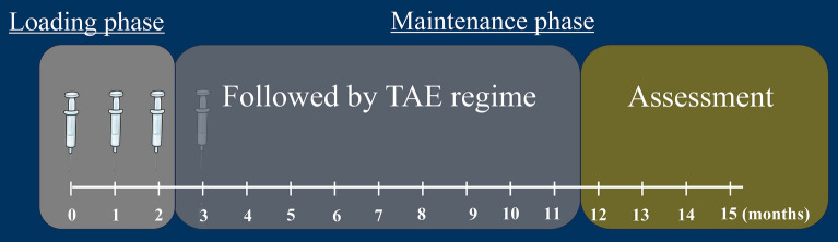
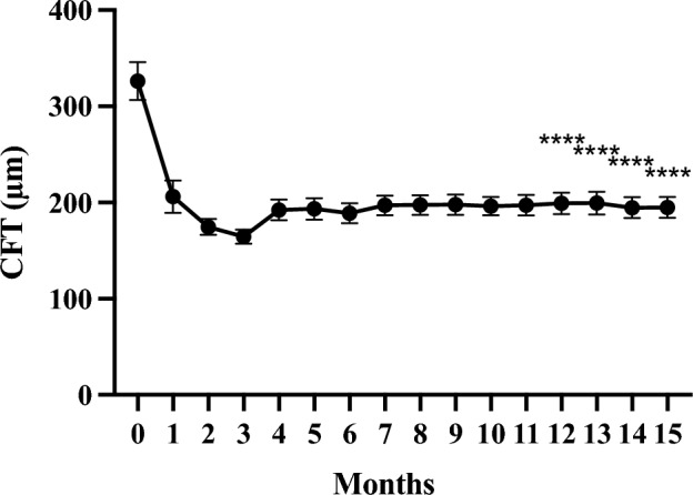
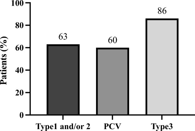
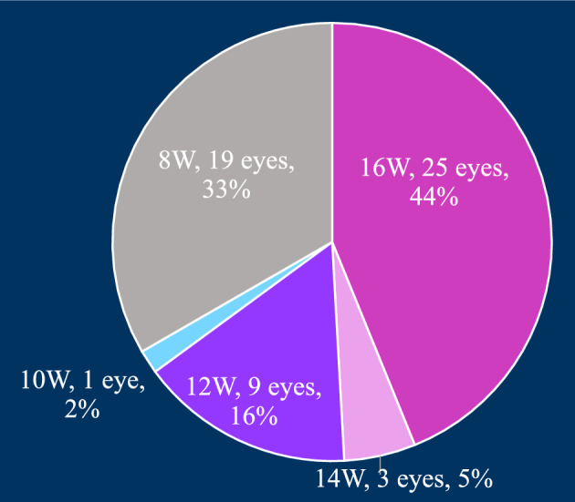
This multicentre retrospective study evaluated the 1-year outcomes and safety profile of faricimab in treatment-naïve patients with neovascular age-related macular degeneration (nAMD). Fifty-five patients (57 eyes) underwent loading therapy comprising three monthly faricimab injections. If dryness was achieved by the third month, subsequent treat-and-extend (TAE) follow-up continued at a minimum 8-week interval thereafter. If wet macula persisted at the third month, a fourth dose was administered, followed by the TAE regimen. After 1 year, improvements in visual acuity (0.44 ± 0.46 [baseline] to 0.34 ± 0.48; p < 0.01) and central foveal thickness (326 ± 149 [baseline] to 195 ± 82 μm; p < 0.0001) were significant. Dry macula, characterised by the absence of intraretinal or subretinal fluid, was achieved in 65% of cases. Treatment intervals varied, ranging from 8 to 16 weeks, with 44% of eyes extending to a 16-week interval, followed by 33% at 8 weeks, 16% at 12 weeks, 5% at 14 weeks, and 2% at 10 weeks. Notably, 50% of the polypoidal choroidal vasculopathy patients exhibited complete regression of polypoidal lesions between 12 and 15 months. Faricimab treatment in nAMD patients induced significant improvements in central vision and retinal morphology. Two cases of retinal pigment epithelial tears and one case of iritis were reported as ocular complications.

Keywords: Choroidal thickness; Faricimab; Neovascular age-related macular degeneration; Retinal morphology; Treat-and-extend regimen; Visual acuity.


Conflict of interest statement

R. Mukai, received lecturing and travel fees from Bayer Pharmaceuticals., Chugai Pharmaceutical., Novartis Pharma, Santen Pharmaceutical., and Senju Pharma.; K. Kataoka, received lecturing and travel fees from Santen Pharmaceutical, Bayer Pharmaceuticals, Senju Pharmaceutical, Japan Beringer Ingelheim, Otsuka Pharmaceutical, Canon Medtech, OMC Chugai Pharmaceutical, and Novartis Pharma.; K. Tanaka, received lecturing and travel fees from Santen Pharmaceutical, Alcon, Bayer Pharmaceuticals, Novartis Pharma, Senju Pharmaceutical, Nihon Tengan Kenkyusho, and Chugai Pharmaceutical.; Y. Miyara, received lecturing and travel fees from Senju Pharmaceutical.; I. Maruko, received lecturing and travel fees from Bayer Pharmaceuticals, Novartis Pharma, Japan Alcon, Santen, Senju Pharmaceutical, Topcon, Chugai Pharmaceutical, Canon, Nidek, and Nikon. He holds a patent.; M. Nakayama, None; Y. Watanabe, None; A. Yamamoto, received lecturing and travel fees from Novartis Pharma, Bayer Pharmaceuticals, and Chugai Pharmaceutical.; Y. Wakatsuki, None; H. Onoe, received lecturing and travel fees from Novartis Pharma, Senju Pharmaceutical, Chugai Pharmaceutical ; S. Wakugawa, received lecturing and travel fees from Senju Pharmaceutical and Novartis Pharma.; N. Terao, received lecturing and travel fees from Novartis Pharma, Bayer Pharmaceuticals, Santen Pharmaceutical, Abbott, Kowa, Topcon, Asahi Kasei Pharma, HOYA, and Chugai Pharmaceutical.; T. Hasegawa, received lecturing and travel fees from Bayer Pharmaceuticals, Novartis Pharma, Alcon Pharma, Santen, Kowa, Senju Pharmaceutical, R.E. Medical, Nikon Health Care Japan, JFC Sales Plan, Otsuka Pharmaceutical, and Japan Beringer Ingelheim.; M. Kawai, None; R. Maruko, received lecturing and travel fees from Kyowa Kirin.; K. Itagaki, received lecturing and travel fees from Novartis Pharma, Bayer, Santen Pharmaceutical, Senju Pharmaceutical, and Chugai Pharmaceutical.; J. Honjo, None; A.A. Okada, Personal fees from Alcon Pharma K.K., Allergan Japan, Apellis Pharmaceuticals Inc., Astellas Pharma Inc., Bayer Australia Ltd., Bayer Healthcare AG, Bayer Yakuhin Ltd., Biocon Biologics Ltd., Chugai Pharmaceutical Co. Ltd., Daiichi-Sankyo Co. Ltd., Kowa Co. Ltd., Mitsubishi Tanabe Pharma Corporation, Novartis Pharma K.K., Otsuka Pharmaceutical Co. Ltd., Santen Pharmaceutical Co. Ltd., and Senju Pharmaceutical Co., Ltd. Departmental research grants from Alcon Pharma K.K., Bayer Yakuhin Ltd., Mitsubishi Tanabe Pharma Corporation, Novartis Pharma K.K., and Santen Pharmaceuticals Co. Ltd.; R. Mori, received lecturing and travel fees from Santen Pharmaceutical, Bayer Pharmaceuticals, Japan Beringer Ingelheim, Kyowa Kirin, Novartis Pharma, Senju Pharmaceutical, and Chugai Pharmaceutical.; H. Koizumi, received grants from Novartis Pharma, Alcon Pharma, Japan Alcon, Bayer Pharmaceuticals, HOYA, Senju Pharmaceutical, Santen Pharmaceutical, AMO, Otsuka Pharmaceutical, Star Japan, First Medical, Pfizer, Kowa, and Nikon, received lecturing and travel fees from Novartis Pharma, Alcon Pharma, Bayer Pharmaceuticals, Senju Pharmaceutical, Santen Pharmaceutical class, Kowa, Japan Alcon, HOYA, AMO, Otsuka Pharmaceutical, Pfizer, Bausch + Lomb Japan, JFC Sales Plan, Canon, Nidek, Abbott, Tomey, Daiichi Sankyo, Chugai Pharmaceutical, and Sanofi, and received consultant fee from Novartis Pharma, Bayer Pharmaceuticals, Chugai Pharmaceutical, Allergan, Japan Beringer Ingelheim.; T. Iida, received grants from Nidek, Topcon, Santen, Novartis Pharma, Senju Pharmaceutical, Japan Alcon, HOYA, and AMO, consultant fee from Bayer Pharmaceuticals, Novartis Pharma, Chugai Pharmaceutical, Japan Beringer Ingelheim, and Janssen Pharma, and received lecturing and travel fees from Bayer Pharmaceuticals, Novartis Pharma, Japan Alcon, Santen, Senju Pharmaceutical, Topcon, Chugai Pharmaceutical, Canon, Nidek, Otsuka Pharmaceutical, Nikon, and Kyowa Kirin. He holds a patent.; T. Sekiryu, received grants from Novartis Pharma, Pfizer, Japan Alcon, Wakamoto Pharmaceutical, HOYA, Bayer Pharmaceuticals, Santen Pharmaceutical, Senju Pharmaceutical, AMO Japan, and Kowa, received lecturing and travel fees from Novartis Pharma, Chugai Pharmaceutical, Akura, Abbott, Alcon Pharma, Santen Pharmaceutical, and Senju Pharmaceutical, and received consultant fee from Allergan Japan.

Figures

Figure 1
Figure 1
Treatment schedule for optimising neovascular age-related macular degeneration management: combining loading dose therapy with a treat-and-extend (TAE) regimen. Patients who achieved macular dryness by the third month were followed-up with a TAE regimen, with intervals ranging from a minimum of 8 weeks to a maximum of 16 weeks. Patients with persistence of wet macula at the third month were administered a fourth dose, followed by the TAE regimen.
Figure 2
Figure 2
Changes in best-corrected visual acuity (BCVA) in 57 eyes treated with faricimab for 1 year. The data were analysed using the last observation carried forward (LOCF) and shown as the mean ± standard error. **p < 0.01, ***p < 0.001.
Figure 3
Figure 3
Changes in average central foveal thickness (CFT) in 57 eyes treated with faricimab injections for 1 year. The data were analysed using the last observation carried forward (LOCF) method and shown as the mean ± standard error. ****p < 0.0001.
Figure 4
Figure 4
Changes in average subfoveal choroidal thickness (SCT) in 57 eyes treated with faricimab injections for 1 year. *p < 0.05, **p < 0.01.
Figure 5
Figure 5
Total number of faricimab injections over 1 year.
Figure 6
Figure 6
The treatment interval for 1 year.
Figure 7
Figure 7
Proportions of patients with dry macula at 1 year after faricimab treatment, categorised by lesion type. A total of 37 out of 57 eyes (65%) achieved dry macula. PCV polypoidal choroidal vasculopathy.
Figure 8
Figure 8
Case 1—A 70-year-old male with type 1 macular neovascularisation (MNV) at baseline (AD). The best corrected Snellen visual acuity was 20/25. (A) Fundus photography (FP) revealed pigment epithelial detachment (PED) with subretinal fluid (SRF) at the fovea. (B) Optical coherence tomography angiography (OCTA) revealed MNV. (C) Late-phase fluorescein angiography revealed leakage from the MNV at the macula. (D) Middle-phase indocyanine green angiography identified vascular network at the fovea. (EH) OCT images at baseline and at loading therapy with faricimab. At baseline, OCT revealed a small PED with SRF. The PED regressed during the loading therapy, and subretinal fluid was absorbed at 1, 2, and 3 months after the first injection of faricimab. (I,J) Fundus photography and OCT at 1 year; FP revealed no recurrence, and OCT showed no recurrence of fluid changes. At 15 months, six faricimab injections were administered, with treatment intervals extending up to a maximum of 16 weeks. His best corrected Snellen visual acuity was 20/16.
Figure 9
Figure 9
Case 2—A 59-year-old male with type 1 and type 2 macular neovascularisation (MNV). The best corrected Snellen visual acuity was 20/63 at baseline (AD). (A) Fundus photography (FP) revealed a yellowish-grey bumped lesion at the fovea with subretinal fluid (SRF), subretinal haemorrhages, and hard exudate. (B) Optical coherence tomography angiography (OCTA) revealed MNV. (C) Late-phase fluorescein angiography detected a type 2 MNV. (D) Middle-phase indocyanine green angiography identified abnormal MNV at the fovea. (EH) OCT at baseline and at loading therapy. (E) OCT revealed an SRF, accompanied by subretinal hyperreflective material, which corresponded to a yellowish-grey bumped lesion at the macula and SRF observed at 3 months. (IK) FP fundus autofluorescence (FAF), and OCT at 1 year; FP showed no subretinal fluid (SRF) and a small scar lesion, FAF revealed a mild atrophic lesion around an original type 2 MNV, and OCT revealed no recurrence of fluid and a small bump lesion. At 15 months, the number of faricimab injections was six, with treatment intervals extending up to a maximum of 16 weeks. His best corrected Snellen visual acuity improved to 20/40.
Figure 10
Figure 10
Case 3—A 74-year-old male with polypoidal choroidal vasculopathy (PCV) at baseline (AD). The best corrected Snellen visual acuity was 20/320. (A) Fundus photography (FP) revealed an orange nodule surrounded by subretinal fluid with hard exudate at the fovea. (B) Optical coherence tomography angiography (OCTA) revealed MNV in the PED. (C) Late-phase fluorescein angiography detected granular leakage from the PED. (D) Middle-phase indocyanine green angiography identified polypoidal lesions at the fovea. (EH) OCT images at baseline and at loading therapy. OCT revealed a sharp peaked pigment epithelial detachment (PED) accompanied by shallow subretinal fluid (SRF) with hard exudate at baseline; at 3 months, the SRF absorbed and the PED regressed. (I,J) FP and OCT at 1 year; FP showed no recurrence of fluid and that the hard exudate was almost absorbed, and OCT revealed no recurrence of fluid. At 15 months, the patient was administered six faricimab injections, with treatment intervals extending up to a maximum of 16 weeks. His best corrected Snellen visual acuity improved to 20/100.
Figure 11
Figure 11
Case 4—A 90-year-old female with type 3 macular neovascularisation (MNV) at baseline (AD). The best corrected Snellen visual acuity was 20/200. (A) Fundus photography (FP) revealed a pigment epithelial detachment (PED) with intraretinal fluid (IRF) and intraretinal haemorrhage at the fovea. (B) Fundus autofluorescence (FAF) revealed no macular atrophy. (C) Late-phase fluorescein angiography detected dye leakage from a type 3 MNV. (D) Middle-phase indocyanine green angiography also identified type 3 neovascularisation. (EH) Optical coherence tomography (OCT) images at baseline and at loading therapy. OCT revealed a large PED with IRF at baseline and just 1 month after the first faricimab injection, the large pigment epithelial detachment (PED) visible at baseline rapidly regressed, and intraretinal fluid (IRF) had completely absorbed. (IK) FP, FAF, and OCT at 1 year; FP showed no recurrence of the IRF, FAF revealed small patchy atrophy, and OCT revealed no recurrence of IRF or intraretinal haemorrhages. The patient was administered six faricimab injections, with treatment intervals extending up to a maximum of 16 weeks at 15 months. Her best corrected Snellen visual acuity improved to 20/100.

References

    1. Teutsch SM, Woodbury B, Welp A. Making Eye Health a Population Health Imperative: Vision for Tomorrow. The National Academies Collection: Reports funded by National Institutes of Health. National Academies Press; 2016. - PubMed
    1. Shirley M. Faricimab: First approval. Drugs. 2022;82:825–830. doi: 10.1007/s40265-022-01713-3. - DOI - PubMed
    1. Inoda S, et al. Cytokine profiles of macular neovascularization in the elderly based on a classification from a pachychoroid/drusen perspective. Graef. Arch. Clin. Exp. Ophthalmol. 2022;260:747–758. doi: 10.1007/s00417-021-05445-0. - DOI - PubMed
    1. Joussen AM, et al. Angiopoietin/Tie2 signalling and its role in retinal and choroidal vascular diseases: A review of preclinical data. Eye (Lond.) 2021;35:1305–1316. doi: 10.1038/s41433-020-01377-x. - DOI - PMC - PubMed
    1. Akwii RG, Sajib MS, Zahra FT, Mikelis CM. Role of angiopoietin-2 in vascular physiology and pathophysiology. Cells. 2019;8:471. doi: 10.3390/cells8050471. - DOI - PMC - PubMed

Publication types

Substances