. 2024 Jun;630(8018):1012-1019.
doi: 10.1038/s41586-024-07458-1. Epub 2024 May 22.

Structural insights into the cross-exon to cross-intron spliceosome switch

Affiliations

Structural insights into the cross-exon to cross-intron spliceosome switch

Zhenwei Zhang et al. Nature. 2024 Jun.

Abstract

Early spliceosome assembly can occur through an intron-defined pathway, whereby U1 and U2 small nuclear ribonucleoprotein particles (snRNPs) assemble across the intron1. Alternatively, it can occur through an exon-defined pathway2-5, whereby U2 binds the branch site located upstream of the defined exon and U1 snRNP interacts with the 5' splice site located directly downstream of it. The U4/U6.U5 tri-snRNP subsequently binds to produce a cross-intron (CI) or cross-exon (CE) pre-B complex, which is then converted to the spliceosomal B complex6,7. Exon definition promotes the splicing of upstream introns2,8,9 and plays a key part in alternative splicing regulation10-16. However, the three-dimensional structure of exon-defined spliceosomal complexes and the molecular mechanism of the conversion from a CE-organized to a CI-organized spliceosome, a pre-requisite for splicing catalysis, remain poorly understood. Here cryo-electron microscopy analyses of human CE pre-B complex and B-like complexes reveal extensive structural similarities with their CI counterparts. The results indicate that the CE and CI spliceosome assembly pathways converge already at the pre-B stage. Add-back experiments using purified CE pre-B complexes, coupled with cryo-electron microscopy, elucidate the order of the extensive remodelling events that accompany the formation of B complexes and B-like complexes. The molecular triggers and roles of B-specific proteins in these rearrangements are also identified. We show that CE pre-B complexes can productively bind in trans to a U1 snRNP-bound 5' splice site. Together, our studies provide new mechanistic insights into the CE to CI switch during spliceosome assembly and its effect on pre-mRNA splice site pairing at this stage.


Conflict of interest statement

The authors declare no competing interests.

Figures

Fig. 1
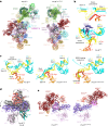
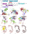
Fig. 1. 3D structure of human CE pre-B complexes.
a, Schematic of the MINX exon RNA. Yn, polypyrimidine tract. b, Left, fit of two monomeric, CE pre-B molecular models into the EM density of the class 1, pre-B dimer. Right, cartoon of the structural organization of the CE pre-B complex dimer. BP, branch point. c, Top, two different views of the molecular architecture of the human CE pre-B complex. Bottom, summary of all modelled proteins and RNAs with colour code.
Fig. 2
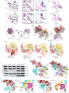
Fig. 2. 3D structure of human CE B-like complexes.
a, Top, two different views of the molecular architecture of the human CE B-like complex. Bottom, summary of all modelled proteins and RNAs with colour code. bd, Interactions of the 5′ss oligo with U5 snRNA loop 1 nucleotides and residues of PRP8 and DIM1. e, Schematic of the base-pairing interactions of the two copies of the 5′ss oligo that are bound in the B-like complex. 5′ss oligo 2 stacks through its 5′ terminal A−3 on the 3′ terminal U+8 of the first 5′ss oligo. f, The ZnF domains of FBP21 and SNU23 stabilize the base-pairing interactions of 5′ss oligo 1 and 5′ss oligo 2, respectively, with U6 and U5 snRNA. Left, coloured EM density. Right, molecular model. Arrowhead indicates missing EM density, which confirms that two distinct 5′ss oligos are bound. g, Schematic of the domain organization of PRP8. HB, helical bundle. h, Repositioning of the PRP8 RT/En and RH domains and BRR2 and PRP6HAT during the conversion of the CE pre-B complex (left) into a B-like complex (right). The 3D structures are aligned through PRP8NTD.
Fig. 3
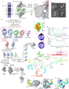
Fig. 3. RNP rearrangements in pre-B triggered by the addition of 5′ss oligo alone or in combination with ATPγS.
a, Molecular architecture of CE pre-B5′ss complexes (left) and pre-B5′ss+ATPγS complexes (right). Notably, PRP28 helicase is still bound to PRP8 in CE pre-B5′ss in essentially the same manner as in CE pre-B (compare with Fig. 1). This is presumably due to the absence of the PRP38A–SNU23–MFAP1 complex, which helps to displace PRP28 during the formation of B complexes and B-like complexes owing to their overlapping (that is, mutually exclusive) binding sites. b, Top, schematic of the U4/U6 snRNA helices and base-pairing interactions of the 5′ss oligo in pre-B5′ss. Bottom, 3D organization showing the position of RBM42. c, 3D organization of the U4/U6 helices and interactions of the 5′ss oligo with U6 snRNA in pre-B5′ss (left), pre-B5′ss+ATPγS (middle) and pre-B5′ssLNG+ATPγS (right). d, 5′ss oligo binding triggers PRP8RT/En movement towards the PRP8NTD and concomitant movement of the PRP8 RH and HB domains and other bound proteins. An overlay of the indicated PRP8 domains in pre-B (grey) and pre-B5′ss (various colours) is shown. Structures are aligned through PRP8NTD. e, Large-scale translocation of BRR2 requires ATP. Comparison of the location of the BRR2 helicase cassettes and the PRP8 RH, JAB1 and RT/En domains in pre-B5′ss versus pre-B5′ss+ATPγS. Aligned through PRP8NTD.
Fig. 4
Fig. 4. B-specific proteins are required for the final positioning of BRR2 and the docking of its U4 snRNA substrate.
a, Schematic of the domain organization of BRR2. PWI, domain with a PWI tri-peptide located within its N-terminal region; SL, separator loop; WH, winged helix. b,c, Multiple B-specific proteins contact BRR2 and tether it to its activation position. Comparison of the location and conformation of BRR2 and its U4 snRNA substrate in pre-B5′ssLNG+ATPγS (b), which is formed in the absence of the B-specific proteins, and the B-like complex (c). In pre-B5’ssLNG+ATPγS, the BRR2NC has a closed, inactive conformation and is located about 15 Å away from its final position observed in the B-like complex. In the presence of B-specific proteins, BRR2NC has an open, active conformation and is bound to the U4 snRNA.
Fig. 5
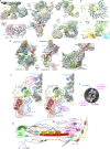
Fig. 5. ATP-dependent transfer in trans of the 5′ss from U1 snRNP to the U6 ACAGA box by PRP28.
a, Structure of purified pre-B complexes incubated with ATP alone (pre-BATP). Fit of the pre-BATP molecular model into the EM density of the pre-BATP dimer. b, Molecular architecture of the pre-BATP complex. The view shown in b is obtained if the structure of protomer 1 in a is rotated 150°. c, Cartoon showing the structural organization of the CE pre-BATP complex dimer. d, The 5′ss of the MINX exon RNA from one pre-B monomer interacts with U5 loop 1 nucleotides and the U6 ACAGA box of the adjacent monomer, forming a short U6/5′ss helix. U4/U6 stem III is still present. Top right, schematic of the base-pairing interactions with the MINX exon RNA near or/at the 5′ss. Nucleotides at the 3′ end of the exon (C−5 to G−1) base pair with nucleotides of U5 loop 1. e, Molecular architecture of a pre-BAMPPNP monomer bound in trans to the U1 snRNP of an adjacent monomer of the pre-BAMPPNP dimeric complex. Right, zoomed-in region that is boxed on the left, showing the molecular architecture of U1 snRNP and its interaction with PRP28. ssRNA, single-stranded RNA. f, Comparison of the corresponding regions shown in e with those of the pre-B complex dimer.
Fig. 6
Fig. 6. Structural model of the conversion of a spliceosomal CE pre-B complex to a CI B complex.
a, Schematic of RNA remodelling (bottom) and rearrangement and repositioning of tri-snRNP proteins (top) during the pre-B to B-like to B complex transition. First, binding of a 5′ss to the U6 ACAGA box leads to the formation of a short U6/5′ss helix and rearrangements in PRP8 to produce a half-closed conformation. Following phosphorylation of PRP6 and PRP31 (indicated by stars), major structural remodelling of the tri-snRNP occurs, including the large-scale movement of the BRR2 helicase cassettes, and the U4/U6 quasi-pseudoknot is dissolved. B-specific proteins are recruited to the remodelled complex and tether BBR2 to its activation position. At the same time, they facilitate the formation of the extended U6/5′ss helix, freeing the U4 nucleotides that are subsequently bound by the BRR2 helicase. b, Model of the conversion of a CE to CI spliceosome, including alternative 5′ss/U1 snRNP choices.
Extended Data Fig. 1
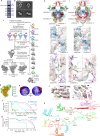
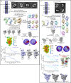
Extended Data Fig. 1. Cryo-EM and image-processing of the cross-exon pre-B complex.
a, RNA composition of purified CE pre-B complex dimers. For gel source data, see Supplementary Fig. 1. Pre-B complexes formed on the MINX exon RNA (Fig. 1a) were affinity-purified and RNA from fractions of the fastest sedimenting peak (typically fractions 14-18) was isolated, separated on a NuPAGE gel, and visualized by staining with SyBr gold. The nucleotide (nts) lengths of the snRNAs and MINX exon RNA are indicated on the right. The RNA composition was analysed from three independent pre-B complex purifications with similar results. The MINX exon RNA was generated from the MINX pre-mRNA, which is a derivative of the Adenovirus Major Late (ADML) pre-mRNA. In the MINX exon RNA, the 5’ exon and adjacent downstream intron nucleotides of the MINX pre-mRNA have been deleted, leaving the 3’ exon (a truncated version of the ADML exon 2) and 64 nts of the 3’ end of the upstream, adjacent ADML intron, which contains an anchoring site followed by the branch site, polypyrimidine tract and 3’ss AG. A 5’ss was introduced at the 5’ end of the truncated exon, by adding the last 6 nts of the wildtype ADML exon 2 plus 22 nts of the adjacent downstream ADML intron, which are followed by a short linker and three RNA stem-loops that bind the MS2 protein (see Fig. 1a). Previous studies in our lab showed that cross-exon A-like complex formation on the MINX exon RNA is enhanced by the presence of the downstream 5’ss. Furthermore, depletion of either U1 or U2 from the nuclear extract also led in each case to substantial reduction in the formation of the cross-exon A-like complex. Thus, the complexes formed on the exon-containing substrate used in this study are exon-defined. b, Close up of the interfaces of the pre-B dimer. An expanded view of the boxed regions at the interfaces of the pre-B dimer is shown at the right. The two protomers contact each other via BRR2 and the RecA domains of PRP28 of one protomer, and the globular density of the other protomer, which contains U1 snRNP bound to the 5’ss and exon-binding proteins (see Extended Data Fig. 2). The functional relevance of these interfaces, as well as dimer formation in general, is currently not known. c, Representative cryo-EM 2D class averages of the pre-B dimers, where the top two represent class 1 dimers and the bottom two, class 2 dimers (see below). d, Cryo-EM computation sorting scheme. All major image-processing steps are depicted. For a more detailed explanation, see the EM data processing section in the Methods. Two major classes of the pre-B dimers are detected. In the class 2 dimer, the structure of only one pre-B complex is well-defined, whereas in class 1 both protomers are well-defined. The poorly resolved protomer in class 2 could potentially be a CE A-like complex. The tri-snRNP core is comprised of all tri-snRNP components excluding the U4 Sm core, U5 Sm core and BRR2. e, Local resolution estimation of the tri-snRNP region of the pre-B complex. f, Orientation distribution plot for the particles contributing to the reconstruction of the tri-snRNP region. g, Fourier shell correlation (FSC) values for the listed parts of the CE pre-B complex indicate a resolution of 3.5 Å for the tri-snRNP core, 4.2 Å for BRR2, 6.1 Å for the U4 core and 12 Å for the U2 snRNP. h, Map versus model FSC curves generated for the tri-snRNP core, BRR2 and U4 core regions of CE pre-B using PHENIX mtriage. i, Schematic of the RNA-RNA interaction network in the human CE pre-B protomer. The U1/5’ss base pairing interaction is inferred from previous biochemical characterization of CE pre-B complexes (previously denoted 37 S exon complexes). A dot between two nucleotides indicates that they do not base pair, but that a helix involving these nucleotides is formed (e.g., the extended U2/BS helix). j-k, U4/U6 stem III and the quasi pseudoknot are present in CE pre-B. Panel j, fit of the U4/U6 stem III and quasi-pseudoknot, as well as RBM42, to the CE pre-B EM density. Panel k, 3D molecular model corresponding to the right view in panel j.
Extended Data Fig. 2
Extended Data Fig. 2. U2 snRNP is connected to the U4/U6.U5 tri-snRNP in CE pre-B by three major bridges, and localization of U1 and the MINX exon.
a, Fit of the pre-B model into the EM density of one pre-B protomer. On the left and right, enlargements of the major U2-tri-snRNP bridges are shown. Upper left, PRP4 kinase (PRP4K) bridges SF3B3WD40B with the HAT repeats of the U5 protein PRP6. Lower left, interaction of an α-helix of SF3A1 (aa 461-473) with the U5 snRNP protein DIM1 and the PRP8 helical bundle (PRP8HB). Right panels, two views of the U2/U6 helix II bridge with SF3B1 and SF3B6 at one end and the U6 Lsm ring at the other. In CI pre-B, the tri-snRNP and U2 snRNP are also connected via the U2/U6 helix II and the SF3A1-DIM1/PRP8 interaction. PRP4K is docked to the tri-snRNP via U4/U6 stem II and PRP6HAT in both complexes, but its interaction with U2 SF3B3WD40B appears to be more flexible in CI pre-B, as EM density connecting the two proteins is observed in only a subset of CI pre-B complexes. Guided by crosslinks (see panel b) we can place the RRM domain of SF3B6 at the C-terminal HEAT repeats of SF3B1 directly adjacent to U2/U6 helix II, consistent with the idea that SF3B6 may help to facilitate the formation of U2/U6 helix II during the initial interaction of the tri-snRNP with the spliceosome, in both the cross-intron and cross-exon assembly pathways. b, Cross-links of SF3B6 with SF3B1. Crosslinked residues are depicted by circles connected by a reddish-brown line, where the numbers indicate the positions of the crosslinked amino acids. c, Crosslinks of SF3B3WD40B with PRP4K. Labeling as in panel b. d, Left and middle, U2 snRNP is connected to the poorly-resolved, globular EM density by a thin density element. Right, the latter protrudes from SF3B1HEAT at the position where the 3’ end of the intron was previously shown to exit the HEAT domain. Thus, this density is predicted to contain intron nucleotides between the BS and 3’ss, and the adjacent, poorly-defined globular domain highly likely contains the MINX exon and bound SR proteins, as well as U1 snRNP. e, Distance constraints exclude an alternative pre-B protomer organization. The distance between U1 and U2 SF3B1HEAT in pre-B protomers organized as shown in Fig. 1 (left), and in an alternatively-organized protomer (right) are shown. For simplicity, only one of the U1 snRNPs is shown in each cartoon. Panel 2d shows that there is continuous density that links U2 SF3B1 of one promoter to the exon/U1 snRNP-containing globular density (GD) that is attached to PRP28 of the other protomer. Following this density, the distance between SF3B1 of one protomer and PRP28 of the other promoter would require ca 30 nts of RNA to cover. A similar RNA length would be required to reach the U1 bound to the 5’ss in the adjacent GD. Thus, our placement of the U1 snRNP is consistent with the 39 nt length of the MINX exon plus ca 10 nts of the PPT/3’ss that extend beyond SF3B1HEAT. In the alternative organization, the same U2 snRNP, but instead the U1 from the other GD, would bind to the same MINX exon RNA substrate (and thus in this case U1 would interact in cis with PRP28). This would require that after exiting SF3B1HEAT, the remaining PPT/3’ss nts plus the MINX exon would wrap back across SF3B1HEAT and extend to PRP28 (and to the adjacent U1) within the same protomer. However, this would require more than 60 nts to cover this distance, without clashing with any tri-snRNP proteins, which is much longer than the length of our exon (39 nts) plus ca 10 nts of the PPT/3’ss that extend beyond SF3B1HEAT. Alternative RNA paths that extend along the other side of the complex would be even longer. Therefore, an exon much longer than 60 nts would be required to form such an intra-protomer complex. Thus, in our CE pre-B complexes, PRP28 cannot interact within the same protomer with the U1 snRNP-bound 5’ss. f, Representative EM 2D class of the CE pre-B complex dimer. The “fuzzy” nature of the globular domains containing U1 and the exon binding proteins is likely due to the transient interaction of various SR proteins with the exon. g, Model of cross-exon, protein-protein interactions that indirectly bridge the U2 and U1 snRNPs in the CE pre-B complex based on protein crosslinking of the CE pre-B complex (see also Supplementary Table S2). Crosslinked residues are connected by black arrows. Although the cryo-EM structure of a cross-exon pre-A complex was recently reported, the nature of the bridge connecting the U2 and U1 snRNPs could not be discerned due to the poor resolution in this region of the complex. SR proteins are likely recruited to the defined exon by exonic splicing enhancers, as well as to the U1 snRNP, and have long been proposed to establish a network of protein-protein interactions across the exon that link the U2 and U1 snRNPs. The number and identity of the SR proteins interacting with the exon likely varies from one cross-exon complex to the next. Thus, there may be different combinations of SR proteins bound compared to those depicted in the model. The RRMs of SRSF1 crosslink with the U1-70K RRM, consistent with previous biochemical studies. Likewise, RBM39 crosslinks to U2AF, and the LUC7L paralogs LUC7L2 and LUC7L3 (labeled LUC7L) crosslink to several SR proteins. The U1-related protein PRPF40A crosslinks to U1-70K, U1-A and LUC7L3, consistent with previous studies revealing similar interactions of PRP40 in the yeast U1 snRNP in early splicing complexes,. With the exception of a crosslink between SF3A1 and PRPF40A, crosslinks between U1 and U2 snRNP proteins are not observed, supporting the conclusion that U2 and U1 do not directly contact one another in cross-exon complexes, as previously proposed. The U1 snRNA stem-loop 4 was previously shown to interact with the U2 SF3A1 protein during the early stages of cross-intron spliceosome assembly. However, it is not clear whether this RNA-protein interaction, which would directly connect U1 and U2 snRNP, contributes to the molecular bridge linking U1 and U2 in cross-exon complexes.
Extended Data Fig. 3
Extended Data Fig. 3. Cryo-EM and image-processing of the cross-exon B-like complex.
a, RNA composition of the purified B-like dimers. B-like complexes were affinity-purified and RNA from the fastest-sedimenting gradient peak was isolated, separated on a NuPAGE gel, and visualized by staining with SyBr gold. For gel source data, see Supplementary Fig. 1. The nucleotide (nts) lengths of the snRNAs and MINX exon RNA are indicated on the right. The RNA composition was analysed from four independent B-like complex purifications with similar results. U1 snRNA, which due to the presence of an excess of the 5’ss oligo should no longer be base paired to the 5’ss of the MINX exon RNA (see panel I) is still present, together with U2, U4, U5 and U6 snRNA, and the MINX exon RNA. It is very likely that, under our low salt purification conditions, U1 snRNP remains bound via protein-protein interactions presumably in the vicinity of the MINX 5’ss (i.e., in the upper poorly-resolved, globular EM density). Substantial amounts of U1 are also observed in all other complexes incubated in the presence of an excess of the 5’ss oligo (see below). b, Representative cryo-EM 2D class averages of the CE B-like dimers. c, Cryo-EM computation sorting scheme for CE B-like complexes. All major image-processing steps are depicted. The tri-snRNP core is comprised of all tri-snRNP components excluding the U4 Sm core, U5 Sm core and BRR2. The U4/U6 part consists of the U4/U6 stems I and II and associated proteins. d, Local resolution estimation of the tri-snRNP core region of the B-like complex. e, Orientation distribution plot for the particles contributing to the reconstruction of the tri-snRNP core region. f, Fourier shell correlation (FSC) values for the listed parts of the B-like complex, indicate a resolution of 3.1 Å for the tri-snRNP core, 4.3 Å for BRR2, 3.3 Å for the U4/U6 snRNP and 12 Å for the U2 snRNP. g, Map versus model FSC curves generated for the tri-snRNP core, BRR2 and U4/U6 regions of B-like using PHENIX mtriage. h-i, Structure of the CE B-like dimers (panel h) and CI B dimers (panel i). Upper panels, overview of the 3D organization of the dimers. Middle and lower panels, expanded views of interface 1 and 2, respectively. Note that the view of the interfaces is from the top of the complexes shown in the upper panels. As in the CI hB dimer, the B-like dimer is organized in a parallel manner with U5 located at the bottom of both protomers. Both dimers contain an upper, poorly defined globular density. Based on the position of the thin density bridges adjacent to SF3B1HEAT, in both dimers the upper globular density appears to contain the MINX exon and associated proteins, and may additionally contain loosely-associated U1 snRNP. The two protomers in the B-like and B dimers are connected in the middle by an interface involving SMU1 and its binding partner RED, the PRP6 N-terminal HAT domains, and SNU66 α−helix200-219. The CI B dimers contain a large globular density element that is located at the very bottom, close to the exit site of the 5’ exon in each protomer, suggesting that this density element is comprised of the 5’ exons and associated proteins. Consistent with this idea, CE B-like dimers, which lack an upstream 5’ exon, do not possess this large globular density. This supports the idea that the poorly-defined globular densities indeed are comprised of exons and exon-interacting proteins. In B-like, the lower interface 2 is comprised of unassigned density that separates SNU114 and MFAP1 from one protomer with SNU114 and MFAP1 from the other protomer. The functional relevance of these interfaces, as well as dimer formation in general, is currently not known. j, Fit of the nucleotides and protein side chains shown in Fig. 2b (left) and 2d (right) into the B-like EM density. k, Schematic of the RNA-RNA interaction network in the B-like complex. Two copies of the 5’ss-containing RNA oligonucleotide (5’ss oligo), which is added in excess, base pair not only with U1 (disrupting the U1/5’ss helix), but also with the U6 snRNA at/near the ACAGA box and with U5 snRNA loop 1. The resulting extended U6/5’ss helix is not as long as the bona fide extended U6/5’ss helix that is formed in the human B complex,. A dot between two nucleotides indicates that they do not base pair, but that a helix involving these nucleotides is formed (e.g., the extended U2/BS or U6/5’ss helices).
Extended Data Fig. 4
Extended Data Fig. 4. Spatial organization of B-specific proteins in B-like complexes, and structural comparisons of pre-B, B-like and CI B.
a, Schematic of the domain organization of SMU1, RED, FBP21, SNU23, MFAP1 and PRP38A. Domains localized in the B-like cryo-EM structure are colored. NTR, N-terminal region; LisH, Lissencephaly type 1-like homology; GAC, globular α-helical core; WD40, β-propeller-like domain comprised of ca 40 amino acids that often contains a C-terminal tryptophan-aspartic acid (W-D) dipeptide; ZnF, Zinc Finger; WW; ca 40 amino acid-long domain containing two tryptophan residues; CTR, C-terminal region. b, U2 snRNP, whose molecular architecture is similar in B-like and B complexes, is stably-attached to the remodeled tri-snRNP, not only via U2/U6 helix II and interactions involving SF3A1, but also by the B-specific proteins SMU1 and RED. The latter form a the hetero-tetrameric SMU1-RED complex, whose domains are located at the same positions in the B-like and CI B complexes. That is, the SF3B3-WD40B domain interacts with the WD40 domain of one of the SMU1 subunits (SMU1-BWD40) and also the SMU1 NTRs, while SMU1-AWD40 is also docked to BRR2 at the interface between both helicase domains. As in the CI human B complex, SMU1 forms a homodimer that forms primarily via the interaction of the LisH domain in the NTR of each SMU1 molecule, and each SMU1 additionally interacts with one copy of the RED protein. The identity of the individual SMU1 NTR domains (A or B) cannot be determined unambiguously. c, In addition to SMU1, several other B-specific proteins, including FBP21, SNU23 and MFAP1, interact with BRR2 at its new position in B-like, as they do in the CI B complex. Aside from a potential role in helping to tether BRR2 to its new activation position, several of these proteins may thus additionally, or instead, regulate BRR2 activity in both B-like and B complexes. Our data do not directly implicate the B-specific proteins in regulating alternative splicing events. However, as our studies indicate that splice site pairing occurs at the B complex stage, proteins that play a role in B complex formation (which include the B-specific proteins) are clearly regulatory candidates. Indeed, RNA-mediated knockdowns of several B-specific proteins have revealed that they modulate multiple alternative splicing events in the cell. For example, Papasaikas et al showed that knockdown of SMU1 and RED leads to alternative splice site usage and exon skipping. In addition, the C. elegans homologue of MFAP1 was shown to affect alternative splicing. d, Comparison of the position of PRP31 and SAD1 in CE pre-B (left), B-like (middle) and CI B (PDB 6AHD) (right) complexes. The structures are aligned via PRP8NTD. Surprisingly, SAD1, which is displaced in the CI B complex concomitant with the translocation of the BRR2 helicase domain from PRP8RT to PRP8EN 19,22, is still stably bound to SNU114 and PRP8RT in the B-like complex. This appears to be due to structural differences in B versus B-like, and might arise due to the slightly different conformation of PRP8En because a 5’ exon binding channel is not formed by UBL5 and MFAP1CTR in B-like complexes due to the absence of a 5’ exon. e, Conformational change in the PRP6 HAT repeats during the conversion of the CE pre-B complex (left) into a B-like complex (middle left). Overlays of the PRP6 HAT repeats in CE pre-B versus B-like (middle right) and B-like versus CI B (right) (PDB 6AHD). The structures were aligned via the C-terminal HAT repeats.
Extended Data Fig. 5
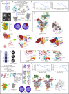
Extended Data Fig. 5. Cryo-EM and image-processing of the pre-B5’ss and pre-B5’ss+ATPγS complexes.
a, RNA composition of pre-B5’ss complexes. Purified pre-B complexes were incubated with an excess of the 5’ss RNA oligonucleotide and subjected to glycerol gradient centrifugation. RNA from the fastest-sedimenting gradient peak was isolated, separated on a NuPAGE gel, and visualized by staining with SyBr gold. For gel source data, see Supplementary Fig. 1. The nucleotide (nts) lengths of the snRNAs and MINX exon RNA are indicated on the right. The RNA composition was analysed from three independent pre-B5’ss purifications with similar results. b, Representative cryo-EM 2D class averages of the pre-B5’ss dimers. c, Cryo-EM computation sorting scheme for pre-B5’ss complexes. All major image-processing steps are depicted. Two major classes of the pre-B5’ss dimers are detected, which are organized in an antiparallel manner and are highly similar to the class 1 and class 2 dimers of the CE pre-B complex (Fig. 1a and Extended Data Fig. 1). In the class 2 dimer, the structure of only one pre-B complex is well-defined, whereas in class 1 both protomers are well-defined. d, Local resolution estimation of the tri-snRNP core region of the pre-B5’ss complex. e, Orientation distribution plot for the particles contributing to the reconstruction of the tri-snRNP core region. f, Fourier shell correlation (FSC) values indicate a resolution of 4.2 Å for the tri-snRNP core in pre-B5’ss. g, Map versus model FSC curves generated for the tri-snRNP core region of pre-B5’ss. h, Comparison of the structure of pre-B5’ss complexes formed by incubating purified pre-B with the 5’ss oligo at 0 °C or 30 °C. An overlay of the EM densities (low-pass filtered to ~20 Å resolution) is shown on the right. Pre-B5’ss complexes with a nearly identical 3D structure were obtained if incubation with the 5’ss oligo was carried out at 0 °C or 30 °C. i, RNA composition of pre-B5’ss+ATPγS complexes. Purified pre-B complexes were incubated with an excess of the 5’ss RNA oligonucleotide followed by ATPγS, and then subjected to glycerol gradient centrifugation. RNA from the fastest-sedimenting gradient peak was isolated, separated on a NuPAGE gel, and visualized by staining with SyBr gold. For gel source data, see Supplementary Fig. 1. The nucleotide (nts) lengths of the snRNAs and MINX exon RNA are indicated on the right. The RNA composition was analysed from two independent pre-B5’ss+ATPγS purifications with similar results. j, Representative cryo-EM 2D class averages of the pre-B5’ss+ATPγS dimers. k, Cryo-EM computation sorting scheme. All major image-processing steps are depicted. l, Local resolution estimation of the tri-snRNP core region of the pre-B5’ss+ATPγS complex. m, Orientation distribution plot for the particles contributing to the reconstruction of the tri-snRNP core region. n, Fourier shell correlation (FSC) values indicate a resolution of 3.1 Å for the tri-snRNP core and 4.0 Å for BRR2. o, Map versus model FSC curves generated for the tri-snRNP core and BRR2 regions of pre-B5’ss+ATPγS. p, Fit of two monomeric pre-B5’ss+ATPγS complex molecular models into the EM density of the pre-B5’ss+ATPγS dimer. As in B-like, the two pre-B5’ss+ATPγS complexes are organized in a parallel manner, with their U5 Sm cores at the bottom. In addition, there is an upper globular density that, like in the B-like dimers, likely contains the exons and associated proteins. However, consistent with the absence of the B-specific proteins, the middle interface present in the B-like dimers that is formed in part by SMU1 and RED, is missing. Furthermore, the lower bridge that contained MFAP1 in B-like complexes, now appears to form mainly between the PRP28 helicase of each protomer. q, Comparison of the structure of complexes formed by incubating purified pre-B with the 5’ss oligo plus ATP (pre-B5’ss+ATP) or ATPγS (pre-B5’ss+ATPγS). An overlay of their EM densities is shown on the right, confirming that they possess a highly similar structure.
Extended Data Fig. 6
Extended Data Fig. 6. Structural changes observed after addition of the 5’ss oligo alone or the 5’ss oligo plus ATPγS.
a, Fit of a single 5’ss oligo to the pre-B5’ss EM density. b, The movement of the PRP8RT/En domain toward the PRP8NTD upon addition of the 5’ss oligo leads to concomitant movements of other tri-snRNP components that interact with PRP8RT/En. Comparison of the molecular architecture of BRR2, PRP8, SAD1 and the U4 Sm core domain in pre-B (left) and pre-B5’ss complexes (middle). Right, overlay of the corresponding surface representations in pre-B (grey) versus pre-B5’ss (various colors), with arrows indicating the concomitant movements of the indicated domains with PRP8RT/En. These movements do not involve any major structural changes such as translocation of BRR2 to its activation position. c,d, Comparison of the molecular architecture of PRP4 kinase, SF3B3, PRP6HAT, SNU66 and the U4 Sm core domain in pre-B (panel c, left) and pre-B5’ss complexes (panel d, left). The boxed regions are expanded and rotated 90° in the corresponding panels shown at the right. Aligned via the PRP8RT/En domain. In pre-B5’ss, PRP4K and SF3B3WD40B are located further apart, and PRP4K is more closely associated with PRP6HAT. Moreover, the U4 Sm core has moved away from PRP8RH in pre-B5’ss (as indicated by the arrow in panel c). e, Overlay of pre-B and pre-B5’ss showing differences in the position of PRP4K. f, In pre-B5’ss, BRR2PWI is detached from SAD1. A comparison of the molecular architecture of SAD1, BRR2CC and BRR2PWI in pre-B (panel f, left) and pre-B5’ss complexes (panel f, middle). An overlay of the structures is shown in panel f (right); alignment on PRP8NTD. g, Interactions of the 5’ss-containing RNA oligonucleotide (5’ss oligo) with U5 snRNA loop 1 nucleotides, and residues of PRP8 and DIM1 in pre-B5’ss+ATPγS. h, Fit of the nucleotides and protein side chains shown in panel g into the pre-B5’ss+ATPγS EM density. i, Rearrangements in the NOP and adjacent coiled-coil domains of PRP31 (aa 52-331) and repositioning of the PRP6 HAT domain and its more N-terminal phosphorylated region upon addition of ATP to pre-B5’ss complexes. The coiled/coil (CC) domains of PRP31 rotate by ca 45° relative to the PRP31 NOP domain after addition of ATP. A comparison of the molecular architecture of the indicated proteins/protein domains in pre-B5’ss (left), pre-B5’ss+ATPγS (middle) and B-like complexes (right). Serine and threonine residues of PRP6 that could be modelled previously, and that were previously shown to be phosphorylated by PRP4K are indicated by green stars. The region of PRP31 that is known to be phosphorylated could not be modelled. j, ATP addition to pre-B5’ss leads to the ca 180° rotation of PRP8RH, which is coordinated with the repositioning of BRR2/PRP8Jab1, PRP6 and SNU66, among others. The β-hairpin loop of PRP8RH is indicated in yellow and by an arrowhead. The SNU66 domain comprised of aa 252-358 is structured first in pre-B5’ss+ATPγS where it is docked to the β-hairpin loop region of the repositioned PRP8RH domain (compare left and middle panels). The C-terminal SNU66630-774 domain, which binds across SNRP27K and the U4 Sm core in pre-B5’ss, is also repositioned in pre-B5’ss+ATPγS.
Extended Data Fig. 7
Extended Data Fig. 7. Structural differences between pre-B5’ss, pre-B5’ss+ATPγS and B-like complexes.
a, BRR2 is anchored to its new position in pre-B5’ss+ATPγS (top) and the B-like complex (bottom) via the PRP8RH-PRP8Jab1- linker whose ends are now stably bound to PRP8En. The boxed regions are shown in expanded form at the right. Jab1 linker, the region of the RH-Jab1 linker proximal to the PRP8 Jab1 domain. RH linker, the region of the RH-Jab1 linker proximal to the PRP8 RH domain. b, Interaction of SNRNP27K with PRP8En in the pre-B (top) and pre-B5’ss (bottom) complexes. c, Rotation of PRP8Jab1 around the RH-Jab1 linker is required to reach its B-like position. An overlay shows the position of the Jab1 domain in pre-B5’ss+ATPγS (purple) and B-like (grey). PRP8Jab1 appears to rotate around the amino acids of the RH-Jab1 liker directly upstream of the Jab1 domain, which may act as a hinge. d, Two different views of an overlay of BRR2 in pre-B5’ss+ATPγS (multi-colored) and B-like (grey), showing that BRR2 in pre-B5’ss+ATPγS must still rotate in order to reach its position in the B-like complex. e, Stepwise opening of the BBR2NC. In pre-B and pre-B5’ss (left panel), the opening of the RNA binding channel is blocked by the BRR2 PLUG domain and C-terminal (CT) tail of the PRP8 Jab1 domain. In pre-B5’ss+ATPγS, pre-B5’ssLNG+ATPγS and the B-like complex (middle and right panels), the BRR2PLUG and PRP8 Jab1 CT tail are displaced. However, in pre-B5’ss+ATPγS, pre-B5’ssLNG+ATPγS (middle left panel), the separator loop (marked by a red arrowhead) of the RecA2 domain contacts an α-helix of the Sec63 domain (blue arrowhead), blocking the RNA binding channel. Thus, in the absence of the B-specific proteins, a new intermediate BRR2NC state (i.e., with a free RNA channel entry site but a blocked RNA channel) is observed. Due to the presence of the U4 Sm domain, U4 snRNA cannot be threaded into the RecA domains. Instead, the latter must to be opened to allow U4 snRNA binding. Downward movement of the RecA1 and RecA2 domains of BRR2NC and the concomitant movement of the separator loop, positions it further away from the BRR2NC Sec63 domain, leading to an open RNA binding channel (middle right panel), allowing the binding of the single-stranded region of U4 snRNA (right panel) (See also Supplementary Video 6). f, PRP6 and PRP31 are phosphorylated in pre-BATP and pre-B5’ss+ATP complexes. Proteins were isolated from the indicated complexes and the phosphorylation status of PRP6 and PRP31 determined by immunoblotting with antibodies specific for phosphorylated PRP6 or PRP31. The less intense signals in the B complex lane obtained with the anti-phosphorylated PRP6 and PRP31 antibodies is due to the fact that these proteins are thiophosphorylated in the B complexes, which were isolated in the presence of ATPγS, and thiophosphorylated proteins are poorly recognized by the anti-phospho antibodies. For blot source data, see Supplementary Fig. 1. Similar blot results where obtained with two independent experiments. g, Comparison of the position of BRR2 and the U4 Sm core in pre-B5’ss+ATPγS (left) compared to the B-like complex (right). The structures are aligned via the PRP8NTD. In pre-B5’ss+ATPγS, the BRR2 helicase domain has not reached its final position. In addition, BRR2NC exhibits a closed, inactive conformation and has not yet bound the U4 snRNA, and the U4 Sm core domain has also not reached its final, B-like position. Although BRR2’s PLUG and PWI domains are not visible, the N-terminal ca 50 amino acids of BRR2 still wrap around PRP8Large at the same position as in the pre-B5’ss complex. These data strongly support the idea that during the translocation of BRR2’s helicase domain across the large domain of PRP8, BRR2 does not dissociate and subsequently reassociate, but rather remains attached via its N-terminal region to the spliceosome. h, BRR2 is translocated, but the U4 Sm core is not docked to BRR2 in pre-B5’ss+ATPγS. Right, zoomed-in view of the boxed region. U6-C37 is inserted into a protein pocket formed by SF3A1 aa 496-521 and PRP6, stabilizing the new position of U4/U6 stem III. Tethering of U6-C37 by SF3A1 and PRP6, may help to stabilize the short U6/5’ss helix after release of RBM42, which appears to contact U6 near C37, and after the repositioning of U4/U6 helix III, which prior to its movement likely constrains the position of U6 nucleotides in this region. In addition, capture of this U6 nucleotide fixes the path of the adjacent U6 snRNA region, which may facilitate the subsequent formation of the extended U6/5’ss helix upon disruption of U4/U6 stem III and release of U6-C37. i, The U4 Sm core contacts BRR2 in pre-B5’ssLNG+ATPγS, and U4/U6 stem III is dissociated by binding of the long 5’ss oligo to U6 snRNA. Right, zoomed-in view of the boxed region. An extended (ext) U6/5’ss helix is formed, and aa 496-521 of SF3A1 are destabilized.
Extended Data Fig. 8
Extended Data Fig. 8. Cryo-EM of the pre-B5’ssLNG+ATPγS and pre-BATP complexes and structural comparisons with the B-like complex.
a, RNA composition of pre-B5’ssLNG+ATPγS complexes. Purified pre-B complexes were incubated with an excess of the long 5’ss RNA oligonucleotide followed by ATPγS, and then subjected to glycerol gradient centrifugation. RNA from the fastest-sedimenting gradient peak was isolated, separated on a NuPAGE gel, and visualized by staining with SyBr gold. For gel source data, see Supplementary Fig. 1. The nucleotide (nts) lengths of the snRNAs and MINX exon RNA are indicated on the right. The RNA composition was analysed twice from the same pre-B5’ssLNG+ATPγS purification with similar results. b, Representative cryo-EM 2D class averages of the pre-B5’ssLNG+ATPγS dimers. All “good” particles show dimerized complexes, in which both protomers are well-defined and aligned in a parallel manner. c, Cryo-EM computation sorting scheme. d, Local resolution estimation of the tri-snRNP core region of the pre-B5’ssLNG+ATPγS complex. e, Orientation distribution plot for the particles contributing to the reconstruction of the tri-snRNP core region. f, Fourier shell correlation (FSC) values indicate a resolution of 3.7 Å for the tri-snRNP core of pre-B5’ssLNG+ATPγS. g, Map versus model FSC curves generated for the tri-snRNP core region of pre-B5’ssLNG+ATPγS. h, Molecular architecture of the pre-B5’ssLNG+ATPγS complex. i, Overlay of BRR2 in pre-B5’ssLNG+ATPγS (grey) and pre-B5’ss+ATPγS (brown), revealing that the position of the BRR2 helicase domain does not change substantially upon addition of the long 5’ss oligonucleotide. Thus, the BRR2 helicase domain must still be rotated to reach its B-like position, even though the U4/U6 helix III is dissolved and an extended U6/5’ss helix has formed. Aligned via PRP8NTD. j, Cartoon showing that the movement and opening of BRR2 are facilitated by SNU23 loop 62-74 and MFAP1 helix 215-255, which are missing in pre-B5’ssLNG+ATPγS but bind in B-like to the BRR2NC Sec63 domain, and also by the binding of FBP21’s long α-helix and SNU23’s α-helix 18-36, which interact both with RecA2 of BRR2NC. SNU23 also tethers BRR2NC to the PRP8NTD, bridging it to the U6/5’ss helix. k, Rotation of BRR2 towards PRP8NTD is likely further facilitated by the SMU1/RED complex which stabilizes the new position of BRR2 in the B-like complex. l-n, Zoomed-in view of the structure of the PRP8 RH-Jab1 linker (designated Jab1Linker) in pre-B5’ssLNG+ATPγS (l) versus B-like (m) complexes, and the interaction of SNU23 with BRR2SEC63 and the Jab1Linker, leading to the stabilization of a hairpin structure (aa 2037-2058) in the linker in B-like. n, An α-helix of RecA2 of the BRR2NC is contacted by both SNU23 α-helix 18-36 and by the long α-helix of FBP21 in B-like. Most of the binding sites of the B-specific proteins are created during or after the translocation of BRR2 to its pre-activation position. For example, MFAP1 and SNU23 are able to interact with BRR2 helicase elements first after BRR2 translocation to its pre-final site. In addition, a binding site for FBP21 is generated by PPIH and an N-terminal region of PRP4 first upon their stabilization during BRR2 translocation/remodeling. o, RNA composition of pre-BATP complexes. As in all other complex purifications, described above, the MINX exon RNA remains intact during incubation of pre-B complexes with ATP. Affinity-purified pre-B complexes were incubated with ATP and then subjected to glycerol gradient centrifugation. RNA from the fastest-sedimenting gradient peak was isolated, and separated on a NUPAGE gel and visualized by staining with Sybr gold. For gel source data, see Supplementary Fig. 1. The nucleotide (nts) lengths of the snRNAs and MINX exon RNA are indicated on the right. The RNA composition was analysed from two independent pre-BATP purifications with similar results. p, Representative cryo-EM 2D class averages of the pre-BATP dimers. q, Cryo-EM computation sorting scheme. r, Local resolution estimation of the tri-snRNP core region of the pre-BATP complex. s, Orientation distribution plot for the particles contributing to the reconstruction of the tri-snRNP core region. t, Fourier shell correlation (FSC) values indicate a resolution of 3.7 Å for the tri-snRNP core of pre-BATP. u, Map versus model FSC curves generated for the tri-snRNP core region of pre-BATP. v, The molecular architecture of the pre-B5’ss+ATPγS (left) and the pre-BATP (right) complex is very similar. One notable exception is that the PRP28 RecA domains are destabilized in pre-BATP, consistent with PRP28 action. In pre-B5’ss+ATPγS, a previously unknown intermediate, spliceosome assembly state in which the U4 Sm core and U4/U6 stem III have moved about 5 nm downwards from their location in pre-B is observed. This intermediate position of U4/U6 stem III is not only observed in pre-B5’ss+ATPγS complexes, but also in pre-BATP complexes, where it is also stabilized by an interaction between U6-C37 and SF3A1 and PRP6NTR. This suggests that the transient docking of stem III at this position also occurs when the U6/5’ss helix is formed by the PRP28-mediated interaction of the U6 ACAGA box with a bona fide 5’ss. w, Fit of the PRP28NTD into the pre-BATP EM density (expanded view of the boxed region in panel h). In pre-BATP aa 273 to 327 of the PRP28NTD remain associated with PRP8NTD presumably due to the absence of MFAP1 whose CTR binds in a mutually exclusive manner with the PRP28NTD.
Extended Data Fig. 9
Extended Data Fig. 9. Cryo-EM and image-processing of the pre-BAMPPNP complex, and localization of the U1 snRNP.
a, Cartoon showing organization of the protomers in the pre-BATP dimers (left). Right, Schematic of the RNA-RNA network in the pre-BATP dimer. The protomers in the pre-B (Fig. 1a) and pre-BATP dimers, are organized in an anti-parallel manner, in contrast to the parallel organization of the protomers in the pre-B5’ss+ATPγS dimers (Extended Data Fig. 5p). Addition of ATP and the 5’ss oligo, thus not only triggers rearrangements within the protomers, foremost the large-scale translocation of BRR2, but also a reorganization of the protomers relative to each other from an anti-parallel orientation to a parallel one. Although the mechanism for this reorganization is not clear, in particular whether it involves partial or complete detachment of the protomers comprising the dimer, it would in both cases likely require the dissociation of protein-protein contacts involving the PRP28 region of the tri-snRNP of one protomer and components of the globular domain encompassing the exon of the adjacent protomer, such as SR proteins and U1 snRNP. During formation of pre-B5’ss+ATPγS, the 5’ss oligo disrupts the U1 base pairing interaction with the 5’ss of the MINX exon RNA and at the same time prevents U6 from interacting with the latter, freeing up the MINX exon RNA. In contrast, in pre-BATP, formation of the base pairing interaction of U6 with the 5’ss of the MINX exon RNA would stabilize the anti-parallel orientation and thus hinder the change in polarity of the dimer during the BRR2 translocation events in the two pre-B protomers. b, RNA composition of pre-BAMPPNP complexes. Purified pre-B complexes were incubated with the non-hydrolyzable ATP analog AMPPNP, and then subjected to glycerol gradient centrifugation. RNA from the fastest-sedimenting gradient peak was isolated, separated on a NuPAGE gel, and visualized by staining with SyBr gold. For gel source data, see Supplementary Fig. 1. The nucleotide (nts) lengths of the snRNAs and MINX exon RNA are indicated on the right. The RNA composition was analysed from two independent pre-BAMPPNP purifications with similar results. c, Representative cryo-EM 2D class averages of the pre-BAMPPNP dimers. d, Cryo-EM computation sorting scheme of the pre-BAMPPNP. e, Local resolution estimation of the tri-snRNP core region of the pre-BAMPPNP complex. f, Orientation distribution plot for the particles contributing to the reconstruction of the tri-snRNP region in of the pre-BAMPPNP. g, Fourier shell correlation (FSC) values indicate a resolution of 4.1 Å for the tri-snRNP core and 6.1 Å for the tri-snRNP plus adjacent U1 snRNP. h, Map versus model FSC curves generated for the tri-snRNP core and tri-snRNP plus adjacent U1 snRNP regions of pre-BAMPPNP. i, Fit into the EM density (low-pass filtered to ~20 Å resolution) of the molecular model of a pre-BAMPPNP complex monomer interacting in trans with a U1 snRNP bound to a second MINX exon RNA. On the right, the molecular architecture of the U1 snRNP and adjacent BRR2 and PRP28 proteins without EM density, as well as a cartoon of the unassigned EM density adjacent to U1, is shown. In pre-BAMPPNP, U1 snRNA stem-loop III is located close to PRP8En of the adjacent protomer, and the U1 Sm domain contacts the PRP28 RecA1 domain. j, Fit into the EM density (low-pass filtered to ~20 Å resolution) of the molecular model of a pre-B complex monomer interacting in trans with PRP28 from the adjacent pre-B monomer. On the right, the molecular architecture of BRR2 and PRP28 without EM density, as well as a cartoon of the unassigned EM density that likely contains the U1 snRNP, is shown. k, Close up of PRP28 docked to U1 of the adjacent monomer in the pre-BAMPPNP dimer. A molecular model of PRP28 with a closed conformation and bound with a single-stranded RNA (based on the crystal structure of Mss116p) can be fit into the pre-BAMPPNP EM density. In pre-BAMPPNP, there is density between the two PRP28 RecA domains that appears to accommodate a single-stranded (ssRNA) but not double stranded RNA. Given this is indeed the case, our data would thus suggest that in the presence of AMPPNP, PRP28 has disrupted the U1/5’ss helix of the adjacent protomer, but due to the lack of ATP hydrolysis, its RecA domains are trapped in a closed conformation. l, EM density fit of PRP28 with an open conformation in the corresponding region of the cross-exon pre-B complex. In the CE pre-B complex, PRP28 adopts an open conformation, and there is no stable docking of U1. m-o, The PRP28 RecA domains undergo a major conformational change. Spatial organization of the PRP28 RecA domains in the closed (m) and open (n) conformations, with an overlay shown in panel o. The open conformation of a DEAD-box helicase such as PRP28 is its default state when it is not bound to a substrate. When the helicase encounters a double-helical RNA in the presence of ATP, it transitions to the closed conformation, where the subsequent closure of the two RecA domains physically separates the two strands, resulting in closed RecA domains bound to a single-stranded RNA (ssRNA). p, Fit of the U6/5’ss helix into the EM density of pre-BATP. q, A U6/5’ss helix is not formed in pre-BAMPPNP. EM density that accommodates a U6/5’ss helix (as seen in panel p) is absent in pre-BAMPPNP, confirming that this helix does not form.

References

    1. Kastner, B., Will, C. L., Stark, H. & Lührmann, R. Structural insights into nuclear pre-mRNA splicing in higher eukaryotes. Cold Spring Harb. Perspect. Biol.10.1101/cshperspect.a032417 (2019). - PMC - PubMed
    1. Robberson BL, Cote GJ, Berget SM. Exon definition may facilitate splice site selection in RNAs with multiple exons. Mol. Cell. Biol. 1990;10:84–94. - PMC - PubMed
    1. Berget SM. Exon recognition in vertebrate splicing. J. Biol. Chem. 1995;270:2411–2414. - PubMed
    1. Black DL. Mechanisms of alternative pre-messenger RNA splicing. Annu. Rev. Biochem. 2003;72:291–336. - PubMed
    1. De Conti L, Baralle M, Buratti E. Exon and intron definition in pre-mRNA splicing. Wiley Interdiscip. Rev. RNA. 2013;4:49–60. - PubMed

MeSH terms

Substances