

Optic disc drusen: Dystrophic calcification, a potential target for treatment
- PMID: 38778137
- PMCID: PMC11306397
- DOI: 10.1038/s41433-024-03138-6
Optic disc drusen: Dystrophic calcification, a potential target for treatment
Abstract
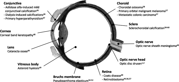
Optic disc drusen (ODD) are calcified, acellular bodies, seen in the optic nerve head of up to 2% of the population. Although seldomly affecting visual acuity, visual field defects are common, and severe, ischemic complications causing irreversible vision loss are known to occur. Different treatment strategies for ODD have been explored, but so far without success. This review focuses on the unique, calcified property of ODD, describing what we know about ODD pathogenesis and previously tried treatment strategies. In this context, we discuss current knowledge about calcium and pathological calcifications, including intracranial and ocular calcifications. We also explore some of the obstacles that must be addressed to develop a therapy centred on the concept of calcification, should calcification be identified as a pathogenic factor contributing to vision loss.
摘要: 视盘玻璃膜疣(ODD)是钙化的无细胞体, 高达2%的人的视盘可见该结构。虽极少影响视力, 但常引起视野缺损, 并且严重的缺血性并发症会导致不可逆的视力损伤。针对ODD的不同治疗策略一直在探索中, 但迄今为止尚未成功。本文聚焦于ODD独特的钙化特性, 阐述了我们对ODD发病机制的认识以及曾尝试过的治疗方法。在此背景下, 我们讨论了有关钙和病理性钙化的现有知识, 其中包括颅内和眼部钙化。我们还探讨了一些亟需解决的问题, 用以开发以钙化为核心的治疗方法, 以及钙化是否应被确定为导致视力损失的致病因素。.
© 2024. The Author(s), under exclusive licence to The Royal College of Ophthalmologists.
Conflict of interest statement
The authors declare no competing interests.
Figures
References
-
- Lorentzen S. Drusen of the optic disk. A clinical and genetic study. Acta Ophthalmol. 1966;90:1–180. - PubMed
Publication types
MeSH terms
LinkOut - more resources
Full Text Sources
