. 2024 May 22;21(1):26.
doi: 10.1186/s12989-024-00585-7.

Combining analytical techniques to assess the translocation of diesel particles across an alveolar tissue barrier in vitro

Affiliations

Combining analytical techniques to assess the translocation of diesel particles across an alveolar tissue barrier in vitro

Gowsinth Gunasingam et al. Part Fibre Toxicol. .

Erratum in

Abstract

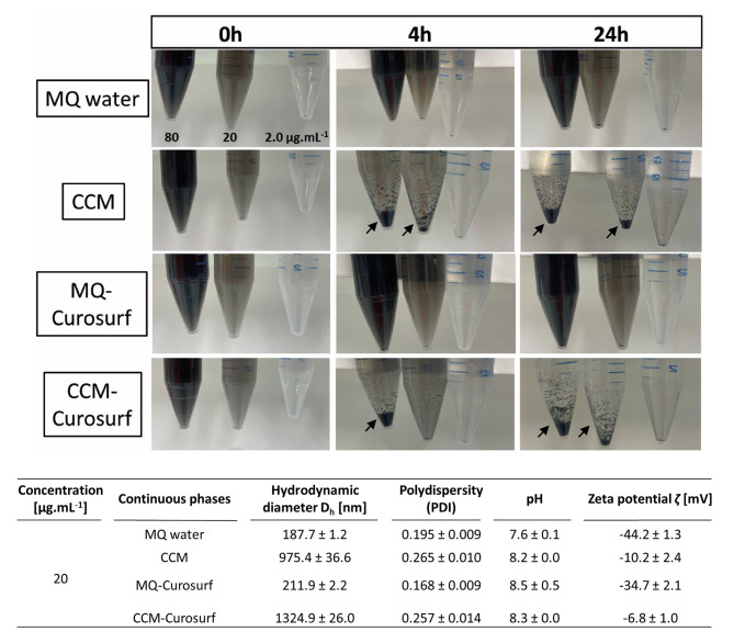
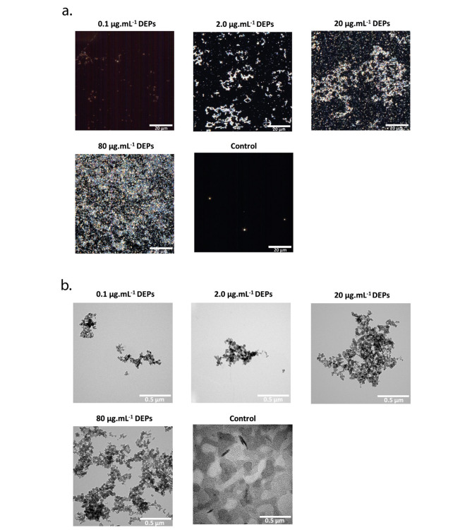
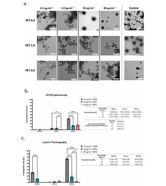
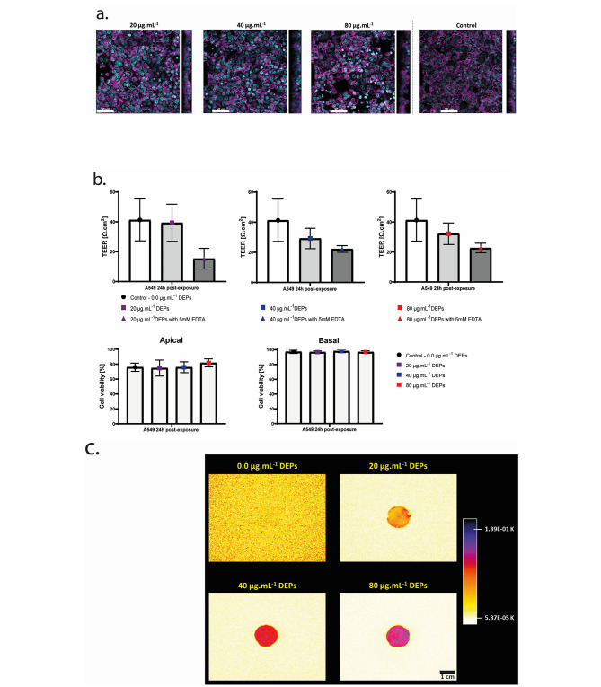
Background: During inhalation, airborne particles such as particulate matter ≤ 2.5 μm (PM2.5), can deposit and accumulate on the alveolar epithelial tissue. In vivo studies have shown that fractions of PM2.5 can cross the alveolar epithelium to blood circulation, reaching secondary organs beyond the lungs. However, approaches to quantify the translocation of particles across the alveolar epithelium in vivo and in vitro are still not well established. In this study, methods to assess the translocation of standard diesel exhaust particles (DEPs) across permeable polyethylene terephthalate (PET) inserts at 0.4, 1, and 3 μm pore sizes were first optimized with transmission electron microscopy (TEM), ultraviolet-visible spectroscopy (UV-VIS), and lock-in thermography (LIT), which were then applied to study the translocation of DEPs across human alveolar epithelial type II (A549) cells. A549 cells that grew on the membrane (pore size: 3 μm) in inserts were exposed to DEPs at different concentrations from 0 to 80 µg.mL- 1 ( 0 to 44 µg.cm- 2) for 24 h. After exposure, the basal fraction was collected and then analyzed by combining qualitative (TEM) and quantitative (UV-VIS and LIT) techniques to assess the translocated fraction of the DEPs across the alveolar epithelium in vitro.

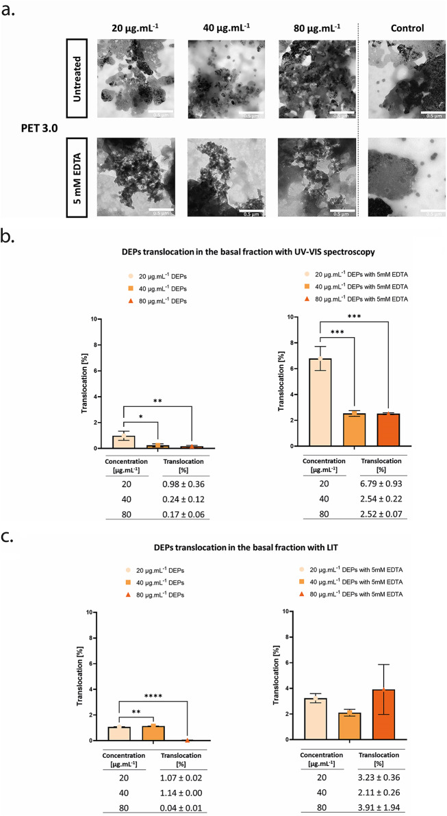
Results: We could detect the translocated fraction of DEPs across the PET membranes with 3 μm pore sizes and without cells by TEM analysis, and determine the percentage of translocation at approximatively 37% by UV-VIS (LOD: 1.92 µg.mL- 1) and 75% by LIT (LOD: 0.20 µg.cm- 2). In the presence of cells, the percentage of DEPs translocation across the alveolar tissue was determined around 1% at 20 and 40 µg.mL- 1 (11 and 22 µg.cm- 2), and no particles were detected at higher and lower concentrations. Interestingly, simultaneous exposure of A549 cells to DEPs and EDTA can increase the translocation of DEPs in the basal fraction.

Conclusion: We propose a combination of analytical techniques to assess the translocation of DEPs across lung tissues. Our results reveal a low percentage of translocation of DEPs across alveolar epithelial tissue in vitro and they correspond to in vivo findings. The combination approach can be applied to any traffic-generated particles, thus enabling us to understand their involvement in public health.

Keywords: A549 cells; Diesel particles (DEPs); Lock-in thermography (LIT); Translocation; Transmission electron microscopy (TEM); Ultraviolet – visible (UV-VIS).


Conflict of interest statement

The authors declare no competing interests.

Figures

Fig. 1
Fig. 1
Characterization of diesel exhaust particles (DEPs, SRM2975) in water-based dispersion and in cell culture medium. DEPs dispersion in Milli Q (MQ) water, MQ water containing surfactant (MQ-Curosurf), cell culture medium (CCM) and CCM containing surfactant (CCM-Curosurf) at 80, 20 and 2.0 µg. mL-1 at 37 °C over time. Agglomerates are indicated with arrows. The physical characteristics including hydrodynamic diameter (Dh), polydispersity, zeta potential (ζ) and pH of 20 µg. mL-1 DEPs in MQ water, MQ-Curosurf, CCM and CCM-Curosurf at 0 h, 37 °C are displayed in the table. The means ± SD from three independent batches were used as technical replicates (n = 3). * represents (p < 0.05), ** (p < 0.01), *** (p < 0.001), **** (p < 0.0001)
Fig. 2
Fig. 2
Visualization of dried DEPs from MQ-Curosurf solution at 0.1, 2.0, 20, and 80 µg.mL-1. a. Enhanced darkfield hyperspectral imaging (EDF-HSI), scale bar: 20 μm, and b. Transmission electron microscope (TEM). The control corresponds to the MQ-Curosurf solution without DEPs, scale bar: 0.5 μm
Fig. 3
Fig. 3
Visualization and quantification of translocated DEPs into the basal fraction across porous PET membranes. (a) TEM images of the translocated DEPs across 0.4, 1.0, and 3.0 μm porous membrane collected from the basal cell culture medium. The applied concentrations of the DEPs on the apical side of the membrane are 0, 0.1, 2.0, 20, and 80 µg. mL-1 in MQ-Curosurf solution, scale bar: 0.5 μm. (b) and (c) Percentage of translocation of DEPs across 0.4, 1.0, and 3.0 μm porous membrane determined with ultraviolet-visible (UV-VIS) absorbance spectroscopy (b) and lock-in thermography (LIT) (c). The means ± SD from three independent experiments were used as technical replicates (n = 3). ** represents (p < 0.01), *** (p < 0.001), **** (p < 0.0001)
Fig. 4
Fig. 4
Effects of deposited DEPs on A549 alveolar cells at 24 h post-exposure. a. 3D – confocal laser scanning images of deposited DEPs at 20, 40, and 80 µg.mL-1 on the A549 monolayer, scale bar: 50 μm. Cell nuclei and F-actin cytoskeleton are stained with DAPI (cyan) and Phalloidin (magenta), respectively. b. A549 monolayer integrity is assessed with transepithelial electrical resistance (TEER) measurement and 5 mM of ethylenediaminetetraacetic acid (EDTA) as a positive control. The viability of the A549 cells is evaluated with the release rate of cytosolic lactate dehydrogenase (LDH) in the apical and basal fractions. The means ± SD of three independent experiments performed in duplicates are displayed (N = 3). c. Thermal emission of deposited DEPs at 20, 40, and 80 µg.mL-1 on the A549 monolayer determined with the LIT, scale bar: 1 cm. * represents (p < 0.05), ** (p < 0.01), *** (p < 0.001), **** (p < 0.0001)
Fig. 5
Fig. 5
Visualization and quantification of translocated DEPs across the alveolar epithelial type II (A549) cells grown on 3.0 μm porous PET membrane into the basal fraction. (a) TEM images of the translocated DEPs across the A549 monolayer in the basal fraction. The applied concentrations on the apical side of the membrane are 20, 40 and 80 µg. mL-1 in CCM-Curosurf solution, with and without 5mM EDTA treatment, scale bar: 0.5 μm. (b) and (c) The percentage of translocation of DEPs across the A549 monolayer was determined with UV-VIS absorbance spectroscopy (b) and LIT (c) techniques. The means ± SD of three independent experiments performed in duplicates are displayed (N = 3)

References

    1. Landrigan PJ, Fuller R, Acosta NJR, Adeyi O, Arnold R, Basu N (Nil), editors. The Lancet Commission on pollution and health. The Lancet. 2018;391(10119):462–512. - PubMed
    1. Liu C, Chen R, Sera F, Vicedo-Cabrera AM, Guo Y, Tong S, et al. Ambient Particulate Air Pollution and Daily Mortality in 652 cities. N Engl J Med. 2019;381(8):705–15. 10.1056/NEJMoa1817364 - DOI - PMC - PubMed
    1. Kelly FJ, Fussell JC. Toxicity of airborne particles—established evidence, knowledge gaps and emerging areas of importance. Philos Trans R Soc Math Phys Eng Sci. 2020;378(2183):20190322. - PMC - PubMed
    1. Kim HB, Shim JY, Park B, Lee YJ. Long-term exposure to Air pollutants and Cancer Mortality: a Meta-analysis of Cohort studies. Int J Environ Res Public Health. 2018;15(11):2608. 10.3390/ijerph15112608 - DOI - PMC - PubMed
    1. Loomis D, Grosse Y, Lauby-Secretan B, Ghissassi FE, Bouvard V, Benbrahim-Tallaa L, et al. The carcinogenicity of outdoor air pollution. Lancet Oncol. 2013;14(13):1262–3. 10.1016/S1470-2045(13)70487-X - DOI - PubMed

Publication types

MeSH terms

LinkOut - more resources