

Cardiovascular disease risk exacerbates brain aging among Hispanic/Latino adults in the SOL-INCA-MRI Study
- PMID: 38778863
- PMCID: PMC11110680
- DOI: 10.3389/fnagi.2024.1390200
Cardiovascular disease risk exacerbates brain aging among Hispanic/Latino adults in the SOL-INCA-MRI Study
Abstract
Background: Cardiovascular disease (CVD) risk factors are highly prevalent among Hispanic/Latino adults, while the prevalence of MRI infarcts is not well-documented. We, therefore, sought to examine the relationships between CVD risk factors and infarcts with brain structure among Hispanic/Latino individuals.
Methods: Participants included 1,886 Hispanic/Latino adults (50-85 years) who underwent magnetic resonance imaging (MRI) as part of the Study of Latinos-Investigation of Neurocognitive Aging-MRI (SOL-INCA-MRI) study. CVD risk was measured approximately 10.5 years before MRI using the Framingham cardiovascular risk score, a measure of 10-year CVD risk (low (<10%), medium (10- < 20%), and high (≥20%)). MR infarcts were determined as present or absent. Outcomes included total brain, cerebral and lobar cortical gray matter, hippocampal, lateral ventricle, and total white matter hyperintensity (WMH) volumes. Linear regression models tested associations between CVD risk and infarct with MRI outcomes and for modifications by age and sex.
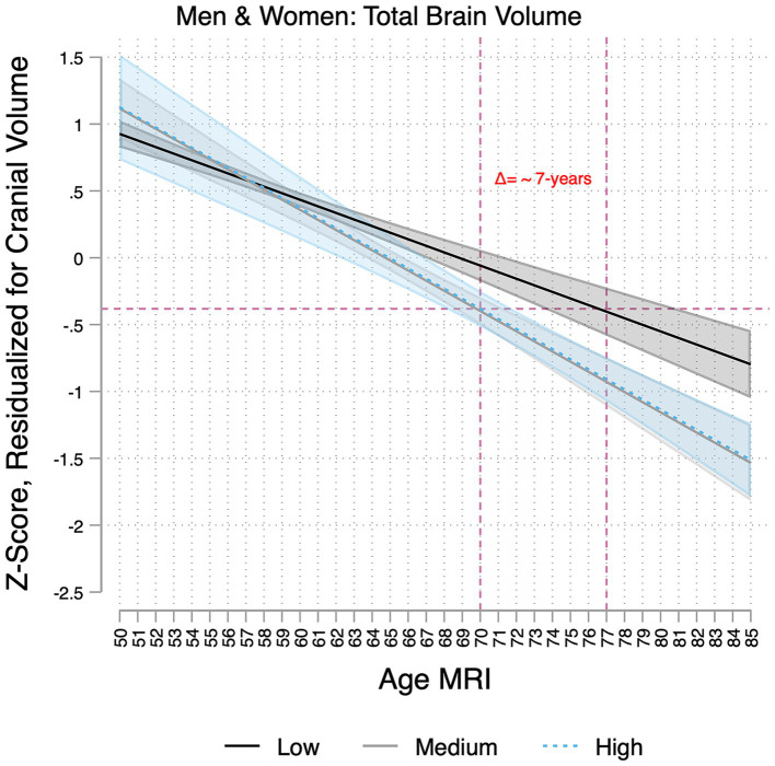
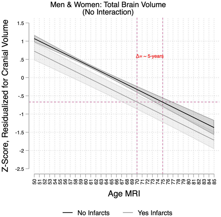
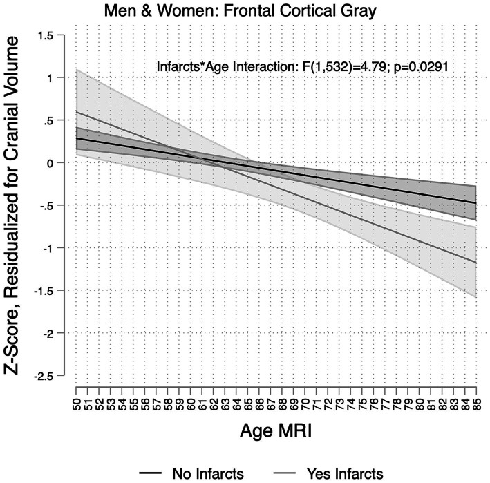
Results: Sixty percent of participants were at medium or high CVD risk. Medium and high CVD risk were associated with lower total brain and frontal gray matter and higher WMH volumes compared to those with low CVD risk. High CVD risk was additionally associated with lower total cortical gray matter and parietal volumes and larger lateral ventricle volumes. Men tended to have greater CVDRF-related differences in total brain volumes than women. The association of CVD risk factors on total brain volumes increased with age, equal to an approximate 7-year increase in total brain aging among the high-CVD-risk group compared to the low-risk group. The presence of infarct(s) was associated with lower total brain volumes, which was equal to an approximate 5-year increase in brain aging compared to individuals without infarcts. Infarcts were also associated with smaller total cortical gray matter, frontal and parietal volumes, and larger lateral ventricle and WMH volumes.
Conclusion: The high prevalence of CVD risk among Hispanic/Latino adults may be associated with accelerated brain aging.
Keywords: Hispanic/Latino heritage; brain aging; brain volumes; cardiovascular disease risk; infarcts.
Copyright © 2024 Stickel, Tarraf, Gonzalez, Paredes, Zeng, Cai, Isasi, Kaplan, Lipton, Daviglus, Testai, Lamar, Gallo, Talavera, Gellman, Ramos, Ivanovic, Seiler, González and DeCarli.
Conflict of interest statement
The authors declare that the research was conducted in the absence of any commercial or financial relationships that could be construed as a potential conflict of interest.
Figures
Similar articles
-
Characterizing age- and sex-related differences in brain structure among middle-aged and older Hispanic/Latino adults in the study of Latinos- investigation of neurocognitive aging magnetic resonance imaging (SOL-INCA MRI).Neurobiol Aging. 2023 Jun;126:58-66. doi: 10.1016/j.neurobiolaging.2023.02.007. Epub 2023 Feb 23. Neurobiol Aging. 2023. PMID: 36933278 Free PMC article.
-
Multimodal Associations of Modifiable Risk Factors on White Matter Injury: The SOL-INCA-MRI Study (HCHS/SOL).Stroke. 2025 May;56(5):1138-1148. doi: 10.1161/STROKEAHA.124.049904. Epub 2025 Mar 24. Stroke. 2025. PMID: 40123500 Free PMC article.
-
Sleep duration and brain MRI measures: Results from the SOL-INCA MRI study.Alzheimers Dement. 2024 Jan;20(1):641-651. doi: 10.1002/alz.13451. Epub 2023 Sep 29. Alzheimers Dement. 2024. PMID: 37772658 Free PMC article.
-
Measures of brain morphology and infarction in the framingham heart study: establishing what is normal.Neurobiol Aging. 2005 Apr;26(4):491-510. doi: 10.1016/j.neurobiolaging.2004.05.004. Neurobiol Aging. 2005. PMID: 15653178
-
Cardiovascular disease risk factors in the Hispanic/Latino population: lessons from the Hispanic Community Health Study/Study of Latinos (HCHS/SOL).Prog Cardiovasc Dis. 2014 Nov-Dec;57(3):230-6. doi: 10.1016/j.pcad.2014.07.006. Epub 2014 Aug 2. Prog Cardiovasc Dis. 2014. PMID: 25242694 Review.
Cited by
-
Association of Lp(a) With Stroke and Cerebral Injury on MRI: Insights From the HCHS/SOL (Hispanic Community Health Study/Study of Latinos) and Investigation of Neurocognitive Aging MRI (SOL-INCA MRI).Stroke. 2025 Jun;56(6):1492-1504. doi: 10.1161/STROKEAHA.124.048439. Epub 2025 Apr 1. Stroke. 2025. PMID: 40166810
-
Cardiovascular Risk Predicts White Matter Hyperintensities, Brain Atrophy and Treatment Resistance in Major Depressive Disorder: Role of Genetic Liability.Acta Psychiatr Scand. 2025 Jun;151(6):709-718. doi: 10.1111/acps.13793. Epub 2025 Feb 27. Acta Psychiatr Scand. 2025. PMID: 40014927 Free PMC article.
References
-
- Aggarwal N. T., Wilson R. S., Bienias J. L., De Jager P. L., Bennett D. A., Evans D. A., et al. . (2010). The association of magnetic resonance imaging measures with cognitive function in a biracial population sample. Arch. Neurol. 67, 475–482. doi: 10.1001/archneurol.2010.42, PMID: - DOI - PMC - PubMed