Review
. 2024 May 8:14:1391293.
doi: 10.3389/fonc.2024.1391293. eCollection 2024.

Current status, challenges and prospects of antifouling materials for oncology applications

Affiliations
Review

Current status, challenges and prospects of antifouling materials for oncology applications

Yingfeng Zhang et al. Front Oncol. .

Abstract

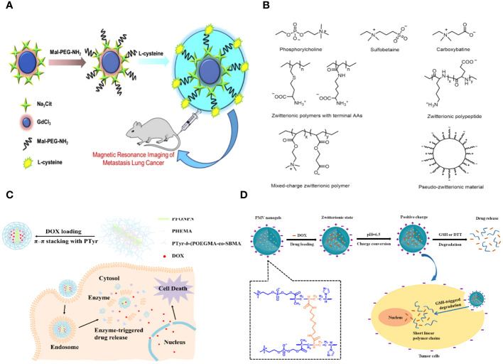
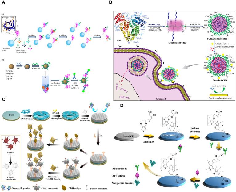
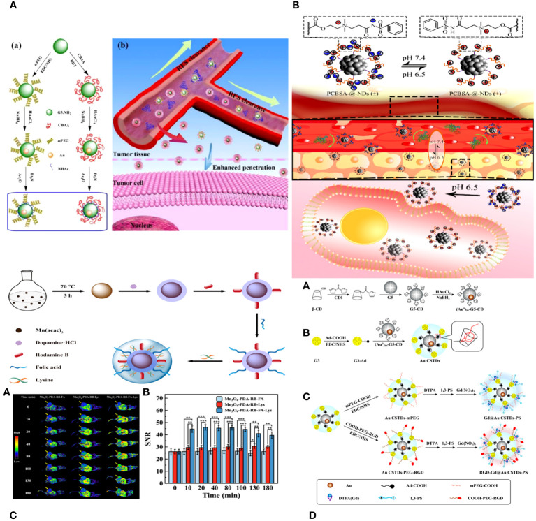
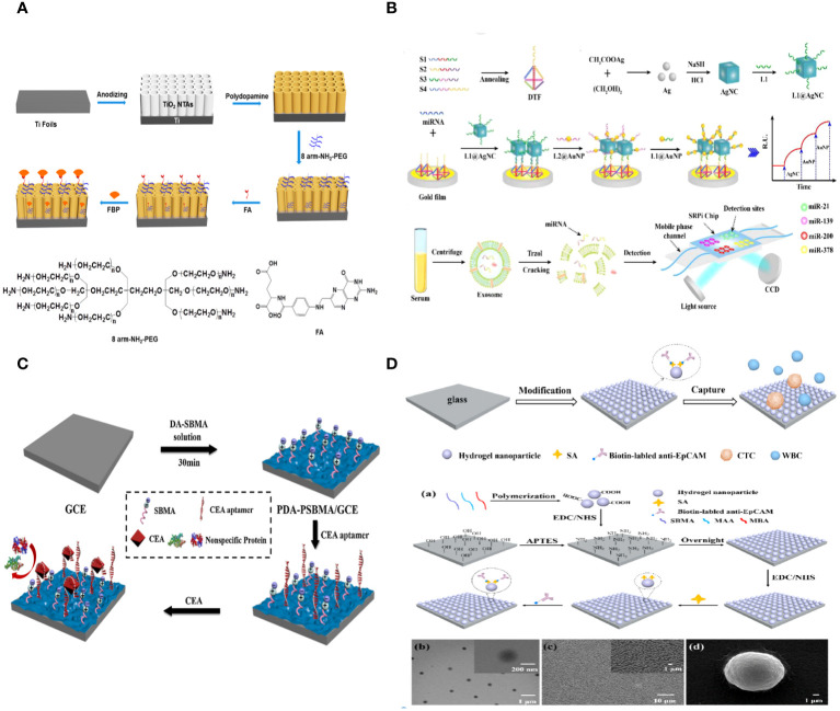
Targeted therapy has become crucial to modern translational science, offering a remedy to conventional drug delivery challenges. Conventional drug delivery systems encountered challenges related to solubility, prolonged release, and inadequate drug penetration at the target region, such as a tumor. Several formulations, such as liposomes, polymers, and dendrimers, have been successful in advancing to clinical trials with the goal of improving the drug's pharmacokinetics and biodistribution. Various stealth coatings, including hydrophilic polymers such as PEG, chitosan, and polyacrylamides, can form a protective layer over nanoparticles, preventing aggregation, opsonization, and immune system detection. As a result, they are classified under the Generally Recognized as Safe (GRAS) category. Serum, a biological sample, has a complex composition. Non-specific adsorption of chemicals onto an electrode can lead to fouling, impacting the sensitivity and accuracy of focused diagnostics and therapies. Various anti-fouling materials and procedures have been developed to minimize the impact of fouling on specific diagnoses and therapies, leading to significant advancements in recent decades. This study provides a detailed analysis of current methodologies using surface modifications that leverage the antifouling properties of polymers, peptides, proteins, and cell membranes for advanced targeted diagnostics and therapy in cancer treatment. In conclusion, we examine the significant obstacles encountered by present technologies and the possible avenues for future study and development.

Keywords: antifouling materials; application status; cancers; challenges; targeted diagnostics and therapy.


Conflict of interest statement

The authors declare that the research was conducted in the absence of any commercial or financial relationships that could be construed as a potential conflict of interest.

Figures

Figure 1
Figure 1
(A) Schematic representation of the synthesis of the Gd2O3- PEG-L-Cys-NPs. The Gd2O3- PEG-L-Cys-NPs in vitro application of MRI of metastasis lung cancer (29). (B) Models and structures of zwitterionic materials. (A) Chemical structures of the common zwitterionic groups. (B) Zwitterionic poly (amino acids) and polypeptide. (C) Mixed-charge zwitterionic polymers that have balanced cationic and anionic groups in different monomer units, and pseudo-zwitterionic materials with equimolar negative and positive charge binding to the same medium. (C) Schematic representation of the synthesis, drug loading, cellular uptake, and rapid enzyme-responsive drug release of the zwitterionic conjugated bottlebrush copolymers (30). (D) Illustration of the preparation, charge-conversion ability and biodegradable behavior of poly (2-methacryloyloxyethyl phosphorylcholine-s-s-vinylimidazole) (PMV) nanogels (31).
Figure 2
Figure 2
(A) Graphical scheme for the design and development of MP-based bioassay for fishing-out of soluble biomarkers. (a) Experimental scheme for selection of phage-displayed peptides with high affinity and specificity for rhTNFa performed on magnetic nickel-coated beads. (b) Graphical representation of integrated system for detection or fishing-out of any soluble biomarker in complex biological medium. This MP-based bioassay consists of selective capturing of target protein due to the presence of a specific binding peptide (previously selected by modified phage display procedure). The binding event is then detected by immunofluorescence measurements (60). (B) Schematic representation of the synthesis of fatty-amine-conjugated cationic BSA nanovehicles formulation, its surface modification with biotin, the capacity for antibiofouling, and successful encapsulation and delivery of anticancer drug Dox to biotin-receptor-positive cancer cells (61). (C) The fabrication process of the electrochemical immunosensor with anti-fouling capability for detection of CD44 (62). (D) Schematic illustration of the fabrication process of the AFP biosensor with PEDOT-HPG (63).
Figure 3
Figure 3
(A) Schematic illustration of the synthesis of CBAA- or PEG-modified Au DENPs (a) and the good antifouling property of CBAA-modified Au DENPs in blood vessels for imaging applications (b) (95). (B) The surface charge-conversional performance of PCBSA-@-NDs and tumor cell uptake under tumor pHe (96). (C) Schematic presentation of the preparation of Mn3O4−PDA−RB−FA−Lys NPs (97). (D) Schematic illustration of the synthesis of RGD-Gd@Au CSTDs-PS (98).
Figure 4
Figure 4
(A) Schematic Representation of the Fabrication of a PEC Biosensor for FBP Detection (112). (B) Schematic illustration of the multiplex exosomal miRNAs detection using SPRi biosensor (113). (C) Schematic illustration of the fabrication process of the PDA-PSBMA based sensing platform (114). (D) Schematic illustration for the capture of circulating tumor cells (CTCs) using the hydrogel nanoparticle substrate. Preparation and characterization of hydrogel nanoparticle substrate (115).
Figure 5
Figure 5
(A) The fabrication, blood cell repellence and tumor cell capture of ligands decorated cell membrane mimetic surface (CMMS-FA-RGD) (117). (B) Overview of working principle in blood from cancer patient; Nanoparticle synthesis and characterization (118).
Figure 6
Figure 6
(A) Illustration of the preparation of PDPA@PEG, PDPA@PCBMA, and PDPA@PCBMA-RGD NPs as well as the low-fouling property and targeting ability of PDPA@PCBMA-RGD NPs. Molecular structures of the monomers [DPA, 2-(diisopropylamino)ethyl methacrylate; PEG-acrylate, poly (ethylene glycol) methyl ether acrylate; CBMA, carboxybetaine methacrylate] and the cross-linker (PEGDMA, polyethylene glycol dimethacrylate) (124). (B) Construction of RGD-CuS DENPs for PA Imaging and PTT/Gene Therapy of Tumors and Tumor Metastasis. After endosomal escape, the pDNAs are dissociated from the polyplexes and enter into the cell nuclei to complete the protein expression to inhibit cancer cell metastasis, while the DENPs enable the PTT of cancer cells (125).
Figure 7
Figure 7
The preparation, hyperthermia-responsiveness, and antitumor therapy of doxorubicin hydrochloride (DOX)-loaded PMEDAPA-Tf nanogels (PMEDAPA-Tf@DOX). PMEDAPA-Tf@DOX shows long blood circulation without inducing the accelerated blood clearance (ABC) phenomenon. F ­urthermore, PMEDAPA-Tf@DOX exhibits enhanced tumor accumulation, penetration, and on-demand drug release with the clinically used microwave heating, leading to improved cancer therapy (127). Upper left: In vivo blood retention profiles of free Cy5, POEGMA, and PMEDAPA nanogels after the first injection (A), second injection (B), and third injection (C) in BALB/c mice. Upper right: Confocal laser scanning microscopy observation. Lower right: Fluorescent imaging of mice bearing HepG2 tumors. Upper left: The preparation, hyperthermia-responsiveness, and antitumor therapy of doxorubicin hydrochloride (DOX)-loaded PMEDAPA-Tf nanogels (PMEDAPA-Tf@DOX).
Figure 8
Figure 8
(A) Schematic diagram of polyprodrug amphiphiles pMPC-b-pHCPT from synthesis, self-assembly, to delivery in vivo (136). (B) Schematic of the Synthesis of Curcumin-Loaded NCPs (Cur@NCPs) and Their Disassembly and Release of Curcumin, which Induces the Apoptosis of Breast Cancer Cells (MDA-MB-231). The diblock copolymer PMPC-b-PserA self-assembles into NCP nanoparticles via complexation of Fe3+ and PserA. Cur@NCPs are produced by partitioning of curcumin into the hydrophobic particle core via an oil-in-water emulsion. Addition of the chelating agent DFO in an acidic solution causes disassembly of the Cur@NCPs, curcumin release, and cell apoptosis (137).
Figure 9
Figure 9
(A) (a) Schematic illustration of the preparation of {(Au0) 25-G5.NH2-PS20}/siRNA polyplexes. (b) The mechanism of {(Au0) 25-G5.NH2-PS20}/siRNA polyplexes for combined endogenous and exogenous sensitization of tumor RT via HIF-1α gene knockdown and Au NPs, respectively (149). (B) (a) Schematic illustration of the preparation of PNS.NHAc-HPAO (131I)-PS. SEM images of (b, c) PNSs and (e, f) PNS.NHAc-HPAO-PS spheres. (d, g) show the size distribution histograms of PNSs and PNS.NHAc-HPAO-PS, respectively (150).
Figure 10
Figure 10
(A) Schematic illustration to show the preparation of multifunctional peptides capped AuNRs and their tumor accumulation in vivo for photothermal therapy (157). (B) Schematic illustration of the synthesis of A) benzaldehyde–thiolated CBT and DOX-DTPA conjugate, B) functionalized CuS DENPs, and C) their therapeutic applications in vivo (158). (C) Schematic Illustration of Enzyme and pH Dual-Sensitive ALA-Conjugated AuNPs for Targeted PDT (159). (D) Construction of RGD-CuS DENPs for PA Imaging and PTT/Gene Therapy of Tumors and Tumor Metastasis. After endosomal escape, the pDNAs are dissociated from the polyplexes and enter into the cell nuclei to complete the protein expression to inhibit cancer cell metastasis, while the DENPs enable the PTT of cancer cells (125).

Similar articles

Cited by

References

    1. Wargo JA, Reuben A, Cooper ZA, Oh KS, Sullivan RJ. Immune effects of chemotherapy, radiation, and targeted therapy and opportunities for combination with immunotherapy. Semin Oncol. (2015) 42:601–16. doi: 10.1053/j.seminoncol.2015.05.007 - DOI - PMC - PubMed
    1. Gazo Hanna E, Younes K, Roufayel R, Khazaal M, Fajloun Z. Engineering innovations in medicine and biology: Revolutionizing patient care through mechanical solutions. Heliyon. (2024) 10:e26154. doi: 10.1016/j.heliyon.2024.e26154 - DOI - PMC - PubMed
    1. Wang W, Chen C, Gu X. Research progress on effect of magnetic nanoparticle composite scaffold on osteogenesis. Zhejiang Da Xue Xue Bao Yi Xue Ban. (2022) 51:102–7. doi: 10.3724/zdxbyxb-2021-0398 - DOI - PMC - PubMed
    1. Nagata T, Aoshi T, Uchijima M, Koide Y. In vivo hierarchy of individual T-cell epitope-specific helper T-cell subset against an intracellular bacterium. Vaccine. (2008) 26:5123–7. doi: 10.1016/j.vaccine.2008.03.061 - DOI - PubMed
    1. Kudruk S, Forsyth CM, Dion MZ, Hedlund Orbeck JK, Luo J, Klein RS, et al. . Multimodal neuro-nanotechnology: Challenging the existing paradigm in glioblastoma therapy. Proc Natl Acad Sci U.S.A. (2024) 121:e2306973121. doi: 10.1073/pnas.2306973121 - DOI - PMC - PubMed