

Case of submandibular schwannoma and review of literature
- PMID: 38779191
- PMCID: PMC11109290
- DOI: 10.1016/j.radcr.2024.04.038
Case of submandibular schwannoma and review of literature
Abstract
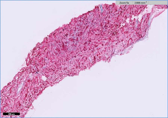
Schwannomas are slow growing, benign tumours arising from Schwann cells. They are usually solitary and are sometimes associated with Neurofibromatosis type 1 and 2. As reported by Okada et al., while approximately 25%-40% of extra-cranial schwannomas occur in the head and neck region, Schwannomas of the oral cavity are very uncommon, accounting for only 1% of all Schwannomas. We report a case of a sublingual schwannoma in a 47-year-old female, discovered incidentally during the workup for tinnitus. The radiological and histopathological findings, along with a literature review, are presented.
Keywords: Floor of mouth; Neuroma; Schwannoma; Sublingual.
© 2024 The Authors. Published by Elsevier Inc. on behalf of University of Washington.
Figures


Similar articles
-
Sublingual Space Schwannoma: An Uncommon Tumour in a Unique Anatomical Location.Indian J Otolaryngol Head Neck Surg. 2024 Jun;76(3):2906-2909. doi: 10.1007/s12070-024-04558-2. Epub 2024 Mar 9. Indian J Otolaryngol Head Neck Surg. 2024. PMID: 38883497 Free PMC article.
-
Gastric Schwannoma as an Important and Infrequent Differential Diagnosis of Gastric Mesenchymal Tumours: A Case Report and Review of Literature.Cureus. 2022 Dec 1;14(12):e32112. doi: 10.7759/cureus.32112. eCollection 2022 Dec. Cureus. 2022. PMID: 36601161 Free PMC article.
-
Schwannoma of floor of the mouth.J Nat Sci Biol Med. 2013 Jul;4(2):487-9. doi: 10.4103/0976-9668.116993. J Nat Sci Biol Med. 2013. PMID: 24082761 Free PMC article.
-
Lingual schwannoma: case report and review of the literature.Acta Otorhinolaryngol Ital. 2013 Apr;33(2):137-40. Acta Otorhinolaryngol Ital. 2013. PMID: 23853407 Free PMC article. Review.
-
Extracranial Schwannomas of the Head and Neck: A Literature Review and Audit of Diagnosed Cases Over a Period of Eight Years.Head Neck Pathol. 2022 Sep;16(3):707-715. doi: 10.1007/s12105-022-01415-y. Epub 2022 Feb 14. Head Neck Pathol. 2022. PMID: 35157211 Free PMC article. Review.
Cited by
-
Submandibular schwannoma - A diagnostic dilemma: A rare case report.Int J Surg Case Rep. 2025 Feb;127:110995. doi: 10.1016/j.ijscr.2025.110995. Epub 2025 Jan 30. Int J Surg Case Rep. 2025. PMID: 39892293 Free PMC article.
References
-
- Okada H, Tanaka S, Tajima H, Akimoto Y, Kaneda T, Yamamoto H. Schwannoma arising from the sublingual gland. Ann Diagn Pathol. 2012;16(2):141–144. - PubMed
Publication types
LinkOut - more resources
Full Text Sources
Research Materials
