

Reporting Pancreatic FNAC using the Papanicolaou System: Still a Diagnostic Challenge
- PMID: 38779600
- PMCID: PMC11108040
- DOI: 10.4103/joc.joc_90_23
Reporting Pancreatic FNAC using the Papanicolaou System: Still a Diagnostic Challenge
Abstract
Introduction: The Papanicolaou Society of Cytopathology System for reporting Pancreaticobiliary Cytology (PSCPC) is a reliable method to classify pancreatic fine needle aspiration cytology (FNAC) smears. However, it is not without practical problems which can diminish the diagnostic accuracy of the cytological diagnosis.
Aims and objectives: To determine the diagnostic pitfalls while reporting cytomorphology of pancreatic lesions according to PSCPC on correlating FNAC findings with histopathology.
Materials and methods: Retrospective analysis of pancreatic FNAC smears received in the Department of Pathology of our tertiary care institute over a period of 2 years was done. The cytological diagnoses were classified according to the Papanicolaou Society of Cytopathology system of reporting pancreaticobiliary cytology and correlated with histopathology. The reasons of cyto-histological discordance were analyzed.
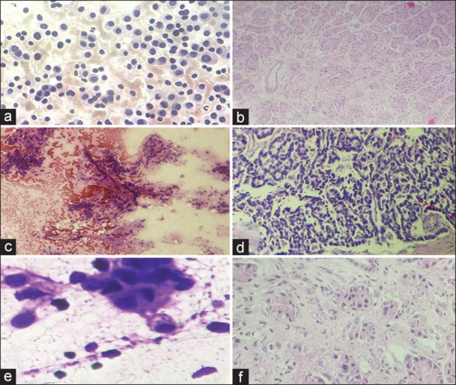
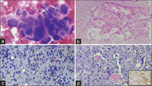
Results: Out of 50 cases in which both FNAC and biopsy of pancreatic lesions were done, 34 cases were positive/malignant (Category VI), eight cases were suspicious for malignancy (Category V), three cases were neoplastic (Category IV), two cases were atypical (Category III), two cases were negative for malignancy (Category II), and one case was non-diagnostic (Category I). Out of 50 cases, histopathology was non-diagnostic due to inadequate material in six cases. The cytological diagnoses were compared with histopathology in the remaining 44 cases. Categories III, IV V, and VI were considered as positive for neoplastic pathology. The sensitivity of FNAC to predict neoplastic pathology was 97.5%, while the specificity was 25%. The positive predictive value was 92.9%. Two cases reported as atypical (Category III) turned out to be adenocarcinoma on histopathology. One case reported as neuroendocrine tumor and two cases reported as adenocarcinoma on cytology displayed features of chronic pancreatitis on histology. One case reported as neoplastic mucinous cyst (Category IV) turned out to be adenocarcinoma on histology (limited concordance).
Conclusion: The cytopathologist needs to be wary of the potential pitfalls to improve the diagnostic accuracy of FNACs.
Keywords: FNAC; Papanicolaou; malignant; neoplastic; pancreatic.
Copyright: © 2024 Journal of Cytology.
Conflict of interest statement
There are no conflicts of interest.
Figures
Similar articles
-
Classification of endoscopic ultrasound guided fine needle aspiration cytology of pancreatic space occupying lesions by Papanicolaou Society of Cytopathology System: A five year study.Diagn Cytopathol. 2023 Feb;51(2):105-116. doi: 10.1002/dc.25058. Epub 2022 Sep 27. Diagn Cytopathol. 2023. PMID: 36165589
-
Update on risk stratification in the Papanicolaou Society of Cytopathology System for Reporting Pancreaticobiliary Cytology categories: 3-Year, prospective, single-institution experience.Cancer Cytopathol. 2020 Jan;128(1):29-35. doi: 10.1002/cncy.22199. Epub 2019 Nov 13. Cancer Cytopathol. 2020. PMID: 31722125
-
Cytologic Categorization with Risk Stratification of Endoscopic Ultrasound-Guided Fine Needle Aspiration from Pancreatic Lesions Based on Guidelines of the Papanicolaou Society of Cytopathology: 12-Year Tertiary Care Experience.Discoveries (Craiova). 2021 Aug 21;9(3):e134. doi: 10.15190/d.2021.13. eCollection 2021 Jul-Sep. Discoveries (Craiova). 2021. PMID: 34816002 Free PMC article.
-
Assessment of Risk of Malignancy of Fine-needle Aspiration Cytology in Salivary Gland Lesions Using the Milan System for Reporting Salivary Gland Cytopathology Categorization: A Systematic Review and Meta-analysis.J Contemp Dent Pract. 2022 Oct 1;23(10):1039-1056. doi: 10.5005/jp-journals-10024-3424. J Contemp Dent Pract. 2022. PMID: 37073919
-
[Needle aspiration cytology of the breast: current perspective on the role in diagnosis and management].Acta Med Croatica. 2008 Oct;62(4):391-401. Acta Med Croatica. 2008. PMID: 19205416 Review. Croatian.
References
-
- Turner BG, Cizginer S, Agarwal D, Yang J, Pitman MB, Brugge WR. Diagnosis of pancreatic neoplasia with EUS and FNA; A report of accuracy. Gastro Endosc. 2010;71:91–8. - PubMed
-
- Pitman MB, Centeno BA, Ali SZ, Genevay M, Stelow E, Mino-Kenudson M, et al. Standardized terminology and nomenclature for pancreatobiliary cytology: The Papanicolaou Society of Cytopathology guidelines. Diagn Cytopathol. 2014;42:338–50. - PubMed
LinkOut - more resources
Full Text Sources