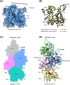
Structural and functional mechanisms of actin isoforms
- PMID: 38779987
- PMCID: PMC11796330
- DOI: 10.1111/febs.17153
Structural and functional mechanisms of actin isoforms
Abstract
Actin is a highly conserved and fundamental protein in eukaryotes and participates in a broad spectrum of cellular functions. Cells maintain a conserved ratio of actin isoforms, with muscle and non-muscle actins representing the main actin isoforms in muscle and non-muscle cells, respectively. Actin isoforms have specific and redundant functional roles and display different biochemistries, cellular localization, and interactions with myosins and actin-binding proteins. Understanding the specific roles of actin isoforms from the structural and functional perspective is crucial for elucidating the intricacies of cytoskeletal dynamics and regulation and their implications in health and disease. Here, we review how the structure contributes to the functional mechanisms of actin isoforms with a special emphasis on the questions of how post-translational modifications and disease-linked mutations affect actin isoforms biochemistry, function, and interaction with actin-binding proteins and myosin motors.
Keywords: actin; actin isoforms; actin‐binding proteins; contractility; cytoskeleton; enzymology; mechanobiology; mutations; myosin; post‐translational modifications.
© 2024 The Authors. The FEBS Journal published by John Wiley & Sons Ltd on behalf of Federation of European Biochemical Societies.
Conflict of interest statement
The authors declare no conflict of interest.
Figures


Similar articles
-
Structural insights into actin isoforms.Elife. 2023 Feb 15;12:e82015. doi: 10.7554/eLife.82015. Elife. 2023. PMID: 36790143 Free PMC article.
-
Tropomyosin and actin isoforms modulate the localization of tropomyosin strands on actin filaments.J Mol Biol. 2000 Sep 22;302(3):593-606. doi: 10.1006/jmbi.2000.4080. J Mol Biol. 2000. PMID: 10986121
-
Insights into Actin Isoform-Specific Interactions with Myosin via Computational Analysis.Molecules. 2024 Jun 23;29(13):2992. doi: 10.3390/molecules29132992. Molecules. 2024. PMID: 38998944 Free PMC article.
-
The complexity and diversity of the actin cytoskeleton of trypanosomatids.Mol Biochem Parasitol. 2020 May;237:111278. doi: 10.1016/j.molbiopara.2020.111278. Epub 2020 Apr 28. Mol Biochem Parasitol. 2020. PMID: 32353561 Review.
-
Actin Post-translational Modifications: The Cinderella of Cytoskeletal Control.Trends Biochem Sci. 2019 Jun;44(6):502-516. doi: 10.1016/j.tibs.2018.11.010. Epub 2019 Jan 2. Trends Biochem Sci. 2019. PMID: 30611609 Review.
Cited by
-
Glycosylation Patterns in Meccus (Triatoma) pallidipennis Gut: Implications for the Development of Vector Control Strategies.Microorganisms. 2025 Jan 1;13(1):58. doi: 10.3390/microorganisms13010058. Microorganisms. 2025. PMID: 39858826 Free PMC article.
-
Identification of housekeeping gene for future studies exploring effects of cryopreservation on gene expression in shrimp.Sci Rep. 2025 Apr 1;15(1):11046. doi: 10.1038/s41598-025-95258-6. Sci Rep. 2025. PMID: 40169849 Free PMC article.
-
β-actin function in platelets and red blood cells can be performed by γ-actin and is therefore independent of actin isoform protein sequence.Mol Biol Cell. 2025 Feb 1;36(2):ar18. doi: 10.1091/mbc.E24-04-0186. Epub 2024 Dec 20. Mol Biol Cell. 2025. PMID: 39705375 Free PMC article.
-
The complete absence of cytoplasmic γ-actin results in no discernible phenotype in mice or primary fibroblasts.FEBS J. 2025 Jul;292(13):3565-3576. doi: 10.1111/febs.70075. Epub 2025 Mar 20. FEBS J. 2025. PMID: 40109120 Free PMC article.
-
Divergent Contribution of Cytoplasmic Actins to Nuclear Structure of Lung Cancer Cells.Int J Mol Sci. 2024 Dec 19;25(24):13607. doi: 10.3390/ijms252413607. Int J Mol Sci. 2024. PMID: 39769373 Free PMC article.
References
-
- Van Troys M, Vandekerckhove J & Ampe C (1999) Structural modules in actin‐binding proteins: towards a new classification. Biochim Biophys Acta 1448, 323–348. - PubMed
Publication types
MeSH terms
Substances
Grants and funding
LinkOut - more resources
Full Text Sources

