. 2024 Jun 4;63(11):1434-1444.
doi: 10.1021/acs.biochem.4c00071. Epub 2024 May 23.

The Light Chain Allosterically Enhances the Protease Activity of Murine Urokinase-Type Plasminogen Activator

Affiliations

The Light Chain Allosterically Enhances the Protease Activity of Murine Urokinase-Type Plasminogen Activator

Constanza Torres-Paris et al. Biochemistry. .

Abstract

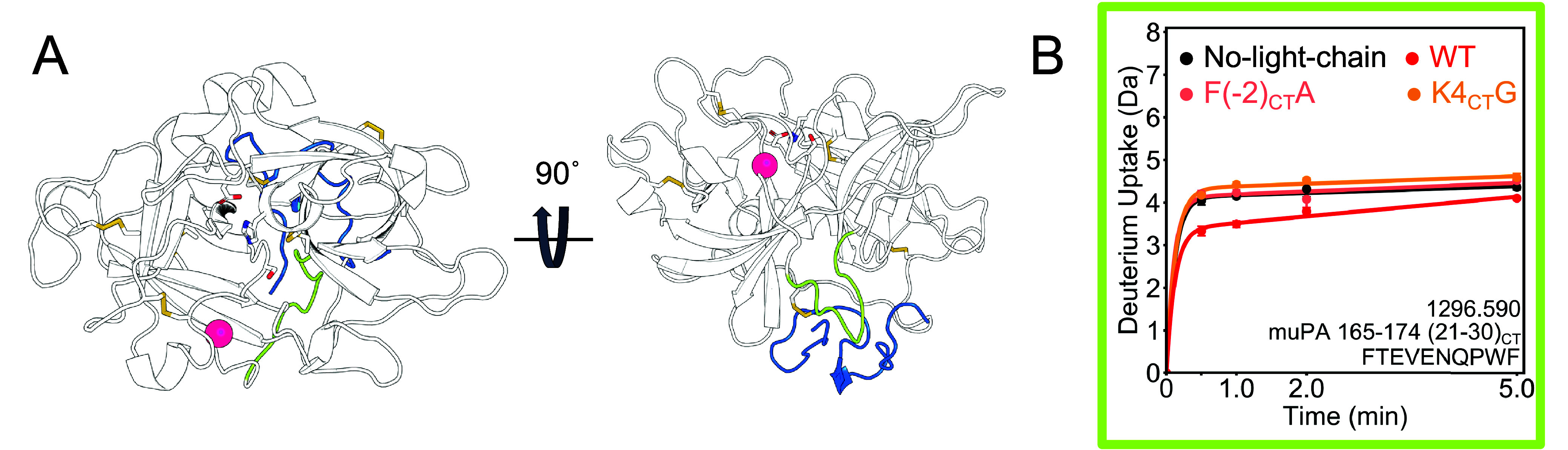
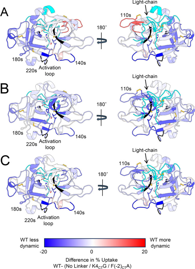
The active form of the murine urokinase-type plasminogen activator (muPA) is formed by a 27-residue disordered light chain connecting the amino-terminal fragment (ATF) with the serine protease domain. The two chains are tethered by a disulfide bond between C1CT in the disordered light chain and C122CT in the protease domain. Previous work showed that the presence of the disordered light chain affected the inhibition of the protease domain by antibodies. Here we show that the disordered light chain induced a 3.7-fold increase in kcat of the protease domain of muPA. In addition, hydrogen-deuterium exchange mass spectrometry (HDX-MS) and accelerated molecular dynamics (AMD) were performed to identify the interactions between the disordered light chain and the protease domain. HDX-MS revealed that the light chain is contacting the 110s, the turn between the β10- and β11-strand, and the β7-strand. A reduction in deuterium uptake was also observed in the activation loop, the 140s loop and the 220s loop, which forms the S1-specificty pocket where the substrate binds. These loops are further away from where the light chain seems to be interacting with the protease domain. Our results suggest that the light chain most likely increases the activity of muPA by allosterically favoring conformations in which the specificity pocket is formed. We propose a model by which the allostery would be transmitted through the β-strands of the β-barrels to the loops on the other side of the protease domain.


Conflict of interest statement

The authors declare no competing financial interest.

Figures

Figure 1
Figure 1
Structure of muPA. A. Schematics of the proteins used in this study. B. Canonical view of the crystal structure of the protease domain of human uPA (huPA) containing part of the light chain (PDB: 1O3P). The N-terminal β-barrel contains six β-strands (β1- β6) and the C-terminal β-barrel contains another six (β7- β12). The catalytic triad residues H206 (57CT), D257 (102CT) and S358 (195CT) (shown as sticks) are in the interface between the two β-barrels. An active site covalent inhibitor is shown in black sticks. The structure is colored according to the secondary structure. C. Canonical view of the crystal structure of huPA showing the loops and their names. D. The sequence of the disordered region of the light chain of murine uPA (muPA) and huPA. The numbering of each residue in the full-length (FL) and chymotrypsin-like (CT) numbering. The conserved residues are shown in red. The side chain of C149 (1CT) makes a disulfide bond with the protease domain. E. Zoomed view of the rotated view of the crystal structure of huPA (PDB: 1O3P) showing the residues in the light chain (blue backbone) and the protease (white backbone) that have been proposed as interactions. All the residue numbers correspond to the chymotrypsin numbering. Here we show the huPA structures because no muPA structures are available with the light chain.
Figure 2
Figure 2
Disordered light chain is very dynamic and does not appear to adopt a defined position on the protease domain. A. Canonical view into the active site of the ensemble of conformations acquired by muPA WT during AMD. The loops are colored in the same color scheme as the crystal structure of human uPA in Figure 1C. B. 180° rotated view of muPA WT showing the light chain ensemble of conformations acquired by AMD. Each light chain conformation is colored in a unique shade. C. Canonical (top) and rotated (bottom) view of a conformation from the ensemble of muPA WT showing the light chain colored in blue with residues 280–286 (121CT −127CT) colored in green. The catalytic triad residues and the disulfide bonds are shown as sticks, and the inhibitor is colored black. D. Deuterium uptake plot of the light chain peptide of muPA WT. Each data point represents the average of three technical replicates and the error bars represent the standard deviation. E. Deuterium uptake plots of the protease peptides which contain either C122 (left) or C122A (right), the site where the light chain covalently binds to the protease.
Figure 3
Figure 3
Effect of the light-chain on the activation loop. A. The activation loop is located downstream of the new N-terminus (fuchsia sphere), at the bottom of the protease domain if looked from the front (left). Residues 165–174 (21CT-30CT)) are part of the activation loop and are colored green. The light chain is shown in blue. The catalytic triad residues and the disulfide bonds are shown as sticks. B. Deuterium uptake plot of residues 165–174 (21CT-30CT) in the activation loop.
Figure 4
Figure 4
Effect of the light chain on the 110s loop. A. A conformation from the AMD ensemble of muPA WT shows that the light chain contacts the N-terminal β-barrel around P198 (49CT), in the turn between β2 and β3, and P273 (114CT) located in the connection between the two β-barrels and the 110s loop. Residues 261–276 (106CT-117CT) covering the 110s loop and part of the connection between the two β-barrels is shown in yellow. The light chain is colored in blue. B. Cartoon representation of the interactions between the light chain and the N-terminal β-barrel. C. Deuterium incorporation spectra of peptide 261–276 (106CT-117CT) sequence LKIRTSTGQCAQPSRS, in the +3 charge state. The x-axis of each plot is m/z and the y axis is the intensity at each peak. Each column represents one muPA variant (WT, no light chain, F(−2CT)A, K4CTG), and each row is a deuteration time point (0–5 min). This region of the protease shows a bimodal deuterium uptake distribution in the protease that is missing the light chain. The envelope of the higher m/conformation is shown in blue, and the one for the lower m/z, in red.
Figure 5
Figure 5
Light chain reduces the dynamics of the β9 strand and does not affect the 180s loop. A. A representative conformation of muPA WT from the AMD simulation shows the β9 strand (teal) and the 180s loop (magenta), which are in the C-terminal β-barrel. The catalytic triad residues are shown as sticks. B. The non-chymotrypsin-like fold of muPA No-light-chain (PDB: 5LHS) has a distorted C-terminal β-barrel. C. Deuterium uptake plot of residues 344−364 (183CT-201CT) in the 180s loop. This peptide encompasses the catalytic S358(195CT). D. Deuterium uptake plot of residues 337−343 (176CT-182CT) in the β9 strand.
Figure 6
Figure 6
Light chain affects the dynamics of the 220s loop and the β10-β11 turn. A. The 220s loop (purple) is in the C-terminal β-barrel of muPA WT. The S1 specificity pocket is formed by two residues in the 220s loop (Gly 379 (216CT) and Gly 389 (226CT), shown as purple spheres) and a residue in the 180s loop (Asp 352 (189CT), shown as a white sphere. The structure in the canonical view of the protein and the structure rotated by 270° on the x axis are shown for comparison. The light-chain is shown in blue, and the peptide analyzed by HDX-MS corresponding to residues 365–391 (202CT-228CT), covering part of the 220s loop, β10 and β11 strands and the turn between those β strands, is shown in purple. B. Cartoon model showing how the light chain would interact with the region corresponding to residues 365–391 (202CT-228CT). C. Deuterium incorporation spectra of peptide 365–391 (202CT-228CT) sequence NIEGRPTLSGIVSWGRGCAEKNKPGVY, in the +5 charge state. The x-axis of each plot is m/z and the y axis is the intensity. Each column represents one muPA variant (WT, no light chain, F(−2)A, K4CTG) and each row a deuteration time point (0–5 min). The envelope of the higher m/z conformation is shown in blue and the one of the lower m/z, in red.
Figure 7
Figure 7
Light chain regulates the dynamics of the 140s loop. A. The 140s loop (colored in cyan and green) is at the “bottom” of the protease as seen from the canonical view (top). A peptide corresponding to residues 295–307 (136CT-148CT) representing the β7 strand and the beginning of the 140s loop is colored in cyan and a peptide corresponding to residues 308–318 (149CT-159CT) representing the last part of the 140s loop and part of the β8 strand is colored in green. B. Deuterium uptake plot of residues 295–307 (136CT-148CT) in the 140s loop. C. Deuterium uptake plot of residues 308–318 (149CT-159CT) in the 140s loop. D. Cartoon model showing where the light chain would interact to affect the 140s loop.
Figure 8
Figure 8
Summary of the HDX-MS changes in the protease domain due to the light-chain and the mutants K4CTG and F(−2CT)A The difference of deuterium uptake between WT and the protease lacking the light chain (A) or the K4CTG mutant (B) or the F(−2CT)A mutant (C) were mapped onto a conformation of the ensemble acquired by muPA WT during the AMD. The peptides are colored blue if the WT is less dynamic or red if its more dynamic that the compared condition. The regions that were not covered in the HDX-MS are shown in black. The regions that were covered only in one condition, or that had a different mutation in each condition and are not directly comparable, are shown in cyan.
Figure 9
Figure 9
Model of how dynamic allostery caused by the light chain interactions is transmitted through the β-strands between loops. A. Dynamic allostery works as a Newton’s cradle, where changes in one side of the device are transmitted through the middle. The interactions of the middle balls do not change, and we can only observe the transmission of energy on the balls in the extremes. B. In the absence of the light chain, loops 1 and 2 are dynamic and connected covalently through a β-strand. When the light chain interacts with loop 1, that energy is transmitted to loop 2, which changes its dynamics and hydrogen bonding patterns. As the light chain is dynamic, it can also move away and stop interacting with loop 1, which returns its dynamics to the original conformation. The lack of interaction is also transmitted through the β-strand and returns the dynamics of loop 2 to its original state as well. The hydrogen bonding network and solvent accessibility of the amides in the β-strand does not change during this process.

References

    1. Smith H. W.; Marshall C. J. Regulation of cell signalling by uPAR. Nat. Rev. Mol. Cell Biol. 2010, 11 (1), 23–36. 10.1038/nrm2821. - DOI - PubMed
    1. Huai Q.; Mazar A. P.; Kuo A.; Parry G. C.; Shaw D. E.; Callahan J.; Li Y.; Yuan C.; Bian C.; Chen L.; et al. Structure of human urokinase plasminogen activator in complex with its receptor. Science 2006, 311 (5761), 656–659. 10.1126/science.1121143. - DOI - PubMed
    1. Huai Q.; Zhou A.; Lin L.; Mazar A. P.; Parry G. C.; Callahan J.; Shaw D. E.; Furie B.; Furie B. C.; Huang M. Crystal structures of two human vitronectin, urokinase and urokinase receptor complexes. Nat. Struct Mol. Biol. 2008, 15 (4), 422–423. 10.1038/nsmb.1404. - DOI - PMC - PubMed
    1. Bdeir K.; Kuo A.; Sachais B. S.; Rux A. H.; Bdeir Y.; Mazar A.; Higazi A. A.; Cines D. B. The kringle stabilizes urokinase binding to the urokinase receptor. Blood 2003, 102 (10), 3600–3608. 10.1182/blood-2003-03-0949. - DOI - PubMed
    1. Tarui T.; Akakura N.; Majumdar M.; Andronicos N.; Takagi J.; Mazar A. P.; Bdeir K.; Kuo A.; Yarovoi S. V.; Cines D. B.; et al. Direct interaction of the kringle domain of urokinase-type plasminogen activator (uPA) and integrin alpha v beta 3 induces signal transduction and enhances plasminogen activation. Thromb Haemost 2006, 95 (3), 524–534. 10.1160/TH05-06-0457. - DOI - PubMed

Publication types

MeSH terms

Substances