Review
. 2024 Nov;34(11):7481-7491.
doi: 10.1007/s00330-024-10792-7. Epub 2024 May 23.

ESR Essentials: using the right scoring system in prostate MRI-practice recommendations by ESUR

Affiliations
Review

ESR Essentials: using the right scoring system in prostate MRI-practice recommendations by ESUR

Andrea Ponsiglione et al. Eur Radiol. 2024 Nov.

Abstract

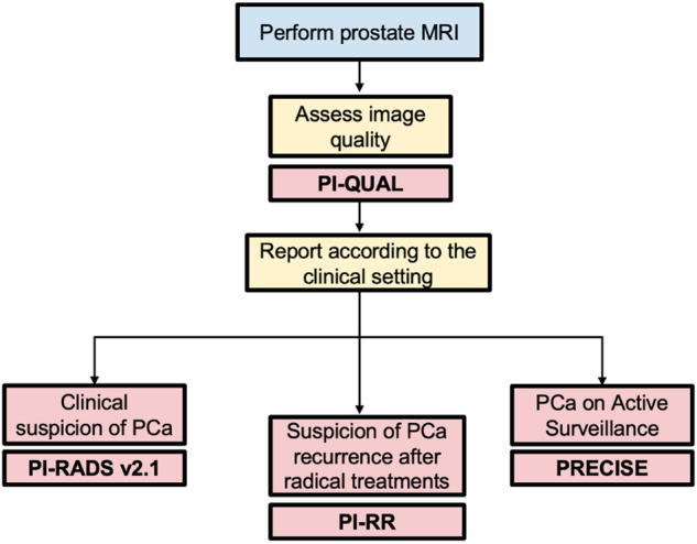
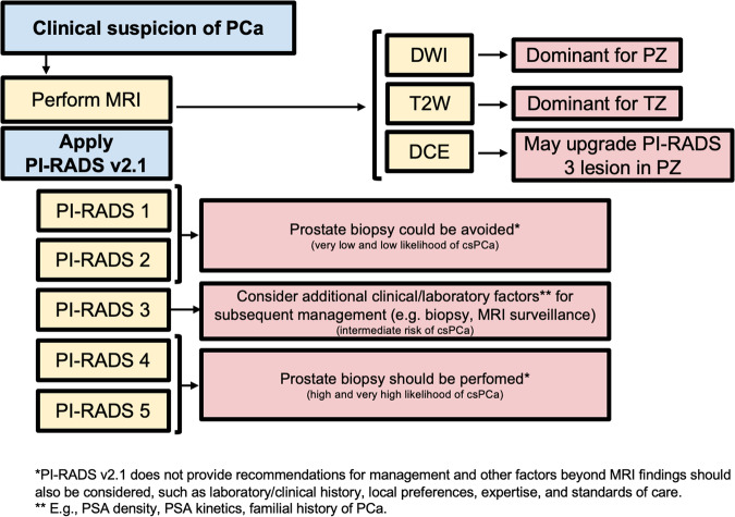
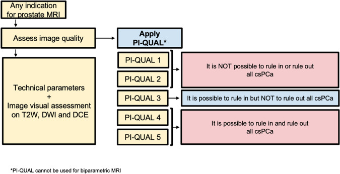
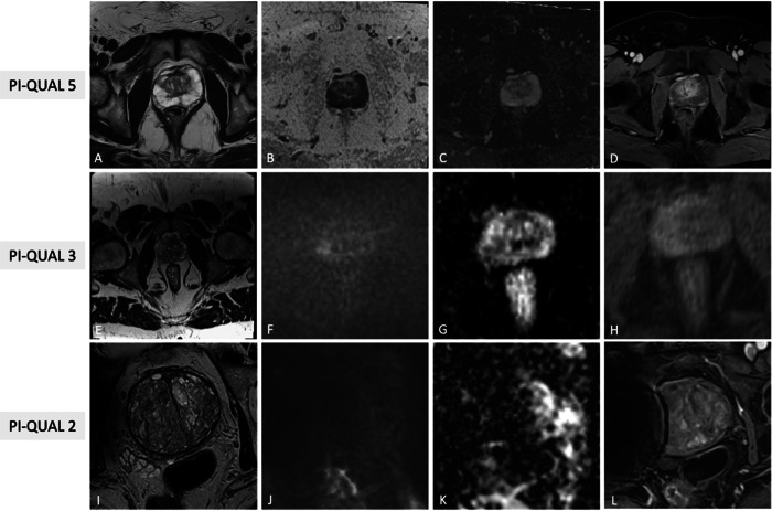
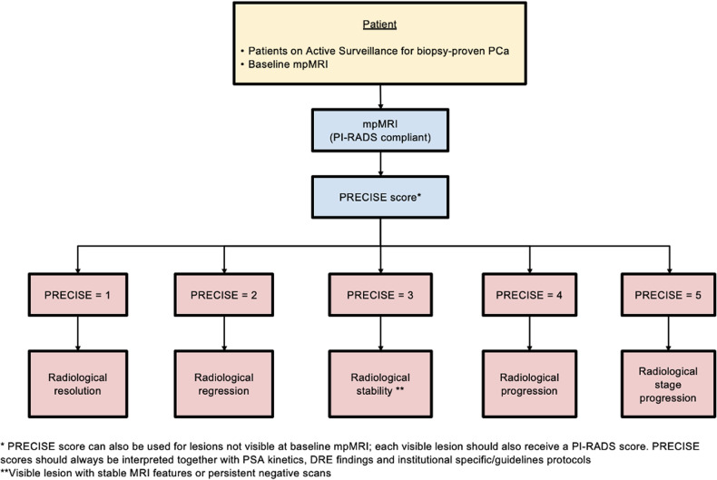
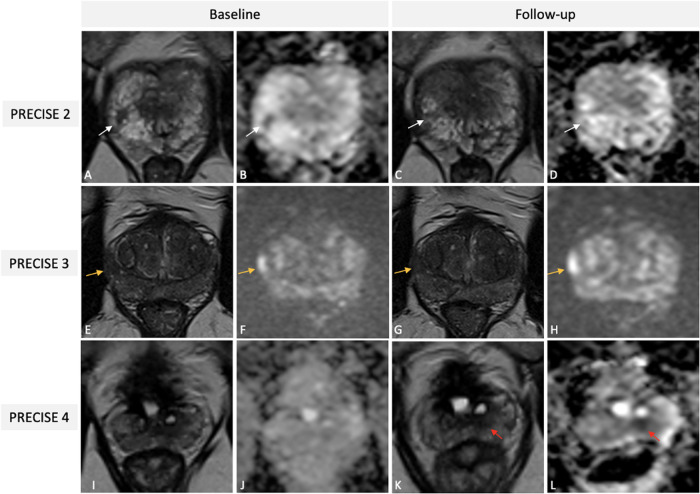
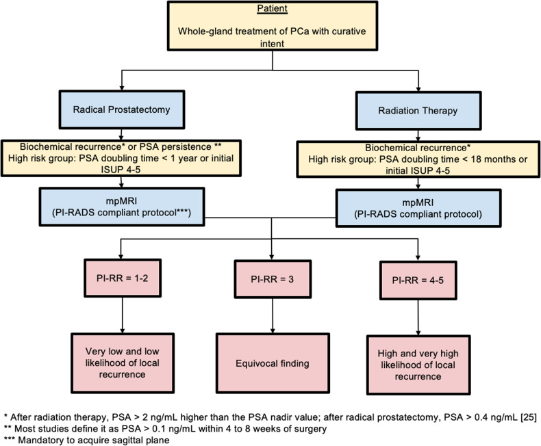
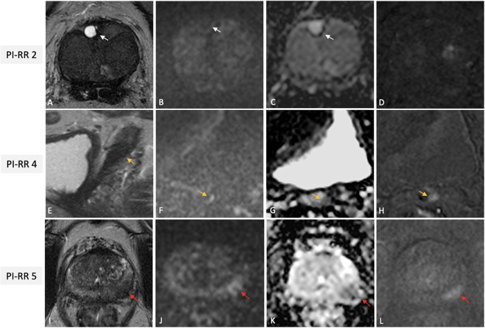
MRI has gained prominence in the diagnostic workup of prostate cancer (PCa) patients, with the Prostate Imaging Reporting and Data System (PI-RADS) being widely used for cancer detection. Beyond PI-RADS, other MRI-based scoring tools have emerged to address broader aspects within the PCa domain. However, the multitude of available MRI-based grading systems has led to inconsistencies in their application within clinical workflows. The Prostate Cancer Radiological Estimation of Change in Sequential Evaluation (PRECISE) assesses the likelihood of clinically significant radiological changes of PCa during active surveillance, and the Prostate Imaging for Local Recurrence Reporting (PI-RR) scoring system evaluates the risk of local recurrence after whole-gland therapies with curative intent. Underlying any system is the requirement to assess image quality using the Prostate Imaging Quality Scoring System (PI-QUAL). This article offers practicing radiologists a comprehensive overview of currently available scoring systems with clinical evidence supporting their use for managing PCa patients to enhance consistency in interpretation and facilitate effective communication with referring clinicians. KEY POINTS: Assessing image quality is essential for all prostate MRI interpretations and the PI-QUAL score represents the standardized tool for this purpose. Current urological clinical guidelines for prostate cancer diagnosis and localization recommend adhering to the PI-RADS recommendations. The PRECISE and PI-RR scoring systems can be used for assessing radiological changes of prostate cancer during active surveillance and the likelihood of local recurrence after radical treatments respectively.

Keywords: Classification; management; Magnetic resonance imaging; Prostate cancer; Standardization.


Conflict of interest statement

Andrea Ponsiglione holds the position of Junior Deputy Editor at European Radiology and is a member of the Scientific Editorial Board of Insights into Imaging, but he has not taken part in the review or selection process of this article. Valeria Panebianco is member of the European Radiology Scientific Editorial Board. She has not taken part in the review or selection process of this article. Renato Cuocolo is member of the European Radiology Scientific Editorial Board. He has not taken part in the review or selection process of this article.

Figures

Fig. 1
Fig. 1
General flowchart of the available prostate MRI-based scoring systems according to clinical indications. MRI, magnetic resonance imaging; PI-QUAL, Prostate Imaging Quality; PCa, prostate cancer; PI-RADS, Prostate Imaging Reporting and Data System; PI-RR, Prostate Imaging for Local Recurrence Reporting; PRECISE, Prostate Cancer Radiological Estimation of Change in Sequential Evaluation
Fig. 2
Fig. 2
Flowchart for how and when PI-RADS v2.1 should be used, with practical implications. PCa, prostate cancer; MRI, magnetic resonance imaging; PI-RADS, Prostate Imaging Reporting and Data System; DWI, diffusion weighted imaging; T2W, T2-weighted; PZ, peripheral zone; TZ, transitional zone; csPCa, clinically significant PCa; PSA, prostate specific agent
Fig. 3
Fig. 3
Illustrative examples of PI-RADS scores 2 (AD), 3 (EH) and 4 (IL), from different patients undergoing mpMRI for clinical suspicion of PCa. Wedge shaped area (white arrows) in the left postero-lateral peripheral zone at mid-gland, hypointense on T2w (A), slightly hyperintense on synthetic high b value DWI (B) and hypointense on ADC (C) without focal early enhancement on the DCE image (D) classified as PI-RADS score 2. Nodular area (orange arrows) in the right anterior peripheral zone at mid-gland, hypointense on T2W (E), moderately hyperintense on synthetic high b value DWI (F) and hypointense on ADC (G) without focal early enhancement on the DCE image (H), scored as PI-RADS 3. Lenticular shaped area (red arrows) in the right postero-lateral peripheral zone at base, hypointense on T2w (I), markedly hyperintense on high b value DWI (J) and markedly hypointense on ADC (K) with focal early enhancement on the DCE image (L), scored as PI-RADS 4 with low probability of extra-prostatic extension, then pathologically confirmed via targeted biopsy as clinically significant PCa (GG2). PI-RADS, Prostate Imaging Reporting and Data System; mpMRI, multiparametric magnetic resonance imaging; PCa, prostate cancer; T2W, T2-weighted; DWI, diffusion weighted imaging; ADC, apparent diffusion coefficient; DCE, dynamic contrast enhanced; GG, grade group
Fig. 4
Fig. 4
Flowchart for how and when PI-QUAL scoring system should be used, with practical implications. MRI, magnetic resonance imaging; PI-QUAL, Prostate Imaging Quality; T2W, T2-weighted; DWI, diffusion weighted imaging; DCE, dynamic contrast enhanced; csPCa, clinically significant prostate cancer
Fig. 5
Fig. 5
Illustrative examples of PI-QUAL scores 5 (AD), 3 (EH) and 2 (IL) from different patients undergoing mpMRI for various purposes. In the first case, T2W (A), synthetic b1500 DWI (B), ADC map (C) and DCE (D) images are of optimal diagnostic quality and meet the technical requirements of PI-RADS v2.1 (PI-QUAL score 5). In the second case, T2W FOV is exceedingly large (E), diffusion images are of good quality (F, G), while DCE shows inadequate in-plan resolution (H); since, at least two mpMRI sequences taken together are of diagnostic quality, the exam has been scored as PI-QUAL 3. In the third case, only T2W (I) shows optimal quality, while high b value DWI (J), ADC map (K) and DCE (L) are suboptimal due to the presence of right hip prostheses (PI-QUAL score 2). PI-QUAL, Prostate Imaging Quality; mpMRI, multiparametric magnetic resonance imaging; T2W, T2-weighted; DWI, diffusion weighted imaging; ADC, apparent diffusion coefficient; DCE, dynamic contrast enhanced; FOV, field of view
Fig. 6
Fig. 6
Flowchart for how and when PRECISE scoring system should be used. PCa, prostate cancer; mpMRI, multiparametric magnetic resonance imaging; PI-RADS, Prostate Imaging Reporting and Data System; PRECISE, Prostate Cancer Radiological Estimation of Change in Sequential Evaluation; PSA, prostate specific agent; DRE, digital rectal examination
Fig. 7
Fig. 7
Illustrative examples of PRECISE scores 2 (AD), 3 (EH), and 4 (IL) from different patients on AS for PCa undergoing mpMRI. Baseline T2W (A) image and ADC map (B) of a 56-year-old patient showing a PI-RADS 4 lesion (white arrows) in the right postero-lateral peripheral zone at the apex, then pathologically confirmed at targeted biopsy as Gleason 3 + 4 PCa (Pattern 4 ≤ 10%). One-year follow-up scan (C, D) demonstrates a reduction in lesion size on axial T2W (white arrow in C) and in conspicuity on ADC map (white arrow in D), scored as PRECISE 2 (PI-RADS score 3). Baseline T2w (E) and high b value DWI (F) images of a 69-year-old patient revealing a PI-RADS 4 lesion (orange arrows) in the right postero-lateral peripheral zone at the mid-gland, then pathologically confirmed at targeted biopsy as Gleason 3 + 3 PCa. A three-year follow-up scan (G, H) indicating stability in both size and conspicuity of the identified lesion, classified as PRECISE score 3 (PI-RADS score 4). Baseline T2w image (I) and ADC map (J) of a 71-year-old patient, with a previous history of TURP, showing absence of suspicious lesions (PI-RADS score 2); systematic biopsy revealed a Gleason 3 + 3 PCa. A two-year follow up scan showed the presence of a small nodular lesion (red arrows in K and L) in the left postero-median peripheral zone at the mid-gland and was classified as PRECISE score 4 (PI-RADS score 4); targeted biopsy yielded upgrade to Gleason score 3 + 4. PRECISE, Prostate Cancer Radiological Estimation of Change in Sequential Evaluation; AS, active surveillance; PCa, prostate cancer; mpMRI, multiparametric magnetic resonance imaging; T2W, T2-weighted; ADC, apparent diffusion coefficient; PI-RADS, Prostate Imaging Reporting and Data System; DWI, diffusion weighted imaging; TURP, transurethral resection of the prostate
Fig. 8
Fig. 8
Flowchart for how and when PI-RR scoring system should be used. PCa, prostate cancer; PSA, prostate specific agent; mpMRI, multiparametric magnetic resonance imaging; PI-RADS, Prostate Imaging Reporting and Data System; PI-RR, Prostate Imaging for Local Recurrence Reporting; ISUP, International Society of Urological Pathology
Fig. 9
Fig. 9
Illustrative examples of PI-RR scores 2 (AD), 4 (EH) and 5 (IL), from different patients undergoing mpMRI for rising PSA values after whole-gland treatment with curative intent for PCa. Images (AD) from a 70-year-old man with serum PSA of 0.31 ng/mL after RT for csPCa (GG2) showing a diffusely hypointense gland on axial T2W (A), with a focal fluid-filled hyperintense nodule in the right anterior transitional zone at prostate base (white arrow), with no restricted diffusion (with arrows in B and C) nor early enhancement on the DCE image (D), scored as PI-RR 2 (residual cystic atrophy). Images of a 72-year-old man with BCR (PSA value = 0.81 ng/mL) after RP for PCa (GG1), showing a masslike (orange arrows) hypointense focus on sagittal T2W, at the origin of the seminal vesicle residues, with focal marked hyperintensity on high–b value DWI (F), hypointense on ADC (G) and with focal early enhancement on the DCE image (H); the case was scored as PI-RR 4 (no data on primary tumor side). Images (IL) of a 64-year-old man with BCR (PSA value = 3.4 ng/mL) 2 years after RT for csPCa (GG2) showing a masslike focus on the left postero-lateral peripheral zone at mid-gland (red arrows) hypointense on axial T2W (I) with focal marked hyperintensity on high–b value DWI (J), hypointense on ADC (K) with focal early enhancement on the DCE image (L) at the same site of the primary tumor, scored as PI-RR 5. PI-RR, Prostate Imaging for Local Recurrence Reporting; mpMRI, multiparametric magnetic resonance imaging; PSA, prostate specific agent; PCa, prostate cancer; RT, radiation therapy; csPCa, clinically significant PCa; GG, grade group; T2W, T2-weighted; DCE, dynamic contrast enhanced; BCR, biochemical recurrence; RP, radical prostatectomy; DWI, diffusion weighted imaging; ADC, apparent diffusion coefficient

References

    1. Padhani AR, Schoots IG (2023) Imaging-based diagnostic and therapeutic strategies for prostate cancer in the coming decades. Radiology 307:e222990. 10.1148/radiol.222990 - PubMed
    1. Turkbey B, Rosenkrantz AB, Haider MA et al (2019) Prostate Imaging Reporting and Data System version 2.1: 2019 update of prostate imaging reporting and data system version 2. Eur Urol. 10.1016/j.eururo.2019.02.033 - PubMed
    1. Giganti F, Allen C, Emberton M et al (2020) Prostate Imaging Quality (PI-QUAL): a new quality control scoring system for multiparametric magnetic resonance imaging of the prostate from the Precision trial. Eur Urol Oncol 3:615–619. 10.1016/j.euo.2020.06.007 - PubMed
    1. Moore CM, Giganti F, Albertsen P et al (2017) Reporting magnetic resonance imaging in men on active surveillance for prostate cancer: the PRECISE recommendations—a report of a European School of Oncology Task Force. Eur Urol 71:648–655. 10.1016/j.eururo.2016.06.011 - PubMed
    1. Panebianco V, Villeirs G, Weinreb JC et al (2021) Prostate magnetic resonance imaging for local recurrence reporting (PI-RR): international consensus -based guidelines on multiparametric magnetic resonance imaging for prostate cancer recurrence after radiation therapy and radical prostatectomy. Eur Urol Oncol 4:868–876. 10.1016/j.euo.2021.01.003 - PubMed

MeSH terms