. 2024 May 23;9(12):e181073.
doi: 10.1172/jci.insight.181073.

Endothelial to mesenchymal Notch signaling regulates skeletal repair

Affiliations

Endothelial to mesenchymal Notch signaling regulates skeletal repair

Sanja Novak et al. JCI Insight. .

Abstract

We present a transcriptomic analysis that provides a better understanding of regulatory mechanisms within the healthy and injured periosteum. The focus of this work is on characterizing early events controlling bone healing during formation of periosteal callus on day 3 after fracture. Building on our previous findings showing that induced Notch1 signaling in osteoprogenitors leads to better healing, we compared samples in which the Notch 1 intracellular domain is overexpressed by periosteal stem/progenitor cells, with control intact and fractured periosteum. Molecular mechanisms and changes in skeletal stem/progenitor cells (SSPCs) and other cell populations within the callus, including hematopoietic lineages, were determined. Notably, Notch ligands were differentially expressed in endothelial and mesenchymal populations, with Dll4 restricted to endothelial cells, whereas Jag1 was expressed by mesenchymal populations. Targeted deletion of Dll4 in endothelial cells using Cdh5CreER resulted in negative effects on early fracture healing, while deletion in SSPCs using α-smooth muscle actin-CreER did not impact bone healing. Translating these observations into a clinically relevant model of bone healing revealed the beneficial effects of delivering Notch ligands alongside the osteogenic inducer, BMP2. These findings provide insights into the regulatory mechanisms within the healthy and injured periosteum, paving the way for novel translational approaches to bone healing.

Keywords: Bone biology; Growth factors; Orthopedics.


Figures
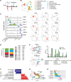
Figure 1
Figure 1. Transcriptional profiling of periosteal nonhematopoietic cells from intact and fractured bones.
(A) Experimental design for mouse treatment aimed at collecting samples for scRNA-seq. Femur fractures were induced in SMACreER/NICD1 and SMACre+ER/NICD1 male mice. To induce overexpression of NICD1, animals were treated with tamoxifen (Tx) on the day of fracture and 2 dpf. Femur samples of intact or injured periosteum were collected, digested, and cells were sorted for CD45 and CD45+. Subsequently, scRNA-seq was performed. (B) Clusters of periosteal CD45 cell populations with (C) violin and feature plots presenting characteristic conserved gene expression for each cluster from integrated intact and fractured Cre and Cre+ samples are shown. (D) Proportion of cells within the control intact and fractured sample of each cluster. (E) Periosteal cells from Cre intact and fractured samples were analyzed for cell cycle phases and cell proportion in the G2M phase. (F) GSEA indicates that MSCs1 is a stem/progenitor cell population within the periosteum and (G) Monocle3 trajectory analysis (https://cole-trapnell-lab.github.io/monocle-release/) shows cell differentiation from the MSCs1 cluster to mature chondrocytes and osteoblasts from integrated intact and fractured Cre and Cre+ samples and (H) trajectory of the clusters in pseudotime.
Figure 2
Figure 2. Periosteal cells from intact and fractured bones have distinct transcriptional profiles.
(A) Heatmaps of EC, MSCs1, MSCs2, satellite cell, and chondrocyte clusters showing differentially expressed genes with significantly increased or decreased expression of Cre control fractured compared with intact samples. Color intensity represents the mean gene expression of all cells within the cluster. (B) Feature plots of the specific genes identified in A that were found to be significantly increased in periosteal cells upon fracture compared with intact samples.
Figure 3
Figure 3. Overexpression of NICD1, during fracture healing, induces osteogenic and IFN signaling gene expression.
(A) Proportion of cells within each cluster in control Cre and Cre+ αSMACreER/NICD1 fractured samples. (B) Volcano plots showing differentially expressed genes in NICD1 fractured periosteal samples compared to NICD1 intact samples of EC, MSCs1, and MSCs2 clusters. (C) Heatmaps with the complete list of differentially expressed genes. Color intensity represents the mean gene expression of all cells within the cluster. (D) Feature plots of αSMACreER/NICD1 fractured Cre and Cre+ samples showing increased osteogenic genes (Ibsp, Alpl) and IFN signaling genes (Isg15, Ifit1).
Figure 4
Figure 4. Notch signaling in fracture healing.
(A) Feature plot of Dll4 expression within periosteal cells. (B) Experimental design of recombination efficiency evaluation by determining mRNA expression of Dll4 and Notch downstream signaling genes (Hes1 and Hey1) in male and female mice. To induce recombination, tamoxifen (Tx) was injected at 0, 2, and 4 dpf, and gene expression was evaluated at 7 dpf. Dll4 was successfully deleted in male mice, with decreased expression of Hes1 and Hey1 in the Cre+ callus (C) and lungs (D). Males: Cre n = 6, Cre+ n = 6; females: Cre n = 5, Cre+ n = 5. (E) Proportion of thymocyte subpopulations in the thymus, where deletion of Dll4 in ECs on day 5 after the first tamoxifen injection induced a small decrease in CD4+ cells and an increase in CD8+ cells, with representative dot plots. (F) Flow cytometry analysis showing dot plots of CD31+ cells within the callus at 5 dpf expressing DLL4. (G) Proportion of callus cells expressing Sca1, CD90, PDGFRα, and CD51 at 5 dpf, with representative overlaid histograms of Sca1 and CD90 cell expression (unstained control, Cre and Cre+ sample). In EG, Cre n = 3, Cre+ n = 4. Unpaired, 2-tailed Student’s t test. *P < 0.05.
Figure 5
Figure 5. Deletion of Dll4 in ECs impairs fracture healing.
(A) Experimental design. Deletion of Dll4 in ECs was induced by injecting tamoxifen (Tx) at 0, 2, and 4 dpf. Fractured bone samples were evaluated on day 4, 7, and 14 by histology and day 21 by microCT and torsion testing. (B) Dll4 deletion led to a decreased callus size at 4 dpf (Cre n = 6, Cre+ n = 9), 7 dpf (Cre n = 9, Cre+ n = 8), and 14 dpf (Cre n = 8, Cre+ n = 10). (C) Dll4 deletion also resulted in decreased proliferation at 4 dpf (Cre n = 5, Cre+ n = 9), which increased by 7 dpf (Cre n = 9, Cre+ n = 8) in Cre+ compared with Cre animals. (D and F) Cre+ mice had significantly less cartilage. Sample numbers at 4 dpf (Cre n = 5, Cre+ n = 9), 7 dpf (Cre n = 9, Cre+ n = 8), and 14 dpf (Cre n = 8, Cre+ n = 10) were analyzed by evaluating Safranin O–stained sections, as observed on representative sections. (E and G) Mineralized area was analyzed by von Kossa staining (Cre n = 8, Cre+ n = 10), as shown on Cre- and Cre+ representative sections. (H) MicroCT analysis showed decreased callus bone mass on day 14 and no difference in callus volume, with representative 3D reconstructions of Cre and Cre+ fractures on the right. At 7 dpf, Cre n = 8, Cre+ n = 6; and at 21 dpf Cre n = 7, Cre+ n = 9. (I) Biomechanical properties were evaluated by torsion testing and are presented as bone strength (maximum torque), stiffness as a measure of torsional rigidity, and toughness as a work to fracture measure, with no change between Cre and Cre+ fractured bones at 21 dpf. Cre n = 9, Cre+ n = 6. (J) Bglap gene expression analysis at 7 dpf (Cre n = 6, Cre+ n = 6). (K) Histological analysis of osteocalcin-stained samples at 14 dpf (Cre n = 8, Cre+ n = 10). (L) Evaluation of CD31+ cells within the periosteal callus at 7 dpf (Cre n = 9, Cre+ n = 12) and 14 dpf (Cre n = 7, Cre+ n = 9). Deletion of Dll4 in ECs with (M) representative magnified images of CD31 staining within the mineralized callus at 14 dpf. Scale bar: 200 μm. (N) Proportion of osterix-stained cells within the periosteal callus (Cre n = 5, Cre+ n = 6). (O) Representative images of osterix-stained fractured femurs with magnified areas of cartilaginous callus (a and b), mineralized callus (a’ and b’), and cortical bone with the area next to the pin insertion (a” and b”). The analyzed periosteal callus is shown by the yellow line. Scale bars: 1 mm (left) and 100 μm (right). PT, pin trace; CB, cortical bone; P, periosteum. Unpaired, 2-tailed Student’s t test. *P < 0.05, **P < 0.01.
Figure 6
Figure 6. Notch ligands improve critical size femoral defect healing.
BMP2 (5 μg), JAG1 (6 μg), and DLL4 (5 μg) alone or in combination were applied to a collagen scaffold (Medtronic). Femurs were evaluated by microCT and bone volume within the defect area was determined. JAG1 in combination with BMP2 resulted in significantly more bone within the defect compared with BMP2 only, with cortical bone formation in the whole length of a defect. On the right, representative 3D reconstructions of healing femoral defects 9 weeks after defect surgery. Saline, n = 5; BMP2, n = 12; JAG1, n = 11; BMP2 + JAG1, n = 10; DLL4, n = 4; BMP2 + DLL4, n = 11. One-way ANOVA with Bonferroni’s post hoc test. ***P < 0.001, statistically different from saline; ##P < 0.01, ###P < 0.001, statistically different from BMP2; †††P < 0.001, statistically different from BMP2 + JAG1; ‡‡‡P < 0.001, statistically different from BMP2 + DLL4.

Similar articles

Cited by

References

    1. Colnot C. Skeletal cell fate decisions within periosteum and bone marrow during bone regeneration. J Bone Miner Res. 2009;24(2):274–282. doi: 10.1359/jbmr.081003. - DOI - PMC - PubMed
    1. Grcevic D, et al. In vivo fate mapping identifies mesenchymal progenitor cells. Stem Cells. 2012;30(2):187–196. doi: 10.1002/stem.780. - DOI - PMC - PubMed
    1. Xu J, et al. PDGFRα reporter activity identifies periosteal progenitor cells critical for bone formation and fracture repair. Bone Res. 2022;10(1):7. doi: 10.1038/s41413-021-00176-8. - DOI - PMC - PubMed
    1. Liu YL, et al. Fibrous periosteum repairs bone fracture and maintains the healed bone throughout mouse adulthood. Dev Cell. 2024;59(9):1192–1209. doi: 10.1016/j.devcel.2024.03.019. - DOI - PubMed
    1. Zanotti S, Canalis E. Notch signaling and the skeleton. Endocr Rev. 2016;37(3):223–253. doi: 10.1210/er.2016-1002. - DOI - PMC - PubMed

MeSH terms

LinkOut - more resources