. 2024 May 23;20(5):e1011821.
doi: 10.1371/journal.ppat.1011821. eCollection 2024 May.

Mitotic deacetylase complex (MiDAC) recognizes the HIV-1 core promoter to control activated viral gene expression

Affiliations

Mitotic deacetylase complex (MiDAC) recognizes the HIV-1 core promoter to control activated viral gene expression

Emmanuelle Wilhelm et al. PLoS Pathog. .

Abstract

The human immunodeficiency virus (HIV) integrates into the host genome forming latent cellular reservoirs that are an obstacle for cure or remission strategies. Viral transcription is the first step in the control of latency and depends upon the hijacking of the host cell RNA polymerase II (Pol II) machinery by the 5' HIV LTR. Consequently, "block and lock" or "shock and kill" strategies for an HIV cure depend upon a full understanding of HIV transcriptional control. The HIV trans-activating protein, Tat, controls HIV latency as part of a positive feed-forward loop that strongly activates HIV transcription. The recognition of the TATA box and adjacent sequences of HIV essential for Tat trans-activation (TASHET) of the core promoter by host cell pre-initiation complexes of HIV (PICH) has been shown to be necessary for Tat trans-activation, yet the protein composition of PICH has remained obscure. Here, DNA-affinity chromatography was employed to identify the mitotic deacetylase complex (MiDAC) as selectively recognizing TASHET. Using biophysical techniques, we show that the MiDAC subunit DNTTIP1 binds directly to TASHET, in part via its CTGC DNA motifs. Using co-immunoprecipitation assays, we show that DNTTIP1 interacts with MiDAC subunits MIDEAS and HDAC1/2. The Tat-interacting protein, NAT10, is also present in HIV-bound MiDAC. Gene silencing revealed a functional role for DNTTIP1, MIDEAS, and NAT10 in HIV expression in cellulo. Furthermore, point mutations in TASHET that prevent DNTTIP1 binding block the reactivation of HIV by latency reversing agents (LRA) that act via the P-TEFb/7SK axis. Our data reveal a key role for MiDAC subunits DNTTIP1, MIDEAS, as well as NAT10, in Tat-activated HIV transcription and latency. DNTTIP1, MIDEAS and NAT10 emerge as cell cycle-regulated host cell transcription factors that can control activated HIV gene expression, and as new drug targets for HIV cure strategies.


Conflict of interest statement

PPMcD is also employed as Executive Director, Research at Insmed Inc. Insmed was however not involved in this work; did not fund it; and does not endorse it, implicitly or otherwise. B.B. owns shares in the biotechnology company Ascioma. Ascioma had no involvement with this research, financial or otherwise.

Figures

Fig 1
Fig 1. Affinity chromatography coupled with mass spectrometry to identify TASHET bound factors.
A) The unknown composition of pre-initiation complexes of HIV (PICH). The HIV LTR with transcription factor binding sites is shown at the top with a close-up of the TATA box and adjacent sequences of HIV essential for Tat trans-activation (TASHET) shown below. The CTGC DNA motifs (blue) have been shown to be essential for Tat activation and recognition by PICH [25]. PICH contain general transcription factors such as TBP, but their complete composition and how they link TASHET to Tat function have remained unknown (question mark). B) TASHET sequence and point mutations used in this study. The HIV-1 TATA box and E-box are shown by the purple and green squares, respectively. CTGC motifs are highlighted in blue and mutated nucleotides figure in white or light grey. The ’ATCC’ mutation was formerly named CTGC5’3’ [25]. “Scr” indicates a scrambled control sequence. C) A representative silver-stained polyacrylamide gel reveals the bands corresponding to the proteins specifically (annotated in black) and non-specifically (annotated in light grey) bound to wild type and mutated TASHET. wt 130 and wt 37 stand for the bands that were cut out for mass spectrometry analysis. D) Identity and mass spectrometry scores of the main candidates identified in the analysed bands (see also S2 Fig and S1 Table for further detailed information).
Fig 2
Fig 2. MIDEAS and DNTTIP1 form a complex that specifically binds to TASHET.
A) Western blot validation of the binding of MIDEAS, DNTTIP1 and HDAC1 from HeLa cell nuclear extracts to wild type TASHET. B) As in A, except with nuclear extracts from Jurkat cells. In the case of NAT10, note that levels in lanes 5 & 6 correspond to background levels that are observed also with random DNA sequences. C) As in A, except with nuclear extracts from PBMC. MiDAC components selectively bind wt TASHET and retain binding for USF1KO mutant. Tat response as assayed in reporter transfected cells is expressed as ‘+’ when > 90% and ‘-’ when <20% response to Tat transactivation [25, 111]. D) As in A, except that the HeLa cells were blocked in the G2/M phase by a 16h treatment with nocodazole before nuclear protein extraction. E) Co-immunoprecipitation showing interactions among MiDAC components, as well as with Cyclin T1 in Jurkat nuclear extracts. Antibodies used for IP are indicated at the top, antibodies used in Western Blot to the left. For visibility, co-IP are compared to 20% (lane 1) or 5% (lane 2) input, except for DNTTIP1, where lane 2 (indicated by *) corresponds to 10% input.
Fig 3
Fig 3. DNTTIP1 interacts directly, with high affinity and specificity to wt TASHET.
A) Upper panel: schematic view of DNTTIP1 protein showing the dimerization domain (DD in orange), AT-hook (purple) and c-terminal DNA binding domain (in pale blue) structurally related to the SKI/SNO/DAC domain [42]. The SKI/SNO/DAC domain contains an embedded motif proposed to have structural similarity to helix-turn-helix (HTH) motifs [47, 48] (dark blue). Lower panel: alignment of the DNTTIP1 consensus sequence (top), the SELEX sequence #12 as published by Kubota et al. [50], and the TASHET sequence. High similarity sequences are shown including the 5’CTGC and TATA motifs (purple box) and around the 3’ CTGC motif (blue box). Vertical lines indicate identities. B) Coomassie staining, and C) Circular Dichroism (CD) spectroscopic spectrum of purified recombinant His-tagged DNTTIP1. D) CD spectroscopy thermal denaturation curves measured at a 222nm wavelength (left) and calculated melting temperatures (Tm) (right) of recombinant DNTTIP1 in absence (blue) or equimolar presence of wt (yellow), or ATCC (green) mutated TASHET, or of a DNTTIP1 binding consensus sequence (sequences in S1 File) [50] (red). D) Anisotropy changes of fluorescein labeled DNA (sequences in Fig 1 and S1 File) in presence of increasing concentration of recombinant DNTTIP1. Measured values have been fitted to a specific binding with Hill slope (left panel: graphic representation), yielding the calculated Kd values with its SD, high R2 (right panel), ΔRmax corresponding to the plateau, n corresponding to the Hill coefficient with its SD.
Fig 4
Fig 4. DNTTIP1 occupies the HIV proviral promoter in cellulo.
A) Western Blot showing the expression level of exogenous DNTTIP1-Flag transgene compared to endogenous DNTTIP1 in a cell population with stable integration of a lentivirus bearing the DNTTIP1-Flag sequence (+) vs an empty lentivirus (-). TBP was used as a loading control. B) Positioning of the qPCR primers used in ChIP qPCR on HIV LTR region. C) Chromatin Immunoprecipitation in NL4.3-Luc infected HeLa cells expressing a stable version of flagged-DNTTIP1. Enrichment obtained with antibodies directed against Pol-II, TBP, Flag-DNTTIP1, HIV1-Tat and HDAC1 vs no antibody (-) on inactive chromatin regions (GDM, bGlob, green bars), an active cellular gene promoter (DDIT3, purple bars), integrated HIV distal (-455, light blue bars) and proximal (-90, blue bars) promoter. The error bars represent the standard deviation of three independent ChIP assays performed in parallel. The chromatin occupancy is expressed in percentage of input.
Fig 5
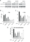
Fig 5. DNTTIP1 and MIDEAS are required for optimal HIV-1 promoter activity in living cells.
The activity of a transfected HIV-1 promoter reporter was measured in HeLa cells after siRNA mediated knock-down of DNTTIP1 (D1 and D2 siRNAs), MIDEAS (M1, M2, and M3 siRNAs) or NAT10 (N1 and N2 siRNAs) compared to a non-targeting siRNA (ctrl-) or mock transfected cells (-). A) Target Knock-Down validation by Western Blot. USF1 protein levels were monitored as a loading control. (B) The percentage of YFP positive cells in the absence (cells co-transfected empty vector control) of HIV Tat. (C) The percentage of YFP positive cells in the presence of HIV Tat (cells co-transfected Tat expressing plasmid) of HIV Tat. P-values were calculated in comparison to the Ctrl siRNA (black bars). * p<0.05, **p<0.01, ***p<0.001. D) Tat transactivation that is expressed as the ratio of mean fluorescence intensity in presence vs that measured in the absence of Tat co-expression, with siRNA ctrl being set to 100%. The error bars in panels B–D represent the standard deviation of three independent experiments performed in parallel.
Fig 6
Fig 6. DNTTIP1 is required for the optimal activity of an integrated provirus in MOLT4 lymphocytes.
The activity of integrated proviral HIV-1 promoter was monitored by luciferase activity after knock-down of DNTTIP1 by three different shRNA lentiviral constructs (#1, #2, #3). A) Target Knock-Down validation by Western Blot. Tubulin was employed a loading control. B) Quantification of relative DNTTIP1 protein expression measured in A. C) The integrated proviral HIV promoter activity (left axis: RLU/μg, the mock control being set to 100%), and the number of HIV proviruses per cell as analyzed by qPCR (right axis, in green) were measured in the same cells. Error bars represent the standard deviation of three independent infections performed in parallel. P-values were calculated in comparison to the Ctrl shRNA (black bars). * p<0.05, **p<0.01.
Fig 7
Fig 7. Latency reversing agents (LRA) acting through the P-TEFb/7SK axis require the DNTTIP1 binding sites of TASHET.
A) A schematic representation of the major LRA pathways acting through NF-κB (green), HDACs (purple) or the P-TEFb/7SK axis (blue). B) Impact of LRAs on reporter viruses. Jurkat cells were infected with single-round reporter viruses bearing mutations in the HIV LTR (indicated on the x-axis), and then treated with LRAs for 16 hours before harvesting cell extracts for luciferase assays. The activity of integrated proviral HIV-1 promoter was monitored by luciferase activity. The results, calculated as RLU/μg of total protein are expressed as fold induction over the DMSO vehicle control for a given virus (y-axis). LRA color codes correspond to those in panel A. C) As in B, with additional LRA treatments and LTR mutations. Error bars represent the standard deviation of three independent experiments performed in parallel.
Fig 8
Fig 8. Working model.
A hypothetical model depicting the binding of the mitotic deacetylase complex (MiDAC) to TASHET of the HIV core promoter. Host cell regulators DNTTIP1, MIDEAS and NAT10 identified in this work are in blue. Multimeric DNTTIP1 recognizes the CTGC motifs flanking the TATA box, likely via its SKI/SNO/DAC domain containing a HTH-like motif, and the TATA box via its AT-hook domain. DNTTIP1 interacts strongly with MIDEAS to recruit HDAC1/2. NAT10 is also recruited via MIDEAS and directly contacts HIV Tat to enhance HIV transcription.

References

    1. Deeks SG, Archin N, Cannon P, Collins S, Jones RB, de Jong M, et al.. Research priorities for an HIV cure: International AIDS Society Global Scientific Strategy 2021. Nat Med. 2021;27(12):2085–98. Epub 20211201. doi: 10.1038/s41591-021-01590-5 . - DOI - PubMed
    1. Barouch DH, Deeks SG. Immunologic strategies for HIV-1 remission and eradication. Science. 2014;345(6193):169–74. Epub 2014/07/12. doi: 10.1126/science.1255512 ; PubMed Central PMCID: PMC4096716. - DOI - PMC - PubMed
    1. Barouch DH. Challenges in the development of an HIV-1 vaccine. Nature. 2008;455(7213):613–9. Epub 2008/10/04. doi: 10.1038/nature07352 ; PubMed Central PMCID: PMC2572109. - DOI - PMC - PubMed
    1. Blancou P, Chenciner N, Ho Tsong Fang R, Monceaux V, Cumont MC, Guetard D, et al.. Simian immunodeficiency virus promoter exchange results in a highly attenuated strain that protects against uncloned challenge virus. J Virol. 2004;78(3):1080–92. Epub 2004/01/15. doi: 10.1128/jvi.78.3.1080-1092.2004 ; PubMed Central PMCID: PMC321388. - DOI - PMC - PubMed
    1. Delannoy A, Poirier M, Bell B. Cat and Mouse: HIV Transcription in Latency, Immune Evasion and Cure/Remission Strategies. Viruses. 2019;11(3). Epub 20190318. doi: 10.3390/v11030269 ; PubMed Central PMCID: PMC6466452. - DOI - PMC - PubMed

MeSH terms

Substances