Review
. 2024 Jun 25;102(12):e209482.
doi: 10.1212/WNL.0000000000209482. Epub 2024 May 23.

Does Surgical Removal of the Thymus Have Deleterious Consequences?

Affiliations
Review

Does Surgical Removal of the Thymus Have Deleterious Consequences?

Henry J Kaminski et al. Neurology. .

Abstract

The role of immunosenescence, particularly the natural process of thymic involution during aging, is increasingly acknowledged as a factor contributing to the development of autoimmune diseases and cancer. Recently, a concern has been raised about deleterious consequences of the surgical removal of thymic tissue, including for patients who undergo thymectomy for myasthenia gravis (MG) or resection of a thymoma. This review adopts a multidisciplinary approach to scrutinize the evidence concerning the long-term risks of cancer and autoimmunity postthymectomy. We conclude that for patients with acetylcholine receptor antibody-positive MG and those diagnosed with thymoma, the removal of the thymus offers prominent benefits that well outweigh the potential risks. However, incidental removal of thymic tissue during other thoracic surgeries should be minimized whenever feasible.


Conflict of interest statement

H.J. Kaminski is principal investigator for the Rare Disease Network (MGNet supported by NIH grant U54NS115054) is a consultant for R43NS12432, Roche, Takeda, Cabaletta Bio, UCB Pharmaceuticals, Canopy EMD Serono, Ono Pharmaceuticals, ECoR1, Gilde Healthcare, and Admirix, Inc., has received research support from Argenix (paid to institution), and has equity interest in Mimivax, LLC, and has equity interest in Mimivax, LLC, and serves as a consultant for GRO Biotechnology, CSL Berhing, Amplo Biotechnology, and Sanofi. G.I. Wolfe has served as vice chair for the International Treatment Consensus panel (funded by the Myasthenia Gravis Foundation of America). The other authors report no relevant disclosures. Go to Neurology.org/N for full disclosures.

Figures

Figure 1
Figure 1. T-Cell Development in the Thymus
Hematopoietic progenitor cells enter the thymus and migrate to the outer cortex as immature double-negative (DN) thymocytes, lacking both CD4 and CD8 coreceptors. The DN population matures as observed by the CD44 and CD25 expression: CD44+CD25 (DN1), CD44+CD25+ (DN2), CD44CD25+ (DN3), and CD44CD25 (DN4). During this time, the DN thymocytes also express the pre-TCR consisting of a TCR β-chain and an invariant preTCR α-chain (pTα) allowing the TCRβ gene rearrangements. Expression of the preTCR enables DN cells with productive TCRβ rearrangements to differentiate into CD4+CD8+ double-positive (DP) thymocytes and the rearrangement of the TCR α-chain to begin. Positive selection through the interaction with the MHCI and MHCII on the cortical thymic epithelial cells (cTECs) determines the fate of the thymocyte during TCRβ and TCR α gene rearrangements. The thymocytes travel to the medulla as single-positive CD4+ or CD8+ to undergo central tolerance through negative selection by interacting with the dendritic cells, medullary thymic epithelial cells (mTECs) and B cells. The recognition of self will direct the cell to an apoptotic pathway or a regulatory T-cell fate. Mature CD4+ and CD8+ thymocytes can then exit the thymus and begin their role in immune regulation.
Figure 2
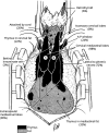
Figure 2. Surgical Anatomy of the Thymus
The illustration depicts the anatomy of the thymus. A and B designate the classic depiction of the thymus with majority of thymus in the cervical mediastinum. However, thymic tissue is frequently found outside these areas extending to the neck and the mediastinum. The black identifies thymus tissue and gray is fat, which may have small amounts of thymic tissue or only microscopic thymus. The percentages estimate the percentage of thymus and fat. Used with permission of Wolters Kluwer Health, Inc. published Jaretzki et al.

References

    1. Ashby KM, Hogquist KA. A guide to thymic selection of T cells. Nat Rev Immunol. 2024;24(2):103-117. doi:10.1038/s41577-023-00911-8 - DOI - PubMed
    1. This S, Rogers D, Mallet Gauthier E, Mandl JN, Melichar HJ. What's self got to do with it: sources of heterogeneity among naive T cells. Semin Immunol. 2023;65:101702. doi:10.1016/j.smim.2022.101702 - DOI - PubMed
    1. Lynch HE, Goldberg GL, Chidgey A, Van den Brink MR, Boyd R, Sempowski GD. Thymic involution and immune reconstitution. Trends Immunol. 2009;30(7):366-373. doi:10.1016/j.it.2009.04.003 - DOI - PMC - PubMed
    1. Steinmann GG, Klaus B, Muller-Hermelink HK. The involution of the ageing human thymic epithelium is independent of puberty. A morphometric study. Scand J Immunol. 1985;22(5):563-575. doi:10.1111/j.1365-3083.1985.tb01916.x - DOI - PubMed
    1. Griffith AV, Fallahi M, Venables T, Petrie HT. Persistent degenerative changes in thymic organ function revealed by an inducible model of organ regrowth. Aging Cell. 2012;11(1):169-177. doi:10.1111/j.1474-9726.2011.00773.x - DOI - PMC - PubMed