Clinical Trial
. 2024 Sep 12;144(11):1153-1167.
doi: 10.1182/blood.2024024104.

Phase 1 study of CAR-37 T cells in patients with relapsed or refractory CD37+ lymphoid malignancies

Affiliations
Clinical Trial

Phase 1 study of CAR-37 T cells in patients with relapsed or refractory CD37+ lymphoid malignancies

Matthew J Frigault et al. Blood. .

Abstract

We report a first-in-human clinical trial using chimeric antigen receptor (CAR) T cells targeting CD37, an antigen highly expressed in B- and T-cell malignancies. Five patients with relapsed or refractory CD37+ lymphoid malignancies were enrolled and infused with autologous CAR-37 T cells. CAR-37 T cells expanded in the peripheral blood of all patients and, at peak, comprised >94% of the total lymphocytes in 4 of 5 patients. Tumor responses were observed in 4 of 5 patients with 3 complete responses, 1 mixed response, and 1 patient whose disease progressed rapidly and with relative loss of CD37 expression. Three patients experienced prolonged and severe pancytopenia, and in 2 of these patients, efforts to ablate CAR-37 T cells, which were engineered to coexpress truncated epidermal growth factor receptor, with cetuximab were unsuccessful. Hematopoiesis was restored in these 2 patients after allogeneic hematopoietic stem cell transplantation. No other severe, nonhematopoietic toxicities occurred. We investigated the mechanisms of profound pancytopenia and did not observe activation of CAR-37 T cells in response to hematopoietic stem cells in vitro or hematotoxicity in humanized models. Patients with pancytopenia had sustained high levels of interleukin-18 (IL-18) with low levels of IL-18 binding protein in their peripheral blood. IL-18 levels were significantly higher in CAR-37-treated patients than in both cytopenic and noncytopenic cohorts of CAR-19-treated patients. In conclusion, CAR-37 T cells exhibited antitumor activity, with significant CAR expansion and cytokine production. CAR-37 T cells may be an effective therapy in hematologic malignancies as a bridge to hematopoietic stem cell transplant. This trial was registered at www.ClinicalTrials.gov as #NCT04136275.


Conflict of interest statement

Conflicts-of-interest disclosure: M.V.M. and I.S. report being inventors on patents describing chimeric antigen receptor-37 (CAR-37) therapy titled “CD37-Targeted Chimeric Antigen Receptor T Cells for Lymphomas/Leukemias” (PCT/US2018/022974); “Anti-CD37 Chimeric Antigen Receptor T Cells for Non-Hodgkin Lymphoma” (PCT/US2019/038518). In addition, M.V.M. reports being an inventor on patents related to adoptive cell therapies, held by Massachusetts General Hospital and the University of Pennsylvania (some licensed to Novartis). M.V.M. reports holding equity in AffyImmune, Century Therapeutics, Oncternal Therapeutics, Neximmune, and TCR2; serving on the board of directors of 2seventy bio; and has served as a consultant for multiple companies involved in cell therapies. M.V.M.’s interests were reviewed and are managed by the Massachusetts General Hospital and Mass General Brigham in accordance with their conflict-of-interest policies. J.F.D. reports holding equity in Magenta Therapeutics and Wugen Inc; receiving research support from MacroGenics, Bioline, and Incyte; and serving as a consultant for Vertex, bluebird bio, SPARC, and RiverVest. J.R. reports receiving research funding from Equillium, Kite/Gilead, Novartis, and Oncternal Therapeutics; and serving on the scientific advisory boards of Akron Biotech, Clade Therapeutics, Garuda, LifeVault Bio, Novartis, Smart Immune, and TScan Therapeutics. The remaining authors declare no competing financial interests.

Figures

None
Graphical abstract
Figure 1.
Figure 1.
Study design and CAR-37 T-cell manufacturing. (A) Design of the lentiviral vector construct used to produce CAR-37 T cells. (B) Study design and (C) CAR-37 manufacturing process. (D) End-product percentage transduction of patient T cells as measured by the percentage EGFR+CD3+CD45+ cells in the live cell population (by 4′,6-diamidino-2-phenylindole staining) via flow cytometry. (E) Copies of CAR-37 vector per microgram of DNA in the final product as measured by PCR. (F) The total number of CAR-37 T cells produced per patient and available for dosing (left y-axis, solid bars) and the fold expansion of the patient’s T cells during manufacturing (x-axis, striped bars), along with the dose of CAR-37 T cells they received (black dots), are shown. (G) Percent of the final product that is CD4+ (colored bar, bottom) vs CD8+ (white bar, top). Cy, cyclophosphamide; DP, drug product; flu, fludarabine; PBMC, peripheral blood mononuclear cell; scFv, single-chain variable fragment.
Figure 2.
Figure 2.
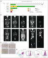
Patient response to CAR-37 T cells. (A) Swimmer plot of patient’s responses to CAR-37 T cells. Two patients received HSCT as a consequence of cytopenia at the time points indicated by the diamonds. Other symbols indicate the cause of death for each patient, as noted. (B) Positron emission tomography (PET) scans of patients with CR, PR, or MR at the indicated time points. (C) Immunohistochemistry for CD37 in lesion biopsies before CAR T-cells infusion (preinfusion) and at the time of relapse by PET scan. Images were taken at 20×. (D) CD19, CD20, and CD37 expression in tumor from patient 1 as measured by flow cytometry performed by the clinical pathology laboratory. The purple dots and histogram represent the aberrant population. CTCL, cutaneous T-cell lymphoma; HGBCL, high-grade B-cell lymphoma; PD, progressive disease; PR, partial response.
Figure 3.
Figure 3.
Toxicity in CAR-37 patients correlates with increased serum cytokines. (A) The timing, grade, and duration of CRS (open bars) and ICANS (dotted bars) after CAR-37 T-cell infusion (day 0) are shown. The bar color indicates grade, as noted. Open/white bars indicate duration with no CRS or ICANS symptoms. (B) As part of the diagnostic workup for CRS and ICANS, ferritin (left y-axis, closed symbols) and c-reactive protein (CRP; right y-axis, open symbols) were measured in patient serum over time. The solid black line indicates the maximum normal ferritin level. The dashed black line indicates the maximum normal CRP level. The underlaying horizontal bars indicate the timing and duration of treatment for CRS or ICANS, as noted by the treatment name in the bar. Bars in the same row represent the same treatment. (C) Cell counts per microliter of blood pre- and post-CAR-37 infusion (day 0). Dashed lines indicate the normal range for each cell type. (D) Hematoxylin and eosin staining (20× original magnification) of bone marrow biopsies taken from patients 4 and 5 on day 27 after CAR-37 T-cell infusion. ANC, absolute neutrophil count; G-CSF, granulocyte colony-stimulating factor; Sil, siltuximab; Toci, tocilizumab; WBC, white blood cell.
Figure 4.
Figure 4.
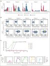
CAR-37 T-cell expansion after infusion. (A) The absolute numbers of neutrophils, monocytes, and lymphocytes per microliter of blood after CAR-37 infusion (day 0) are shown. (B) Flow cytometry dot plots of CD3+EGFR+ CAR-37 T cells in peripheral blood are shown on day −5 (before CAR-37 T-cell infusion) and day 14 after CAR-37 T-cell infusion. (C) The percentage of CD3+ cells that were EGFR+ as detected in patient peripheral blood by flow cytometry over time after CAR-37 T-cell infusion on day 0 is shown. (D) The number of CAR-37 T cells per microliter of peripheral blood is shown as calculated from the percentage of CD3+EGFR+ cells detected by flow cytometry and the absolute lymphocyte count (left y-axis, closed symbols), as well as the VCN, reported as copies per microgram of DNA (right y-axis, open symbols), over time after CAR-37 T-cell infusion. PE, phycoerythrin.
Figure 5.
Figure 5.
Cetuximab treatment to deplete CAR-37 T cells via tEGFR. (A) Flow cytometry dot plots of CD3+EGFR+ cells in peripheral blood at the indicated time points. Patients 4 and 5 who experienced bone marrow aplasia at day 27 after CAR-37 therapy were treated with cetuximab on day 28 (patient 5) or day 35 (patient 4) to deplete their CAR-37 T cells (indicated by the arrows). On day 35 (patient 5) or day 43 (patient 4), the patients were treated with transplant conditioning chemotherapy and underwent a HSCT (indicated by the dashed line). The post-HSCT flow plots are also shown. (B) The percentage of CD3+EGFR+ cells in peripheral blood (left axis, bar graphs) in comparison with the VCN, reported as copies per microgram of DNA, detected by PCR in peripheral blood (right y-axis, line graphs) at the indicated time points is shown. Arrows indicate the timing of cetuximab treatment. The dashed line indicates the timing of the transplant conditioning treatment as shown in panel A.
Figure 6.
Figure 6.
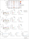
Peripheral cytokine levels in patients with and without cytopenia treated with CAR-37 in comparison with CAR-19. (A) Serum protein levels were measured using the Luminex at the indicated time points. Circle size corresponds to the log10 scaled concentration of the analyte in comparison with the minimum concentration (conc) of the analyte across patients. Circle color corresponds to the log2 fold change of the analyte in comparison with the day 5 time point for each patient. (B-D) Banked serum samples from patients treated at our institution with a licensed CD19 CAR for lymphoma were analyzed using a custom kit measuring (B) IL-8, (C) IFN-γ, and (D) IL-18 on the Ella, bio-techne and were compared with serum samples from our CAR-37 patients measured with the same kit. Patients were classified as having prolonged cytopenia if they had an ANC <0.5 x10³ cells per μL blood at ≥28 days after CAR T-cell infusion. Patients were excluded if they had another cause of cytopenia, such as myelodysplasia or constitutional bone marrow failure. We compared CD19 CAR patients without cytopenia (n = 12) and cytopenia (n = 12) with CAR-37 patients without cytopenia (n = 1) and with cytopenia (n = 4). (E) IL-18 BP levels were measured on the Ella (Bio-Techne) for CAR-37 patients. Absolute values, AUC, and peak fold change from baseline are shown. (F) The ratio of IL-18 to IL-18 BP is shown over time after infusion. P values were calculated using a 1-way analysis of variance test with Tukey multiple comparisons test.

Comment in

References

    1. Locke FL, Miklos DB, Jacobson CA, et al. Axicabtagene ciloleucel as second-line therapy for large B-cell lymphoma. N Engl J Med. 2022;386(7):640–654. - PubMed
    1. Abramson JS, Solomon SR, Arnason J, et al. Lisocabtagene maraleucel as second-line therapy for large B-cell lymphoma: primary analysis of the phase 3 TRANSFORM study. Blood. 2023;141(14):1675–1684. - PMC - PubMed
    1. Kamdar M, Solomon SR, Arnason J, et al. Lisocabtagene maraleucel versus standard of care with salvage chemotherapy followed by autologous stem cell transplantation as second-line treatment in patients with relapsed or refractory large B-cell lymphoma (TRANSFORM): results from an interim analysis of an open-label, randomised, phase 3 trial. Lancet. 2022;399(10343):2294–2308. - PubMed
    1. Neelapu SS, Locke FL, Bartlett NL, et al. Axicabtagene ciloleucel CAR T-cell therapy in refractory large B-cell lymphoma. N Engl J Med. 2017;377(26):2531–2544. - PMC - PubMed
    1. Schuster SJ, Bishop MR, Tam CS, et al. Tisagenlecleucel in adult relapsed or refractory diffuse large B-cell lymphoma. N Engl J Med. 2019;380(1):45–56. - PubMed

Publication types

MeSH terms

Associated data