

Deciphering Unexpected Vascular Locations of Scedosporium spp. and Lomentospora prolificans Fungal Infections, France
- PMID: 38781681
- PMCID: PMC11138966
- DOI: 10.3201/eid3006.231409
Deciphering Unexpected Vascular Locations of Scedosporium spp. and Lomentospora prolificans Fungal Infections, France
Abstract
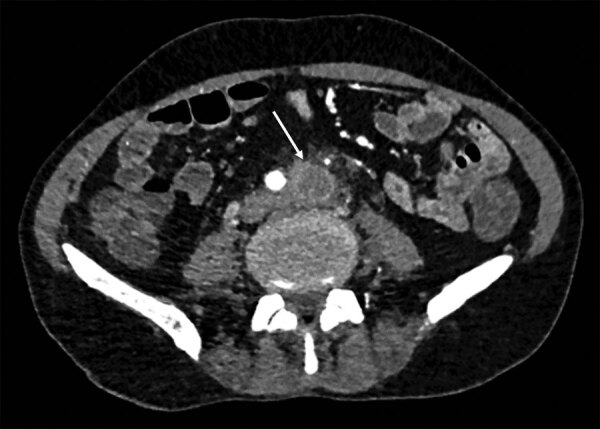
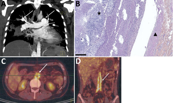
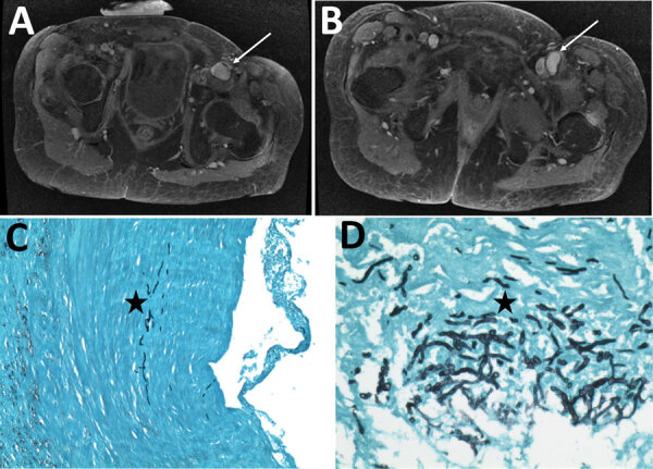
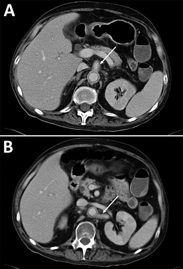
Scedosporium spp. and Lomentospora prolificans are emerging non-Aspergillus filamentous fungi. The Scedosporiosis/lomentosporiosis Observational Study we previously conducted reported frequent fungal vascular involvement, including aortitis and peripheral arteritis. For this article, we reviewed 7 cases of Scedosporium spp. and L. prolificans arteritis from the Scedosporiosis/lomentosporiosis Observational Study and 13 cases from published literature. Underlying immunosuppression was reported in 70% (14/20) of case-patients, mainly those who had solid organ transplants (10/14). Osteoarticular localization of infection was observed in 50% (10/20) of cases; infections were frequently (7/10) contiguous with vascular infection sites. Scedosporium spp./Lomentospora prolificans infections were diagnosed in 9 of 20 patients ≈3 months after completing treatment for nonvascular scedosporiosis/lomentosporiosis. Aneurysms were found in 8/11 aortitis and 6/10 peripheral arteritis cases. Invasive fungal disease--related deaths were high (12/18 [67%]). The vascular tropism of Scedosporium spp. and L. prolificans indicates vascular imaging, such as computed tomography angiography, is needed to manage infections, especially for osteoarticular locations.
Keywords: France; Lomentospora; Lomentospora prolificans; Scedosporium; aortitis; arteritis; fungi; lomentosporiosis; mycotic aneurysm; scedosporiosis; vascular infections.
Figures
Similar articles
-
Prognostic factors in 264 adults with invasive Scedosporium spp. and Lomentospora prolificans infection reported in the literature and FungiScope®.Crit Rev Microbiol. 2019 Feb;45(1):1-21. doi: 10.1080/1040841X.2018.1514366. Epub 2019 Jan 10. Crit Rev Microbiol. 2019. PMID: 30628529
-
Scedosporiosis/lomentosporiosis observational study (SOS): Clinical significance of Scedosporium species identification.Med Mycol. 2021 May 4;59(5):486-497. doi: 10.1093/mmy/myaa086. Med Mycol. 2021. PMID: 33037432
-
Fusarium species,Scedosporium species, and Lomentospora prolificans: A systematic review to inform the World Health Organization priority list of fungal pathogens.Med Mycol. 2024 Jun 27;62(6):myad128. doi: 10.1093/mmy/myad128. Med Mycol. 2024. PMID: 38935914 Free PMC article.
-
Invasive Scedosporium spp. and Lomentospora prolificans infections in pediatric patients: Analysis of 55 cases from FungiScope® and the literature.Int J Infect Dis. 2020 Mar;92:114-122. doi: 10.1016/j.ijid.2019.12.017. Epub 2019 Dec 19. Int J Infect Dis. 2020. PMID: 31863876
-
Invasive bone and joint infections from the French Scedosporiosis/lomentosporiosis Observational Study (SOS) cohort: no mortality with long-term antifungal treatment and surgery.Med Mycol. 2023 Mar 2;61(3):myad023. doi: 10.1093/mmy/myad023. Med Mycol. 2023. PMID: 36813259 Review.
References
-
- Lackner M, de Hoog GS, Yang L, Ferreira Moreno L, Ahmed SA, Andreas F, et al. Proposed nomenclature for Pseudallescheria, Scedosporium and related genera. Fungal Divers. 2014;67:1–10. 10.1007/s13225-014-0295-4 - DOI
-
- Blez D, Bronnimann D, Rammaert B, Zeller V, Delhaes L, Hustache L, et al. Invasive bone and joint infections from the French Scedosporiosis/lomentosporiosis Observational Study (SOS) cohort: no mortality with long-term antifungal treatment and surgery. Med Mycol. 2023;61:myad023. - PubMed
-
- Seidel D, Meißner A, Lackner M, Piepenbrock E, Salmanton-García J, Stecher M, et al. Prognostic factors in 264 adults with invasive Scedosporium spp. and Lomentospora prolificans infection reported in the literature and FungiScope®. Crit Rev Microbiol. 2019;45:1–21. 10.1080/1040841X.2018.1514366 - DOI - PubMed
Publication types
MeSH terms
Substances
Supplementary concepts
LinkOut - more resources
Full Text Sources
Medical
Miscellaneous
