. 2024 Jul:73:103197.
doi: 10.1016/j.redox.2024.103197. Epub 2024 May 16.

Nobiletin regulates intracellular Ca2+ levels via IP3R and ameliorates neuroinflammation in Aβ42-induced astrocytes

Affiliations

Nobiletin regulates intracellular Ca2+ levels via IP3R and ameliorates neuroinflammation in Aβ42-induced astrocytes

Sanjay et al. Redox Biol. 2024 Jul.

Abstract

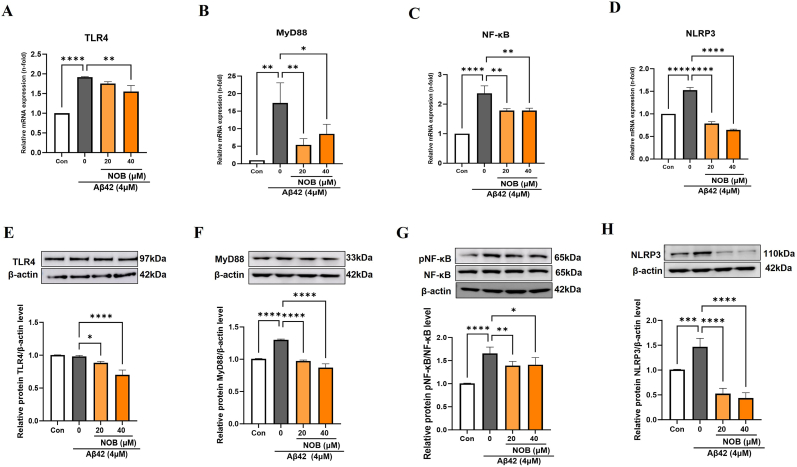
Astrocytes are the major glial cells in the human brain and provide crucial metabolic and trophic support to neurons. The amyloid-β peptide (Aβ) alter the morphological and functional properties of astrocytes and induce inflammation and calcium dysregulation, contributing to Alzheimer's disease (AD) pathology. Recent studies highlight the role of Toll-like receptor (TLR) 4/nuclear factor kappa-light-chain-enhancer of activated B cells (NF-κB) signaling in inflammation. Reactive oxygen species (ROS) generated due to Aβ, induce apoptosis in the brain cells worsening AD progression. Astrocytic cell surface receptors, such as purinergic receptors (P2Y1 and P2Y2), metabotropic glutamate receptor (mGLUR)5, α7 nicotinic acetylcholine receptor (α7nAChR), and N-methyl-d-aspartate receptors (NMDARs), have been suggested to interact with inositol trisphosphate receptor (IP3R) on the endoplasmic reticulum (ER) to induce Ca2+ movement from ER to cytoplasm, causing Ca2+ dysregulation. We found that the citrus flavonoid nobiletin (NOB) protected primary astrocytes from Aβ42-induced cytotoxicity and inhibited TLR4/NF-κB signaling in Aβ42-induced primary rat astrocytes. NOB was found to regulate Aβ42-induced ROS levels through Keap1-Nrf2 pathway. The receptors P2Y1, P2Y2, mGLUR5, α7nAChR, and NMDARs induced intracellular Ca2+ levels by activating IP3R and NOB regulated them, thereby regulating intracellular Ca2+ levels. Molecular docking analysis revealed a possible interaction between NOB and IP3R in IP3R regulation. Furthermore, RNA sequencing revealed various NOB-mediated biological signaling pathways, such as the AD-presenilin, AD-amyloid secretase, and Wnt signaling pathway, suggesting possible neuroprotective roles of NOB. To conclude, NOB is a promising therapeutic agent for AD and works by modulating AD pathology at various levels in Aβ42-induced primary rat astrocytes.

Keywords: Alzheimer's disease; Calcium dysregulation; Citrus flavonoid; Neuroinflammation; Nobiletin.


Conflict of interest statement

Declaration of competing interest The authors declare that they have no known competing financial interests or personal relationships that could have appeared to influence the work reported in this paper.

Figures

Image 1
Graphical abstract
Fig. 1
Fig. 1
Nobiletin protects primary rat astrocytes from Aβ42-induced cytotoxicity and regulates inflammation. (A–C) Primary rat astrocytes were treated with different concentrations of Aβ42 (0.5, 1, 2, and 4 μM), NOB (5, 10, 20, and 40 μM), and 4 μM Aβ42 in the presence or absence of different concentrations of NOB (0–40 μM) for 24 h, and cell viability was observed using the CCK-8 assay. (D–G) Primary astrocytes were treated with 4 μM Aβ42 in the presence and absence of 20 or 40 μM NOB for 24 h and mRNA expressions of pro-inflammatory markers IL-1β, TNF-α, IL-6, and iNOS were assessed through RT-PCR. Con, control; Aβ42, amyloid beta-42; NOB, nobiletin; IL, interleukin; TNF-α, tumor necrosis factor-α; iNOS, inducible nitric oxide synthase. *p < 0.05, ***p < 0.001, ****p < 0.0001.
Fig. 2
Fig. 2
Nobiletin regulates neuroinflammation through TLR4/NF-κB in Aβ42-induced primary rat astrocytes. (A–D) Primary astrocytes were treated with 4 μM Aβ42 in the presence and absence of 20 or 40 μM NOB for 24 h and mRNA expression levels of TLR4, MyD88, NF-κB, and NLRP3 were assessed by RT-PCR. (E–H) Protein expression levels of TLR4, MyD88, pNF-κB/NF-κB, and NLRP3 were assessed by Western blot. Con, control; Aβ42, amyloid beta-42; NOB; nobiletin; TLR4, toll-like receptor 4; MyD88, myeloid differentiation primary-response protein 88; NF-κB, nuclear factor kappa B; NLRP3, NOD-, LRR- and pyrin domain-containing protein 3. *p < 0.05, **p < 0.01, ***p < 0.001, ****p < 0.0001.
Fig. 3
Fig. 3
Nobiletin attenuates Aβ42-induced ROS through Keap1-Nrf2 antioxidant pathway in primary rat astrocytes. (A–B) Primary astrocytes were treated with 4 μM Aβ42 in the presence and absence of 20 or 40 μM NOB for 24 h and relative ROS levels were measured by microplate reader and visualized by fluorescence microscopy using DCF-DA assay. (C–H) Primary astrocytes were treated with 4 μM Aβ42 in the presence and absence of 20 or 40 μM NOB for 24 h and mRNA expression levels of Keap1, Nrf2, HO-1, CAT, GPx and SOD were observed by RT-PCR. Con, control; Aβ42, amyloid beta-42; NOB, nobiletin; ROS, reactive oxygen species; Keap1, Kelch-like ECH-associated protein 1; Nrf2, nuclear factor erythroid 2-related factor 2; HO-1, heme-oxygenase-1; CAT, catalase; GPx, glutathione peroxidase; SOD, superoxide dismutase. *p < 0.05, **p < 0.01, ***p < 0.001, ****p < 0.0001.
Fig. 4
Fig. 4
Nobiletin regulates Aβ42-induced increase in intracellular Ca2+ levels in primary rat astrocytes. (A) Primary rat astrocytes were treated with 4 μM Aβ42 for different time intervals (0–60 min) and (B) 4 μM Aβ42 in the presence or absence of different concentrations of NOB (0, 5, 10, 20, and 40 μM) for 50 min. Intracellular Ca2+ levels were observed using the Fura-2 calcium flux assay. Con, control; Aβ42, amyloid beta-42; NOB, nobiletin. *p < 0.05, **p < 0.01, ***p < 0.001, ****p < 0.0001.
Fig. 5
Fig. 5
Nobiletin regulates mRNA expression levels of P2Y1, P2Y2, α7nAChR, mGLUR5, and NMDARs in Aβ42-induced primary astrocytes. (A–F) Primary astrocytes were treated with 4 μM Aβ42 in the presence and absence of 20 or 40 μM NOB for 50 min, and mRNA expressions of P2Y1, P2Y2, α7nAChR, mGLUR5, NR1, and NR2 were observed using RT-PCR. Con, control; Aβ42, amyloid beta-42; NOB, nobiletin; P2Y1/2, purinergic receptors; NR1/2, N-methyl-d-aspartate receptors; mGLUR5, metabotropic glutamate receptor 5; α7nAChR, α7 nicotinic acetylcholine receptor. *p < 0.05, **p < 0.01, ***p < 0.001, ****p < 0.0001.
Fig. 6
Fig. 6
Nobiletin regulates protein expression levels of P2Y1, P2Y2, α7nAChR, mGLUR5, and NMDARs in Aβ42-induced primary astrocytes. (A–F) Primary astrocytes were treated with 4 μM Aβ42 in the presence and absence of 20 or 40 μM NOB for 50 min, and protein expressions of P2Y1, P2Y2, α7nAChR, mGLUR5, NR1, and NR2 were observed using Western blot. Con, control; Aβ42, amyloid beta-42; NOB, nobiletin; P2Y1/2, purinergic receptors; NR1/2, N-methyl-d-aspartate receptors; mGLUR5, metabotropic glutamate receptor 5; α7nAChR, α7 nicotinic acetylcholine receptor. *p < 0.05, **p < 0.01, ***p < 0.001, ****p < 0.0001.
Fig. 7
Fig. 7
Nobiletin regulates intracellular Ca2+ levels by regulating P2Y1, P2Y2, α7nAChR, mGLUR5, and NMDAR in Aβ42-induced primary astrocytes. (A–D) Primary rat astrocytes were exposed to 4 μM Aβ42 alone and in combination with purinergic antagonist (PPADS, 100 μM), NMDAR antagonist (D-AP5, 50 μM), mGLUR antagonist (MPEP, 50 μM), and α7nAChR antagonist (MLA, 5 μM) for 50 min. (E–F) Primary astrocytes were treated with 4 μM Aβ42 in the absence and presence of 40 μM NOB alone or in combination with NMDAR agonist (TZG, 10 μM), purinergic agonist (ATP disodium salt, 10 μM), mGluR5 agonist (CHPG sodium salt, 10 μM), and αnAChR agonist (PNU 282987, 10 μM) for 50 min, and intracellular calcium levels were observed using the Fura-2A assay. Con, control; Aβ42, amyloid beta-42; NOB, nobiletin; PPADS, pyridoxalphosphate-6-azophenyl-2′,4′-disulfonic acid; MPEP, 2-methyl-6-(phenylethynyl)-pyridine hydrochloride; MLA, methyllycaconitine citrate; TZG, D,L-(tetrazol-5-yl)glycine. *p < 0.05, **p < 0.01, ***p < 0.001, ****p < 0.0001.
Fig. 8
Fig. 8
Nobiletin regulates intracellular Ca2+ levels by regulating IP3R receptor. (A) Primary astrocytes were treated with 4 μM Aβ42 alone or in combination with the endoplasmic resident Ca2+ ATPase inhibitor (TG, 0.5 μM) and the IP3R receptor blocker (2-APB, 200 μM) for 50 min. Intracellular calcium levels were observed using the Fura-2A assay. (B) Primary astrocytes were treated with 4 μM Aβ42 in the presence and absence of 20 and 40 μM NOB for 50 min, and protein expression of IP3R was observed using western blotting. (C–D) Primary astrocytes were treated with 4 μM Aβ42 in the presence of 40 μM NOB with/without 200 μM 2-APB for 50 min, and Ca2+ imagining was performed using Indec Imaging Workbench. (E) Docked pose and interactions of IP3 and Nobiletin with IP3R. Docked poses are shown on the left, and interactions are shown on the right through LigPlot+. Con, control; Aβ42, amyloid beta-42; NOB, nobiletin; TG, thapsigargin; 2-APB, 2-aminoethoxydiphenyl borate; IP3R, inositol 1,4,5-trisphosphate receptor. *p < 0.05, **p < 0.01, ***p < 0.001, ****p < 0.0001.
Fig. 9
Fig. 9
Nobiletin protects primary neurons from Aβ42-induced cytotoxicity. (A) The primary neurons were isolated and neuron-like morphology was observed under a microscope. (B–C) The primary neurons were treated with the media isolated from Aβ42-treated primary rat astrocytes (4 μM Aβ42 alone treated and 4 μM Aβ42 and 20 or 40 μM NOB co-treated for 50 min) for 24 h. Cell viability was assessed by CCK-8 assay and by using neuron-specific marker NeuN by western blotting. Con, control; Aβ42, amyloid beta-42; NOB, nobiletin. *p < 0.05, **p < 0.01, ***p < 0.001, ****p < 0.0001.
Fig. 10
Fig. 10
Gene expression and functional enrichment analysis. (A–C) MA, Volcano plot, and heat maps are shown for Aβ42 + NOB 20 μM, 50 min group and (D–F) Aβ42 + NOB 40 μM, 50 min group. (G) Venn diagram showing common upregulated and downregulated genes in Aβ42-induced astrocytes treated with NOB. Aβ42, amyloid beta-42; NOB, nobiletin
Scheme 1
Scheme 1
Nobiletin mediates neuroprotection in primary rat astrocytes. Nobiletin regulates Aβ42-induced neuroinflammation through TLR4/NF-κB signaling, intracellular calcium through P2Y1, P2Y2, mGLUR5, α7nAChR, NR1 and NR2 and regulates intracellular ROS through Keap1-Nrf2 pathway in Aβ42-induced primary rat astrocytes. In addition, RNA seq. analysis revealed that NOB influenced various biological pathways such as AD-presenilin, AD-amyloid, inflammation, apoptosis, Wnt signaling and IL-signaling pathways. Aβ42, beta amyloid 42; TLR4, toll like receptor 4; NF-κB, nuclear factor kappa-light-chain-enhancer of activated B cells; MyD88, myeloid differentiation primary response 88; NLRP3, NOD-, LRR- and pyrin domain-containing protein 3; ROS, reactive oxygen species; Keap1, Kelch-like ECH-associated protein 1; Nrf2, nuclear factor erythroid 2-related factor 2; ARE, antioxidative response element; HO-1, heme-oxygenase-1; CAT, catalase; GPx, glutathione peroxidase; SOD, superoxide dismutase; P2Y1/2, purinergic receptors; NR1/2, N-methyl-d-aspartate receptors; mGLUR5, metabotropic glutamate receptor 5; α7nAChR, α7 nicotinic acetylcholine receptor; IP3R, inositol trisphosphate receptor; ER, endoplasmic reticulum.

References

    1. Gobom J., Brinkmalm A., Brinkmalm G., Blennow K., Zetterberg H. Alzheimer's disease biomarker analysis using targeted mass spectrometry. Mol. Cell. Proteomics. 2024;23(2) - PMC - PubMed
    1. Murphy M.P., LeVine H. 3rd, Alzheimer's disease and the amyloid-beta peptide. J Alzheimers Dis. 2010;19(1):311–323. - PubMed
    1. Herculano‐Houzel S. The glia/neuron ratio: how it varies uniformly across brain structures and species and what that means for brain physiology and evolution. Glia. 2014;62(9):1377–1391. - PubMed
    1. Khakh B.S., McCarthy K.D. Astrocyte calcium signaling: from observations to functions and the challenges therein. Cold Spring Harbor Perspect. Biol. 2015;7(4):a020404. - PMC - PubMed
    1. Devaraju P., Sun M.-Y., Myers T.L., Lauderdale K., Fiacco T.A. Astrocytic group I mGluR-dependent potentiation of astrocytic glutamate and potassium uptake. J. Neurophysiol. 2013;109(9):2404–2414. - PubMed

MeSH terms

LinkOut - more resources