. 2024 Jul;300(7):107402.
doi: 10.1016/j.jbc.2024.107402. Epub 2024 May 21.

Glial fibrillary acidic protein is pathologically modified in Alexander disease

Affiliations

Glial fibrillary acidic protein is pathologically modified in Alexander disease

Ni-Hsuan Lin et al. J Biol Chem. 2024 Jul.

Abstract

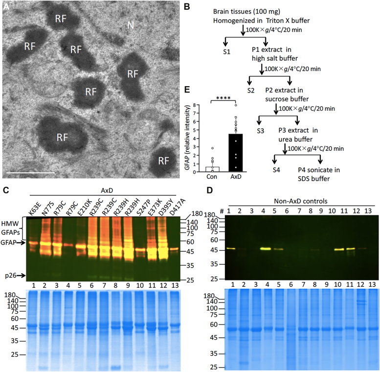
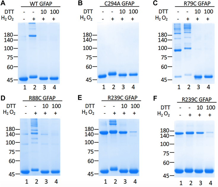
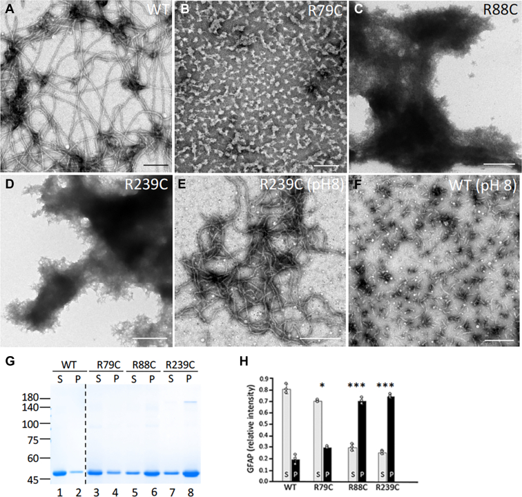
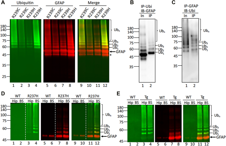
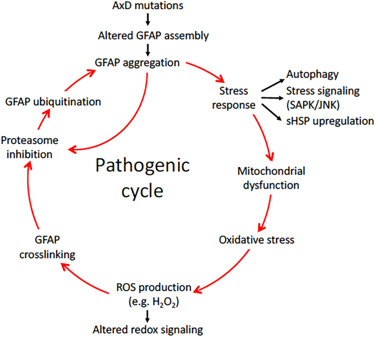
Here, we describe pathological events potentially involved in the disease pathogenesis of Alexander disease (AxD). This is a primary genetic disorder of astrocyte caused by dominant gain-of-function mutations in the gene coding for an intermediate filament protein glial fibrillary acidic protein (GFAP). Pathologically, this disease is characterized by the upregulation of GFAP and its accumulation as Rosenthal fibers. Although the genetic basis linking GFAP mutations with Alexander disease has been firmly established, the initiating events that promote GFAP accumulation and the role of Rosenthal fibers (RFs) in the disease process remain unknown. Here, we investigate the hypothesis that disease-associated mutations promote GFAP aggregation through aberrant posttranslational modifications. We found high molecular weight GFAP species in the RFs of AxD brains, indicating abnormal GFAP crosslinking as a prominent pathological feature of this disease. In vitro and cell-based studies demonstrate that cystine-generating mutations promote GFAP crosslinking by cysteine-dependent oxidation, resulting in defective GFAP assembly and decreased filament solubility. Moreover, we found GFAP was ubiquitinated in RFs of AxD patients and rodent models, supporting this modification as a critical factor linked to GFAP aggregation. Finally, we found that arginine could increase the solubility of aggregation-prone mutant GFAP by decreasing its ubiquitination and aggregation. Our study suggests a series of pathogenic events leading to AxD, involving interplay between GFAP aggregation and abnormal modifications by GFAP ubiquitination and oxidation. More important, our findings provide a basis for investigating new strategies to treat AxD by targeting abnormal GFAP modifications.

Keywords: Alexander disease; aggregation; glial fibrillary acidic protein; intermediate filament; ubiquitination.


Conflict of interest statement

Conflicts of interest The authors declare that they have no conflicts of interests with the contents of this article.

Figures

Figure 1
Figure 1
Ultrastructural and biochemical analyses of Rosenthal fibers.A, EM showed electron-dense GFAP aggregates similar to Rosenthal fibers (RF) in the perinuclear region of the brain tissue from an AxD case carrying R239C GFAP mutation. N, nucleus. Bar represents 500 nm. B, schematic diagram of the biochemical extraction protocol used in this study. The RF-enriched fractions from AxD brain tissues (C, with GFAP mutations indicated on the top) and non-AxD controls (D, #1–13) were analyzed by immunoblotting using a monoclonal anti-GFAP antibody SMI-21 that recognized the N-terminal part of GFAP (green channel) and a polyclonal anti-GFAP antibody that recognized the C-terminal part of GFAP (red channel). Merged immunoblot showed the superimposition of both the green and red signals. Protein samples were loaded at 1 μg per lane and total protein profiles of each lane were visualized by Coomassie blue staining (C and D, bottom panel). GFAP and high molecular weight (HMW) species are indicated on the right. Molecular weight markers (in kDa) are indicated on either left or right side of the gel. E, quantification of GFAP levels in AxD samples compared to non-AxD controls. Data are mean ± SD. ∗∗∗∗p < 0.0001 (two-tailed t test). Each white dot represents a sample from a brain tissue (n = 13). GFAP, glial fibrillary acidic protein; p26, 26 kDa GFAP degradation product.
Figure 2
Figure 2
GFAP oxidation and disulfide bond formation in vitro. Purified recombinant WT (A) and mutant (BF) GFAP were either untreated (AF, lane 1) or treated with 10 mM H2O2 for 15 min (AF, lane 2) as indicated. Peroxide-treated GFAPs were subsequently treated with 10 mM (AF, lane 3) or 100 mM (AF, lane 4) DTT for 15 min. After treatments, WT and mutant GFAPs were analyzed by SDS-PAGE under either nonreducing (AE) or reducing (F) conditions, followed by Coomassie blue staining. Molecular weight markers (in kDa) are indicated on the left. GFAP, glial fibrillary acidic protein.
Figure 3
Figure 3
Assemblyproperties of cysteine-generating GFAP mutants. Purified WT (A, B), R79C (C), R88C (F), and R239C (D, E) GFAP at a concentration of 0.25 mg/ml were assembled in vitro. Before assembly was completed, WT (A) and R239C (D) GFAP in low ionic strength buffer (pH 8) were fixed and processed for subsequent negative staining. After assembly, GFAPs were processed by negative staining followed by EM (B, C, E, and F). Note that R239C mutant showed a greatly increased tendency to polymerize even under preassembly conditions (D, pH 8), under which WT GFAP (A, pH 8) remained mostly as unit length filament-like structures. Bar represents 500 nm, except in (E) and (F), which were 1 μm. G, WT and mutant GFAPs were subjected to a low-speed sedimentation assay and the resulting supernatant (S) and pellet (P) fractions were analyzed by reducing SDS-PAGE, followed by Coomassie blue staining. Under these assay conditions, WT (lane 1) and R79C (lane 3) GFAP remained mainly in the supernatant fraction. In contrast, R88C (lane 6) and R239C (lane 8) mutants sedimented more efficiently into the pellet fraction. Molecular weight markers (in kDa) are indicated on the left. Dashed line indicated that samples were run on different gels. H, quantification of GFAP mutants in the supernatant and pellet fractions were compared to WT controls. Data are mean ± SD. ∗p < 0.05, ∗∗∗p < 0.001 (two-tailed t test). Each white dot represents a biological replicate (n = 3). GFAP, glial fibrillary acidic protein.
Figure 4
Figure 4
Cystine-generating GFAP mutants formed cross-linked GFAP species in astrocytes. Primary astrocytes derived from GFAP KO rats were transduced with indicated GFAP expression constructs. At 72 h after transduction, cells were extracted, and the total (A, lanes 1–5) and pellet (A, lanes 6–10) fractions were analyzed by either nonreducing (A, top panels) or reducing (A, bottom panels) immunoblotting using an anti-GFAP antibody. Note that high molecular weight (HMW) GFAP bands were absent in cells expressing cysteine-deficient C294A GFAP (A, lanes 5 and 10). In-gel staining was shown to visualize total protein profiles and to assist comparison of equal protein loading in each sample (Fig. S4A). Molecular weight markers (in kDa) are indicated on the left. HMW GFAPs are indicated by black arrows, and an immunopositive band above the monomeric GFAP is indicated by red arrows. B, mutant GFAPs in the pellet fraction relative to total GFAP were quantified and compared to WT GFAP. Data are mean ± SD. For all two-tailed t test, ∗p < 0.05, ∗∗p < 0.01, and ∗∗∗p < 0.001. Each white dot represents a biological replicate (n = 3). C, the pellet fractions from cystine-generating mutants-transduced KO astrocytes were subjected to immunoprecipitation using a mouse monoclonal anti-GFAP antibody SMI21, followed by immunoblotting using a rabbit polyclonal anti-panGFAP (lanes 1–3) and a mouse monoclonal anti-ubiquitin (lanes 4–6) antibodies. GFAP and its ubiquitinated form (Ub-GFAP), as well as the heavy chain (HC) of the capture antibody are indicated on the right. Molecular weight markers (in kDa) are indicated on the left. In-gel staining and full-length blots were shown in Fig. S4B. DG, GFAP-KO astrocytes transduced with R239C GFAP were double stained with anti-GFAP (D, red channel) and anti-ubiquitin (E, green channel) antibodies. Cells were counterstained with DAPI (F, blue channel) to reveal nuclei. A merged image was shown (G), with white lines indicating the edge of GFAP-transduced cells. Note that ubiquitin labeling was prevalent in astrocytes but did not always colocalize with GFAP, which may reflect accumulation of other ubiquitinated proteins. Bar represents 10 μm. Representative images were shown from astrocyte cultures prepared from three GFAP-KO rats. GFAP, glial fibrillary acidic protein.
Figure 5
Figure 5
GFAP is pathologically ubiquitinated in human AxD brains and rat AxD model.A, RF-enriched fractions from four AxD cases carrying either R239C or R239H mutations were analyzed by immunoblotting using anti-ubiquitin (lanes 1–4) and anti-panGFAP (lanes 5–8) antibodies. Immunoblots with merged signals showing high molecular weight smears of GFAP species (Ub1-3, and Ubn) were immunopositive for both ubiquitin and GFAP. Total protein profiles of the RF-enriched fraction were visualized by in-gel staining with trichloroethanol (Fig. S5A). B and C, RF-enriched fraction from an AxD patient with R239C mutation was subjected to immunoprecipitation using either anti–ubiquitin (B) or anti–GFAP (C) antibody. The inputs (In, lane 1) and immunoprecipitates (IP, lane 2) were analyzed by immunoblotting using anti-panGFAP (B) and anti-ubiquitin (C) antibodies. Ubiquitinated GFAPs were indicated on the right, with Ub1-3, are presumably mono-, di-, and tri-ubiquitinated GFAP and Ubn represents polyubiquitinated GFAP. Molecular weight markers (in kDa) are shown on the left. Similar accumulation of ubiquitinated GFAP species were also observed in different brain regions of AxD rodent models. D, RF-enriched fractions prepared from hippocampus (Hip) and brain stem (BS) of WT and R237H rat brains were analyzed by immunoblotting using anti-ubiquitin (lanes 1–4) and anti-GFAP (lanes 5–8) antibodies. Merged images showed ubiquitinated GFAP (lanes 9–12) in the indicated brain regions of R237H rat. Representative immunoblots were shown from samples prepared from three WT and R237H rats at 8 weeks of age. Dashed line indicated that lanes were run on the same gel but were noncontiguous. E, immunoblotting analysis of RF-enriched fractions from WT and GFAPTg mice for ubiquitin (1, 2, 3, 4) and GFAP (lanes 5–8) in the hippocampus (Hip) and brain stem (BS). A merged image showed ubiquitinated GFAP in the indicated brain regions (lanes 9–12). Each lane represents samples prepared from individual animals at 8 weeks of age (n = 3). Molecular weight markers (in kDa) are indicated on the left. In-gel staining was shown (Fig. S5, C and D) to assist comparison of equal protein loading. GFAP, glial fibrillary acidic protein; RF, Rosenthal fiber.
Figure 6
Figure 6
Effect of arginine on GFAP solubility and ubiquitination.A, SW13 (Vim-) cells were transduced with E373K GFAP in the presence of 10 mM or 50 mM arginine as indicated. At 48 h after transduction, cells were extracted the resulting supernatant (A, lanes 1–3) and pellet (A, lanes 4–6) fractions, as well as the total lysates (B) were analyzed by immunoblotting using anti-ubiquitin (green channel) and anti-GFAP (red channel) antibodies. Merged immunoblot was shown (A, and B lanes 7–9). Molecular mass markers are shown on the left, and the positions of GFAP and ubiquitinated GFAP species are indicated on the right. In-gel staining was shown (Fig. S6C) to assist comparison of equal protein loading of each lane. Quantification of GFAP and ubiquitin levels in the pellet (C) and total (D) fractions of arginine-treated cells compared to untreated controls. Data are mean ± SD. For all two-tailed t test, ∗p < 0.05, ∗∗∗p < 0.001, and ∗∗∗∗p < 0.0001. Each white dot represents a biological replicate (n = 3). EH, purified recombinant E373K GFAP at a concentration of 0.25 mg/ml was assembled in vitro in the absence (E) or presence (F) of 50 mM arginine. Assembled GFAPs were negatively stained and visualized by transmission electron microscopy. Bar represents 1 μm. The extent of GFAP aggregation was assessed by a low-speed sedimentation assay (G). The E373K GFAP assembled in vitro in the absence (lanes 1 and 2) or presence (lanes 3 and 4) of 50 mM arginine was subjected to a low speed centrifugation, and the resulting supernatant (lanes 1 and 3) and pellet (lanes 2 and 4) fractions were analyzed by SDS-PAGE and visualized by Coomassie blue staining. Molecular weight markers (in kDa) are indicated on the left. H, quantification of arginine-treated mutant GFAP in the supernatant and pellet fractions compared to untreated controls. Data are mean ± SD. ∗∗∗p < 0.001 (two-tailed t test). Each white dot represents a biological replicate (n = 3). GFAP, glial fibrillary acidic protein; HMW, high molecular weight.
Figure 7
Figure 7
We hypothesize a potential two-step mechanism, in which GFAP aggregation induced by AxD mutations through altered filament assembly may represent an initiation event that triggers subsequent GFAP oxidation and ubiquitination, leading to proteasome inhibition and further aggregation. This eventually would lead to a pathogenic cycle that emerge with further disease progression in the pathogenesis of AxD. GFAP, glial fibrillary acidic protein.

Similar articles

Cited by

References

    1. Sofroniew M.V. Astrogliosis. Cold Spring Harb. Perspect. Biol. 2014;7 - PMC - PubMed
    1. Hol E.M., Capetanaki Y. Type III intermediate filaments desmin, glial fibrillary acidic protein (GFAP), vimentin, and peripherin. Cold Spring Harb. Perspect. Biol. 2017;9 - PMC - PubMed
    1. Eliasson C., Sahlgren C., Berthold C.H., Stakeberg J., Celis J.E., Betsholtz C., et al. Intermediate filament protein partnership in astrocytes. J. Biol. Chem. 1999;274:23996–24006. - PubMed
    1. Thomsen R., Pallesen J., Daugaard T.F., Borglum A.D., Nielsen A.L. Genome wide assessment of mRNA in astrocyte protrusions by direct RNA sequencing reveals mRNA localization for the intermediate filament protein nestin. Glia. 2013;61:1922–1937. - PubMed
    1. Jing R., Wilhelmsson U., Goodwill W., Li L., Pan Y., Pekny M., et al. Synemin is expressed in reactive astrocytes in neurotrauma and interacts differentially with vimentin and GFAP intermediate filament networks. J. Cell Sci. 2007;120:1267–1277. - PubMed

Publication types

MeSH terms

Substances

LinkOut - more resources