. 2024 May 23;12(5):e008817.
doi: 10.1136/jitc-2024-008817.

Dose escalation study of a personalized peptide-based neoantigen vaccine (EVX-01) in patients with metastatic melanoma

Affiliations

Dose escalation study of a personalized peptide-based neoantigen vaccine (EVX-01) in patients with metastatic melanoma

Sofie Kirial Mørk et al. J Immunother Cancer. .

Abstract

Background: Neoantigens can serve as targets for T cell-mediated antitumor immunity via personalized neopeptide vaccines. Interim data from our clinical study NCT03715985 showed that the personalized peptide-based neoantigen vaccine EVX-01, formulated in the liposomal adjuvant, CAF09b, was safe and able to elicit EVX-01-specific T cell responses in patients with metastatic melanoma. Here, we present results from the dose-escalation part of the study, evaluating the feasibility, safety, efficacy, and immunogenicity of EVX-01 in addition to anti-PD-1 therapy.

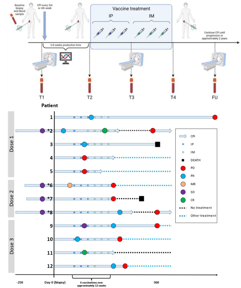
Methods: Patients with metastatic melanoma on anti-PD-1 therapy were treated in three cohorts with increasing vaccine dosages (twofold and fourfold). Tumor-derived neoantigens were selected by the AI platform PIONEER and used in personalized therapeutic cancer peptide vaccines EVX-01. Vaccines were administered at 2-week intervals for a total of three intraperitoneal and three intramuscular injections. The study's primary endpoint was safety and tolerability. Additional endpoints were immunological responses, survival, and objective response rates.

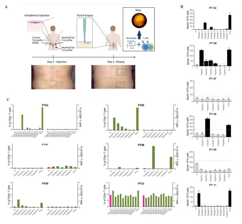
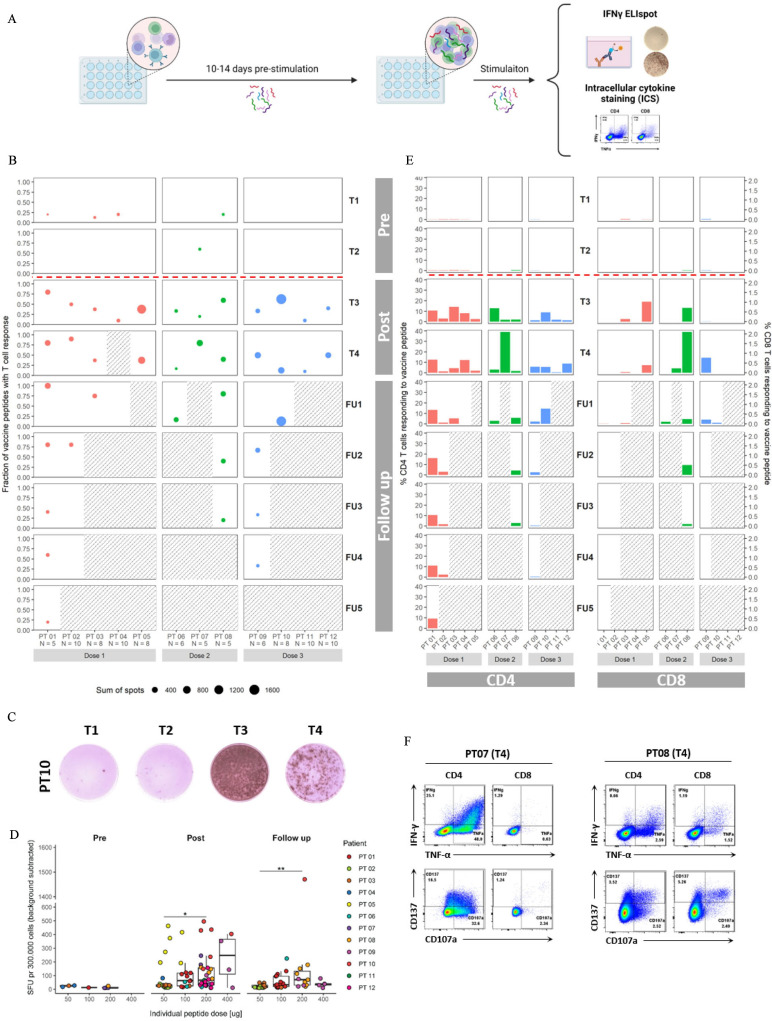
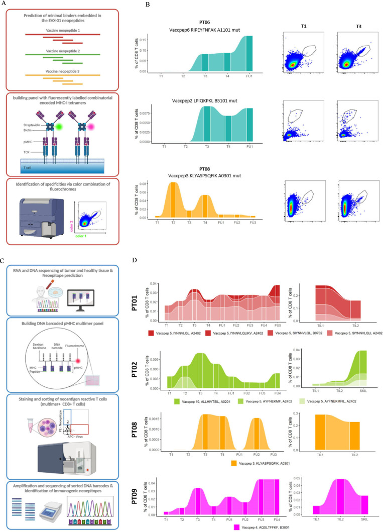
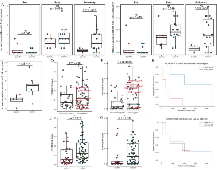
Results: Compared with the base dose level previously reported, no new vaccine-related serious adverse events were observed during dose escalation of EVX-01 in combination with an anti-PD-1 agent given according to local guidelines. Two patients at the third dose level (fourfold dose) developed grade 3 toxicity, most likely related to pembrolizumab. Overall, 8 out of the 12 patients had objective clinical responses (6 partial response (PR) and 2 CR), with all 4 patients at the highest dose level having a CR (1 CR, 3 PR). EVX-01 induced peptide-specific CD4+ and/or CD8+T cell responses in all treated patients, with CD4+T cells as the dominating responses. The magnitude of immune responses measured by IFN-γ ELISpot assay correlated with individual peptide doses. A significant correlation between the PIONEER quality score and induced T cell immunogenicity was detected, while better CRs correlated with both the number of immunogenic EVX-01 peptides and the PIONEER quality score.

Conclusion: Immunization with EVX-01-CAF09b in addition to anti-PD-1 therapy was shown to be safe and well tolerated and elicit vaccine neoantigen-specific CD4+and CD8+ T cell responses at all dose levels. In addition, objective tumor responses were observed in 67% of patients. The results encourage further assessment of the antitumor efficacy of EVX-01 in combination with anti-PD-1 therapy.

Keywords: immunotherapy; vaccine.


Conflict of interest statement

Competing interests: MD has received honoraria for lectures from Roche and Novartis (past 2 years). IMS has received honoraria for lectures and consultancies from Novartis, Roche, MSD, BMS, and Pierre Fabre. KB has received honoraria (institutional) for lectures and advisory boards of MSD, Pierre Fabre, and BMS. CCIT-DK has been granted economic support for personal wages from Evaxion Biotech A/S, Denmark. SRH is a cofounder of PokeACell and coinventor of several licensed patents. ABS, DK-K, AJ, NVP, NT and JK are employees of Evaxion Biotech A/S and have a financial interest in the company. All other authors have declared that they have no conflict of interest.

Figures

Figure 1
Figure 1
Clinical setup and response. (A) Clinical setup; biopsy, PET/CT scan, and blood samples were collected at baseline (T1). Treatment with aPD1 was either initiated around the first biopsy (group A) or had already been initiated for at least 4 months before the biopsy (group B: *). EVX-01 vaccination was administered approximately at weeks 6–8 and every second week for six vaccinations in total (3 IM+3 IP). Tumor biopsies were performed (if possible) at T2 and T4. In addition, radiographic imaging was done every 12 weeks. Blood samples were collected from T1 to T4, and thereafter every 12 weeks. Figure created with BioRender.com. (B) Overview of patients included in trial: checkpoint inhibitor (CPI) initiation, baseline biopsy (day 0), vaccine treatment, and follow-up information of the twelve patients at three different dose levels. Small blue and green dots indicate either IP vaccinations or IM vaccinations, respectively. The depiction of disease condition and patient status are indicated in various colors. CR, complete response (green); IM, intramuscular; IP, intraperitoneal; MR, mixed response (salmon); PD, progressive disease (red); PR, partial response (blue); SD, stable disease (purple); PET, positron emission tomography.
Figure 2
Figure 2
EVX-01 reactive SKIL derived T cell. (A) DTH skin test was done approximately 2 weeks after the last vaccination. Two intradermal injections of the EVX-01 peptides and one control (DSMO) injection were administered on day 0 (approximately 2 weeks from the last vaccination). After 48 hours, 5 mm punch biopsies were taken from the three injections site. The tissue was then transported to the laboratory for SKILs culture. Minimally expanded SKILs were expanded from tissue fragments for 3–6 weeks. Created with BioRender.com. EVX-01-specific T cell responses in SKILs (skin-test infiltrating lymphocytes) in patients 2 (dose level 1), 6, 7, 8 (dose level 2), and 9, 11 (dose level 3). (B) Elispot responses were examined in SKILs at T4 (after six vaccinations) from six patients. SKILs were co-cultured with EVX-01 peptide pool and individual EVX-01 peptides. Black bars represent significant responses, gray bars are not significant. The dotted line indicated the threshold value for a significant response; background (irrelevant peptide) plus 3×SD of the background and at least 10 spots over background response (C) T cells specific toward EVX-01 peptides were identified in SKILs, which were restimulated with EVX-01 peptide pool (green), single-vaccine peptides (green) or irrelevant peptide (pink) for 8 hours. The T cell reactivity was defined as the percentage of live CD8+ or CD4+ T cells staining positive for at least two of four markers (TNF-α, IFNγ, CD107a, CD137).
Figure 3
Figure 3
EVX-01-specific PBMC derived T cell responses analyzed by Elispot and ICS. (A) Experimental setup for detection of T cells responses. PBMCs were prestimulated with EVX-01 neopeptide pool for 10–14 days. Prestimulated PBMCs were hereafter stimulated with EVX-01 neopeptides in an ELISpot assay or intracellular cytokine staining assay. (B) An overview of patient IFN-γ responses detected by Elispot grouped by dose levels and time points. Red dots represent dose level 1, green dots dose level 2, and blue dots dose level 3. The height of the dots on the y-axis represents the fraction of single vaccine peptides with a T cell response out of total neopeptides per patient. A total number of single peptides (N) are stated under the patient number at the X-axis. The size of the dots represents the sum of the spot after subtraction of the background (Irrelevant peptide). This only includes spots from single peptide stimulation, which induces positive responses. Gray scattered boxes indicate time points which were not analyzed. (C) Representative ELIspot wells for patient 10 pre vaccination (T1 and T2), and post vaccination (T3 and T4). (D) The individual vaccine dose of the single EVX-01 peptides versus delta spots for peptides with T cell response (background stimulated with irrelevant peptide has been subtracted). Responses have been divided in pre vaccination (T1 and T2), postvaccination (T3 and T4) and follow-up (all FU time points). Each dot represents a single-peptide response detected with Elispot. Means were compared between peptide dose groups using Kruskal-Wallis test. Significant difference is indicated with asterisks (*p<0.05, **p<0.01). (E) T cell responses was tested after restimulation with pool vaccine peptides and stained intracellularly for IFN-γ, TNF-α, CD107a and CD137. The percentages of T cells which are positive for at least two of mentioned markers are shown in an overview of EVX-01-specific CD4+ and CD8+ T cells in all patient responses from all three dose levels and all time points. Red bars represent dose level 1, green bars dose level 2, and blue bars dose level 3. Bars show specific CD4+ and CD8+ T cells. Background response has been subtracted. Gray scattered boxes indicate time points which were not analyzed. (F) Flow cytometry dotplots for CD4+ and CD8+ T cells stained for IFN-γ, TNF-α, CD107a and CD137. The dotplots are shown for patient seven with the highest CD4+T cell response at T4, and patient eight with the highest CD8+T cell response at T4. ICS, intracellular cytokine staining; PBMC, peripheral blood mononuclear cell.
Figure 4
Figure 4
EVX-01 prestimulated PBMCs and ex vivo PBMC derived CD8+T cells screened for EVX-01 and other CD8+T cell neoepitopes. (A) A schematic overview of the experimental setup for detection of VaccNARTs in prestimulated PBMCs. Minimal peptides embedded in the EVX-01 neopeptides were predicted and loaded on tetramers conjugated to fluorochromes. Specific combinations of two fluorochromes were used for identification of the specificities of detected VaccNARTs. (B) The frequency of selected VaccNART with only one specificity, from patients 6, 8 and 10. EVX-01 peptide number (Vaccpep), short peptide sequence, HLA, and whether the short peptide includes the mutated region of the Vaccpep (mut) is shown above the graphs. dotplots for time point T1 (baseline) and T3 (after three IP vaccinations) are shown for each peptide. (C) A schematic overview of experimental setup used for detection of NARTs, VaccNARTs and VARTs in ex vivo PBMCs, expanded TILs and SKILs. CD8+T cells reactive toward predicted neoepitopes, including vaccine embedded neoepitopes, and virus epitopes were screened with DNA barcoded pMHC multimers. (D) The frequency VaccNARTs shown for selected patients, showing the single populations dynamics over time. EVX-01 peptide number (Vaccpep), short peptide sequence and HLA is shown next to the graphs. IP, intraperitoneal; PBMC, peripheral blood mononuclear cell.
Figure 5
Figure 5
Correlation between clinical responses, neoepeptide immunogenicity and quality scores. Patient clinical BOR was grouped in responders. Immune responses detected before vaccination (Pre), during and after vaccination (Post) and in follow-up samples are grouped and compared with the clinical outcomes of the patients. (A) The number of EVX-01 peptides with T cell responses detected by Elispot was compared with the patients BOR. (B) The fraction of EVX-01 peptides with T cell responses detected by Elispot was compared with the patients BOR. (C) The number of de-novo responses per patient compared with the patients BOR. (D, F) PIONEER quality score for EVX-01 peptides inducing functional responses detected by Elispot (immunogenic) compared with non-immunogenic EVX-01 peptides. Prediction scored for both PIONEER2 and PIONEER4 is shown. EVX-01 responses detected by the pMHC-I multimers in ex vivo PBMCs are marked in red. A t-test was used to test the difference between immunogenic and non-immunogenic EVX-01 peptides’ prediction scores (*p<0.05, **p<0.01). (E, G) The PIONEER quality scores for EVX-01 peptides (both from PIONEER2 and PIONEER4) was compared between patients BOR. Means were compared between response groups using t-test. Note that measurement is not completely internal independent as they are confounded based on patient response, hence caution should be taken when interpretating the p values. Significant difference is indicated with asterisks (*p<0.05, **p<0.01). (H) PIONEER quality score impact on progression-free survival (PFS). Kaplan-Meier curves were generated by grouping patients based on the median PIONEER4.0 quality score of administered neoepeptides, in two balanced groups (high and low-quality score) (I) TMB impact on PFS. Kaplan-Meier curves were generated by grouping patients based on tumor mutational burden (TBM) (balanced high/low) calculated from NGS data using FDA guidelines (https://www.accessdata.fda.gov/cdrh_docs/pdf17/P170019B.pdf). BOR, best overall response; CR, clinical response; PD, progressive disease; PR, partial response; SD, stable disease.

References

    1. Bagchi S, Yuan R, Engleman EG. Immune Checkpoint inhibitors for the treatment of cancer: clinical impact and mechanisms of response and resistance. Annu Rev Pathol Mech Dis 2021;16:223–49. 10.1146/annurev-pathol-042020-042741 - DOI - PubMed
    1. Twomey JD, Zhang B. Cancer Immunotherapy update: FDA-approved Checkpoint inhibitors and companion diagnostics. AAPS J 2021;23:39. 10.1208/s12248-021-00574-0 - DOI - PMC - PubMed
    1. Esfahani K, Roudaia L, Buhlaiga N, et al. . A review of cancer Immunotherapy: from the past, to the present, to the future. Curr Oncol 2020;27:S87–97. 10.3747/co.27.5223 - DOI - PMC - PubMed
    1. Snyder A, Makarov V, Merghoub T, et al. . Genetic basis for clinical response to CTLA-4 blockade in Melanoma. N Engl J Med 2014;371:2189–99. 10.1056/NEJMoa1406498 - DOI - PMC - PubMed
    1. Rizvi NA, Hellmann MD, Snyder A, et al. . Cancer immunology. mutational landscape determines sensitivity to PD-1 blockade in non-small cell lung cancer. Science 2015;348:124–8. 10.1126/science.aaa1348 - DOI - PMC - PubMed

Publication types