Review
. 2024 May;34(3):e2543.
doi: 10.1002/rmv.2543.

SARS-CoV-2, periodontal pathogens, and host factors: The trinity of oral post-acute sequelae of COVID-19

Affiliations
Review

SARS-CoV-2, periodontal pathogens, and host factors: The trinity of oral post-acute sequelae of COVID-19

Joel Schwartz et al. Rev Med Virol. 2024 May.

Abstract

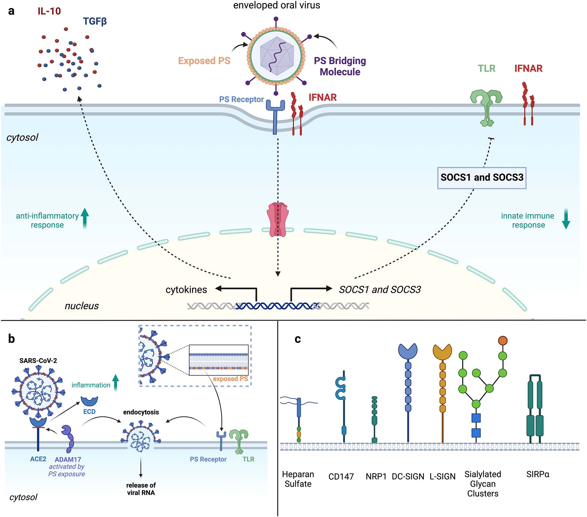
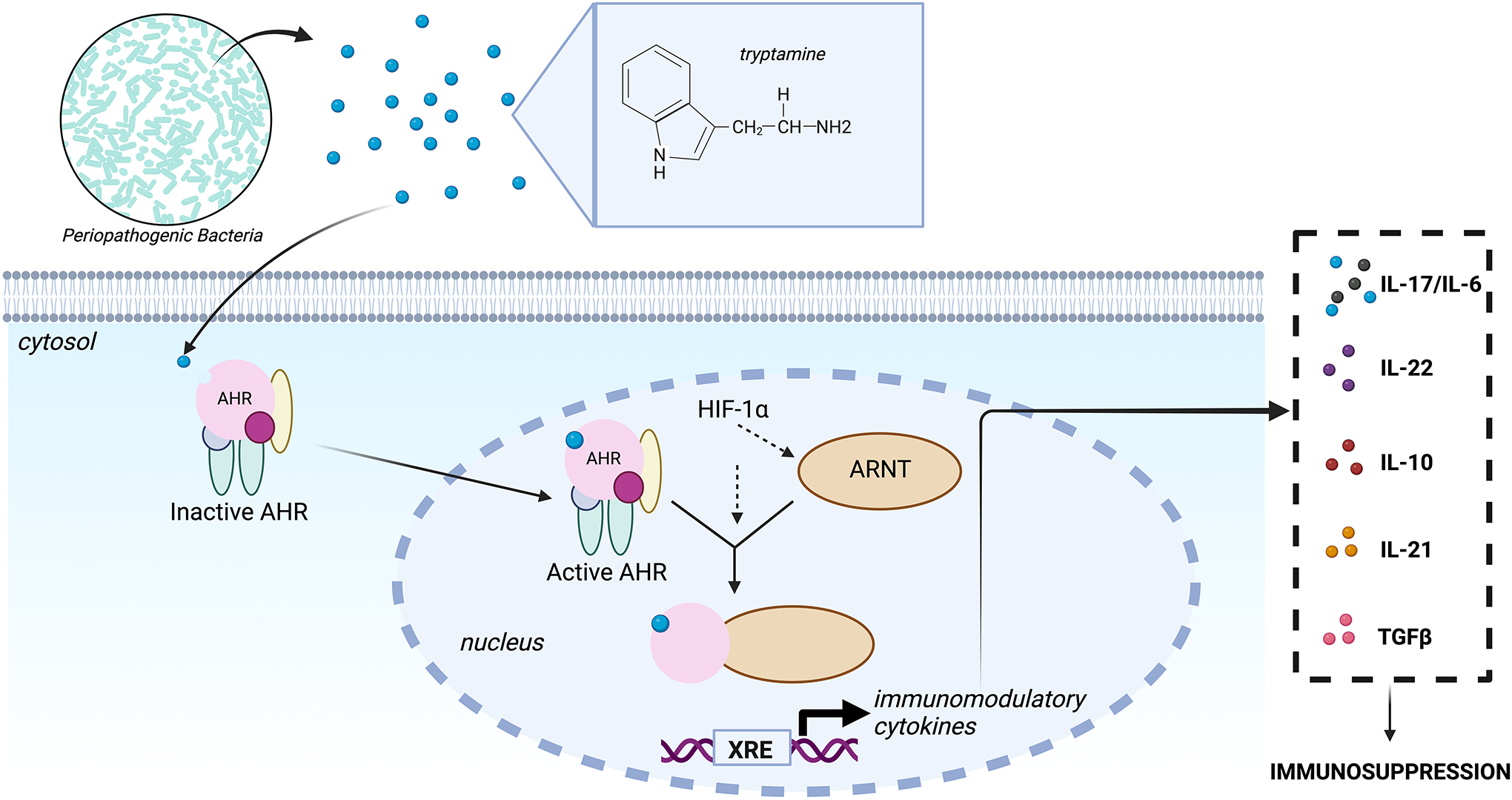
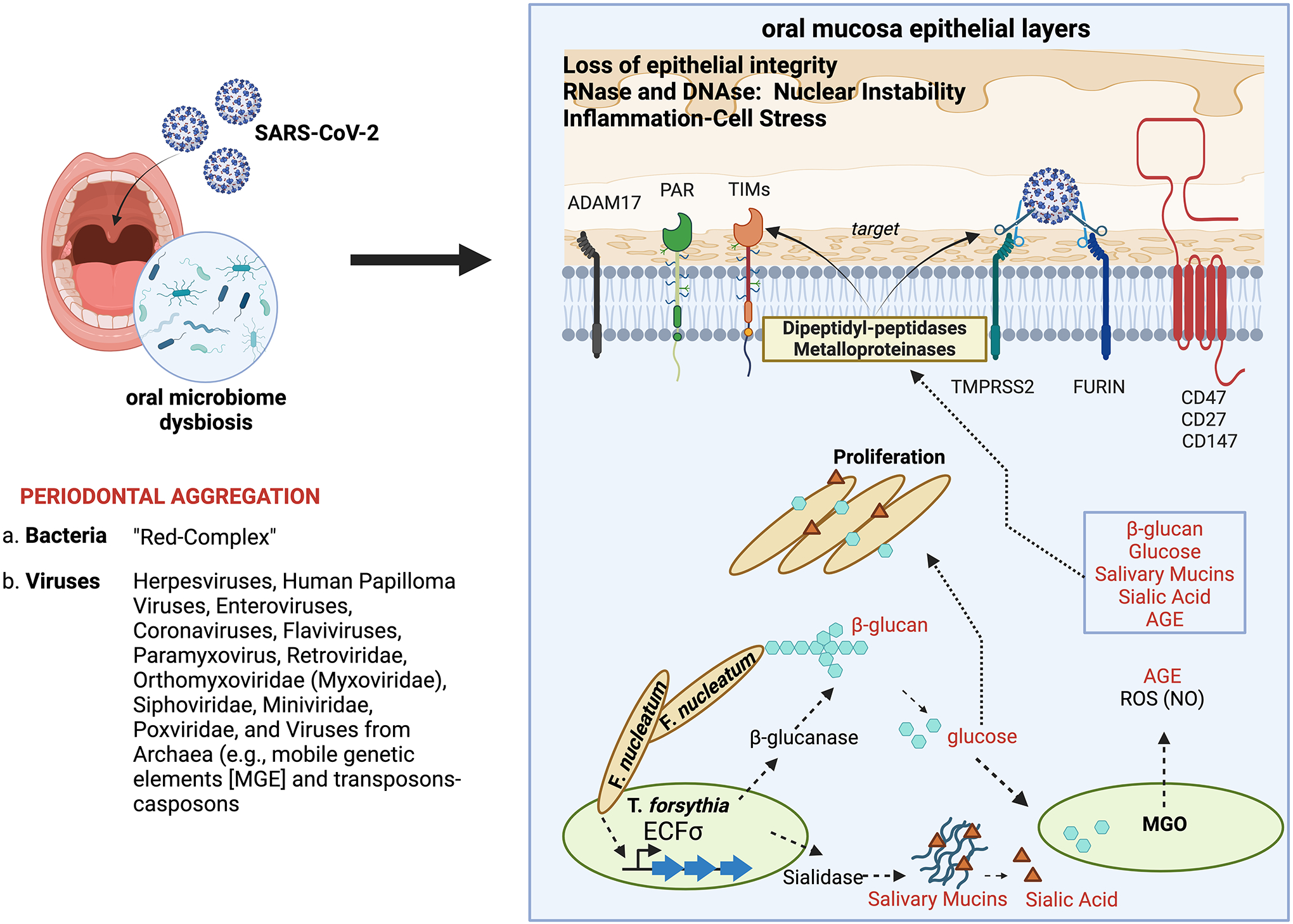
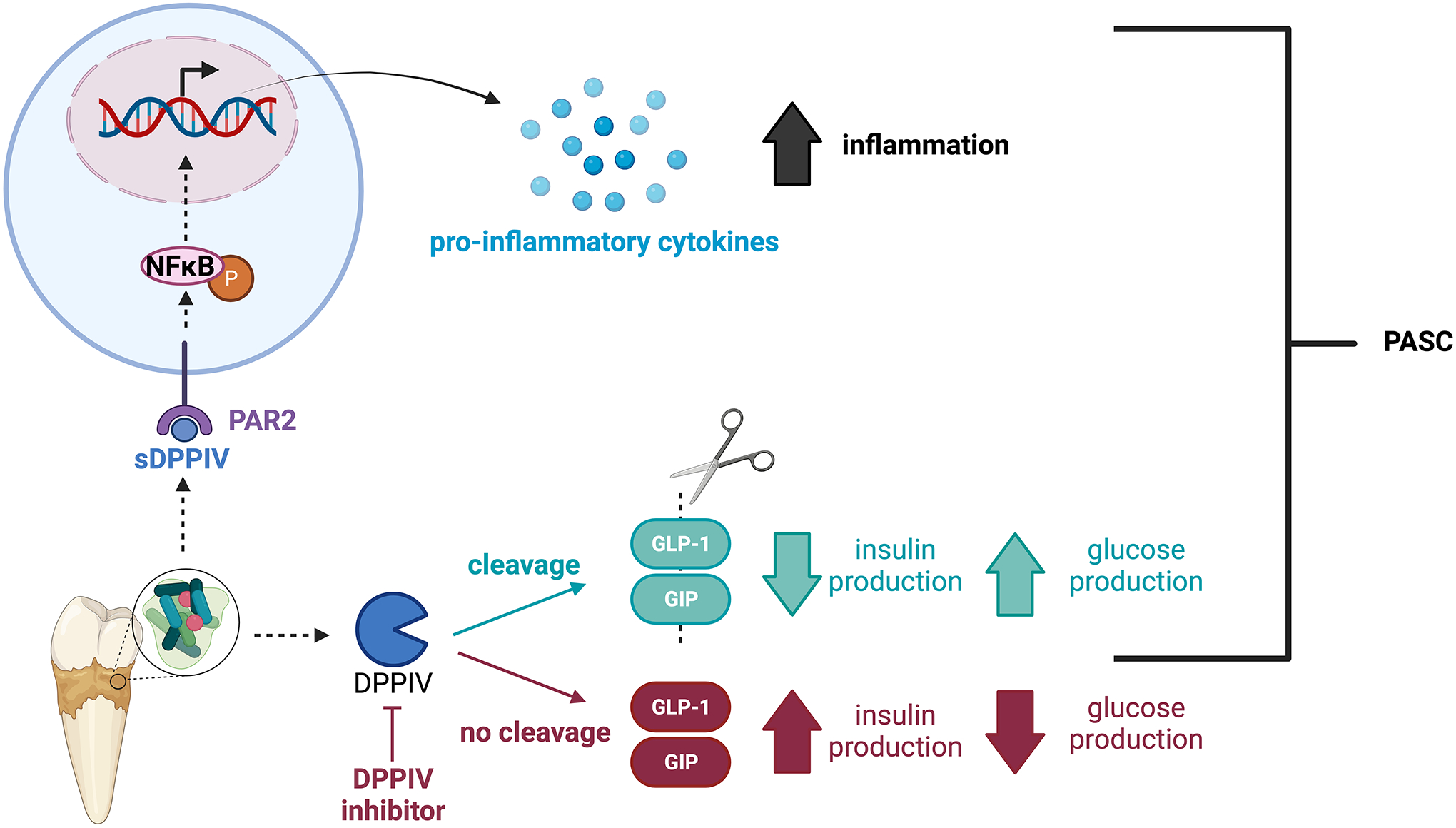
COVID-19 as a pan-epidemic is waning but there it is imperative to understand virus interaction with oral tissues and oral inflammatory diseases. We review periodontal disease (PD), a common inflammatory oral disease, as a driver of COVID-19 and oral post-acute-sequelae conditions (PASC). Oral PASC identifies with PD, loss of teeth, dysgeusia, xerostomia, sialolitis-sialolith, and mucositis. We contend that PD-associated oral microbial dysbiosis involving higher burden of periodontopathic bacteria provide an optimal microenvironment for severe acute respiratory syndrome coronavirus 2 (SARS-CoV-2) infection. These pathogens interact with oral epithelial cells activate molecular or biochemical pathways that promote viral adherence, entry, and persistence in the oral cavity. A repertoire of diverse molecules identifies this relationship including lipids, carbohydrates and enzymes. The S protein of SARS-CoV-2 binds to the ACE2 receptor and is activated by protease activity of host furin or TRMPSS2 that cleave S protein subunits to promote viral entry. However, PD pathogens provide additional enzymatic assistance mimicking furin and augment SARS-CoV-2 adherence by inducing viral entry receptors ACE2/TRMPSS, which are poorly expressed on oral epithelial cells. We discuss the mechanisms involving periodontopathogens and host factors that facilitate SARS-CoV-2 infection and immune resistance resulting in incomplete clearance and risk for 'long-haul' oral health issues characterising PASC. Finally, we suggest potential diagnostic markers and treatment avenues to mitigate oral PASC.

Keywords: PASC; SARS‐CoV‐2; oral biology; oral microbiome; oral mucosal immunity; oral virology; ‘red complex’.


Conflict of interest statement

CONFLICTS OF INTEREST

The authors declare no conflicts of interest.

Figures

Fig, 1.
Fig, 1.
Surface lipid or carbohydrate display by SARS-CoV-2 and host cells is modulated by periodontal bacteria to promote viral tropism, suppress host immune responses and provide parallel adhesion molecules for the virus A. PS display by a perioopathogen (shown is an enveloped oral virus) allows the pathogen to bind to PS ligands on the surface of cells and enter host cells through apoptotic mimicry. After the PS/PS receptor interaction, the pathogen is engulfed and endocytosed into the host cell, initiating the production of anti-inflammatory cytokines such as IL-10 and TGFβ. The enveloped virus binds to bridging molecules (e.g., GAS6), promoting the activation of tyrosine protein kinase receptor 3 (TYRO3)-AXL-MER (TAM) family receptors. In turn, the TAM receptors heterodimerizes with type 1 interferon receptor (IFNAR), promoting SOCS1 (suppressor of cytokine signaling 1) and SOCS3 expression. SOCS1 and SOCS3 then inhibit IFNAR and TLR signaling, reducing the innate immune response. B. Similarly, SARS-CoV-2 can uptake and externalize PS. This PS display allows SARS-CoV-2 to enter host cells. Additionally, PS exposure can activate ADAM17, which can then cleave and release the extracellular domain (ECD) of ACE2 into the extracellular environment. The precise function of the ACE2 ECD is unknown but reportedly upregulates inflammation. C. Parallel adhesion molecules between periopathogens and SARS-CoV-2. Created with Biorender.com.
Fig. 2.
Fig. 2.
Bacterial metabolite tryptamine activates AHR signaling and contributes to immunosuppression against SARS-CoV-2. As an agonist for the aryl hydrocarbon receptor (ArhR), tryptamine binds to and activates AHR. AHR then forms a complex with the aryl hydrocarbon receptor nuclear translocator (ARNT). The AHR-ARNT heterodimer binds to the XRE (xenobiotic response element) sequence in the promoter region of AHR target genes, inducing the transcription and subsequent release of immunosuppressive cytokines. Under hypoxic conditions, HIF-1α translocates to the nucleus and competes with AHR for binding to ARNT. HIF-1α and ARNT interactions induce the transcription of hyoxia resopnse element-containing genes. Created with Biorender.com.
Fig. 3.
Fig. 3.
Aggregation and proliferation of intracellular periopathogens in dental biofilms foster metabolic activities that target oral mucosal epithelium receptors to facilitate SARS-CoV-2 adhesion and entry. Members of Bacteroidaceae, Tannellaceae, and Lactobacillaceae form a “Red Complex” of periodontal disease causing-pathogens, such as Porphyromonas gingivalis (FimA II and FimA IV genotype (Pg), Actinobacillus actinomycetemcomitans (Aa), Tannerella forsythia (Tf), Fusobacteria nulceatum (Fn), and Treponema denticola (Td). Various families of oral viruses will also present as oral disease such as vesicles, atrophy or ulceration of oral epithelial mucosa and may contribute to periodontitis. Fn and Tf aggregate and cooperate to synthesize β-glucan, mucins, sialic acid-sialylated clusters, glucose, and AGE (advanced glycation end-products). Specifically, Fn triggers the release of β-glucanase from Tf by prompting the activation of an ECF σ-factor (extracytoplasmic function sigma factor) that triggers the glcA operon in Tf. Consequently, the secreted β-glucanase plays a role in breaking down dietary β-glucans into glucose fragments. In turn, glucose serves as a carbon source for Fn and a precursor for generating methylglyoxal (MGO) within Tf. MGO then covalently modifies host proteins, resulting in the formation of AGE. Additionally, Tf-secreted sialidase enzyme may liberate sialic acid from salivary mucins, which Fn could employ to adorn its cell surface with this sugar. Altogether, β-glucan, mucins, sialic acid-sialylated clusters, glucose, and AGE products can combine with proteases and metalloproteinases to target oral mucosal epithelium receptors. This interaction facilitates SARS-CoV-2 adhesion and entry by supporting the expression of TMPRSS2, ADAM17, and Furin or expand inflammatory receptor expressions (TIMs, PAR, CD47, CD27, CD147). Additionally, AGE can bind to RAGE and produce ROS nitric oxide. Accumulation of ROS and overt inflammation result in the loss of viability, integrity, and inflammation of oral epithelial mucosal cells. Figure adapted from https://www.researchgate.net/figure/Model-of-T-forsythia-F-nucleatum-interbacterial-interactions-in-dental-biofilm-and_fig1_338899965. Created with Biorender.com.
Fig. 4.
Fig. 4.
Dipeptidyl peptidases (DPP) produced by periodontopathogens may contribute to oral PASC by increasing inflammation and exacerbating diabetes mellitus. Periodontopathogen (e.g., Pg, Tf, and Pi) derived DPPIV cleaves and inactivates incretins GLP-1 and GIP. As a result, the insulinotropic effects of these incretins are shortened, decreasing insulin production, and increasing glucose production. Glucose metabolism influences glycation/AGE-RAGE, producing NO. Thus, the increase in glucose levels during diabetes, accelerates NO production, causing mucosal epithelial cell stress. Pharmacological inhibition can help prevent DPPIV-mediated inactivation of incretins. Soluble DPPIV also activates NFκB signaling through PAR2 (protease-activated receptor 2), consequently promoting the secretion of pro-inflammatory cytokines (e.g., TNFα, IL-1, IL-6, IL-8). Created with Biorender.com.

Similar articles

Cited by

References

    1. Chen T, Yu WH, Izard J, Baranova OV, Lakshmanan A, Dewhirst FE. The Human Oral Microbiome Database: a web accessible resource for investigating oral microbe taxonomic and genomic information. Database. 2010;2010(0):baq013–baq013. doi:10.1093/database/baq013 - DOI - PMC - PubMed
    1. Ohara-Nemoto Y, Shimoyama Y, Nakasato M, et al. Distribution of dipeptidyl peptidase (DPP) 4, DPP5, DPP7, and DPP11 in human oral microbiota – potent biomarkers indicating presence of periodontopathic bacteria. FEMS Microbiology Letters. Published online September 10, 2018. doi:10.1093/femsle/fny221 - DOI - PubMed
    1. Overmyer KA, Rhoads TW, Merrill AE, et al. Proteomics, Lipidomics, Metabolomics, and 16S DNA Sequencing of Dental Plaque From Patients With Diabetes and Periodontal Disease. Molecular & Cellular Proteomics. 2021;20:100126. doi:10.1016/j.mcpro.2021.100126 - DOI - PMC - PubMed
    1. Petersen PE. The World Oral Health Report 2003: continuous improvement of oral health in the 21st century – the approach of the WHO Global Oral Health Programme. Comm Dent Oral Epid. 2003;31(s1):3–24. doi:10.1046/j..2003.com122.x - DOI - PubMed
    1. Sedghi LM, Bacino M, Kapila YL. Periodontal Disease: The Good, The Bad, and The Unknown. Front Cell Infect Microbiol. 2021;11:766944. doi:10.3389/fcimb.2021.766944 - DOI - PMC - PubMed

MeSH terms

Substances