. 2024 May 23;15(1):4350.
doi: 10.1038/s41467-024-48555-z.

Development of a nucleoside-modified mRNA vaccine against clade 2.3.4.4b H5 highly pathogenic avian influenza virus

Affiliations

Development of a nucleoside-modified mRNA vaccine against clade 2.3.4.4b H5 highly pathogenic avian influenza virus

Colleen Furey et al. Nat Commun. .

Abstract

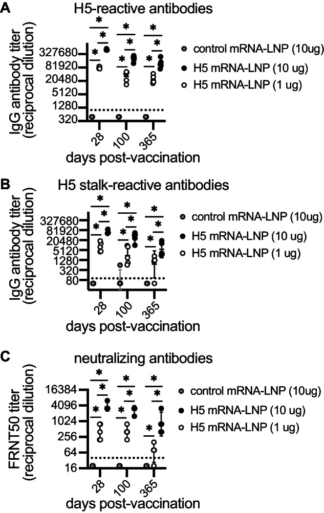
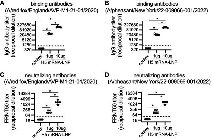
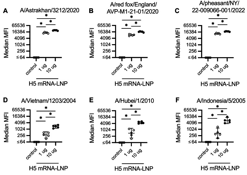
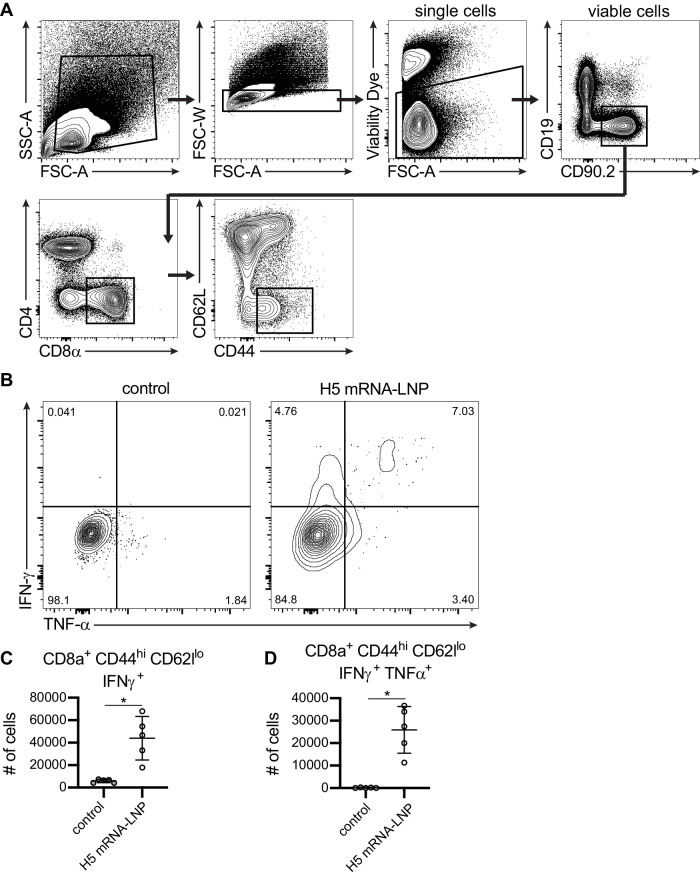
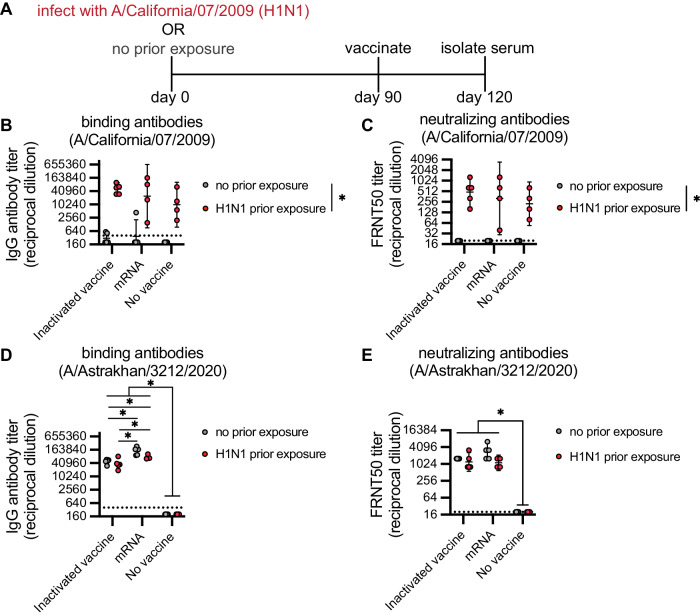
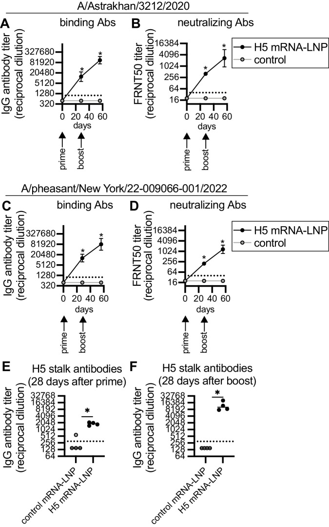
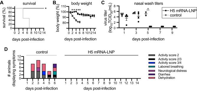
mRNA lipid nanoparticle (LNP) vaccines would be useful during an influenza virus pandemic since they can be produced rapidly and do not require the generation of egg-adapted vaccine seed stocks. Highly pathogenic avian influenza viruses from H5 clade 2.3.4.4b are circulating at unprecedently high levels in wild and domestic birds and have the potential to adapt to humans. Here, we generate an mRNA lipid nanoparticle (LNP) vaccine encoding the hemagglutinin (HA) glycoprotein from a clade 2.3.4.4b H5 isolate. The H5 mRNA-LNP vaccine elicits strong T cell and antibody responses in female mice, including neutralizing antibodies and broadly-reactive anti-HA stalk antibodies. The H5 mRNA-LNP vaccine elicits antibodies at similar levels compared to whole inactivated vaccines in female mice with and without prior H1N1 exposures. Finally, we find that the H5 mRNA-LNP vaccine is immunogenic in male ferrets and prevents morbidity and mortality of animals following 2.3.4.4b H5N1 challenge. Together, our data demonstrate that a monovalent mRNA-LNP vaccine expressing 2.3.4.4b H5 is immunogenic and protective in pre-clinical animal models.


Conflict of interest statement

S.E.H. and D.W. are co-inventors on patents that describe the use of nucleoside-modified mRNA as a platform to deliver therapeutic proteins and as a vaccine platform. S.E.H. reports receiving consulting fees from Sanofi, Pfizer, Lumen, Novavax, and Merck. S.H.Y.F. is an employee of Acuitas Therapeutics, a company focused on the development of lipid nanoparticulate nucleic acid delivery systems for therapeutic applications. The authors declare no other competing interests.

Figures

Fig. 1
Fig. 1. Clade 2.3.4.4b H5 HA mRNA-LNP vaccine elicits long-lasting antibody responses in mice.
Five mice were included per experimental group. Mice were vaccinated i.m. with 1 or 10 μg A/Astrakhan/3212/2020 HA mRNA-LNP (H5 mRNA-LNP) or 10 µg Ovalbumin mRNA-LNP (control mRNA-LNP). Serum samples were collected from mice at 28, 100, and 365 days after vaccination and serum IgG reactive to the A/Astrakhan/3212/2020 recombinant full-length HA protein (A) or ‘headless’ H5 stalk protein (B) were quantified by ELISA. C A/Astrakhan/3212/2020 neutralizing antibodies were also quantified by a 50% Foci reduction neutralization test (FRNT50); reciprocal dilutions of serum required to inhibit 50% virus infection are shown. All data are shown as geometric means ± 95% confidence intervals. Data were compared using two-way ANOVA with Tukey’s multiple comparisons test. Values were log-transformed before statistical analysis. Data are representative of 2 independent experiments. A all *P < 0.0001; (B) Day 365 control vs. 1 µg H5 mRNA-LNP *P = 0.0009, all others *P < 0.0001; (C) Day 28 1 µg H5 mRNA-LNP vs. 10 µg H5 mRNA-LNP *P = 0.0015, day 100 1 µg H5 mRNA-LNP vs. 10 µg H5 mRNA-LNP *P = 0.0002, day 365 1 µg H5 mRNA-LNP vs. 10 µg H5 mRNA-LNP *P = 0.0018, all other comparisons *P < 0.0001.Source data are provided as a Source Data file.
Fig. 2
Fig. 2. Clade 2.3.4.4b H5 HA mRNA-LNP vaccine elicits antibodies that bind and neutralize different clade 2.3.4.4b H5 viruses.
Five mice were included per experimental group. Mice were vaccinated i.m. with 1 or 10 μg A/Astrakhan/3212/2020 HA mRNA-LNP (H5 mRNA-LNP) or 10 µg Ovalbumin mRNA-LNP (control). A, B Serum samples collected 28 days after vaccination were tested by ELISA to quantify IgG reactive to A/red fox/England/AVP-M1-21-01/2020 or A/pheasant/New York/22-009066-001/2022 recombinant HA proteins. C, D A/red fox/England/AVP-M1-21-01/2020 and A/pheasant/New York/22-009066-001/2022 neutralizing antibodies were quantified by 50% Foci reduction neutralization test (FRNT50) using serum samples collected 28 days after vaccination; reciprocal dilution of serum amounts required to inhibit 50% virus infection are shown. All data are shown as geometric means ± 95% confidence intervals. Data were compared using one-way ANOVA with Tukey’s post hoc test. Values were log-transformed before statistical analysis. Data are representative of 2 independent experiments. *P < 0.0001. Source data are provided as a Source Data file.
Fig. 3
Fig. 3. Clade 2.3.4.4b H5 HA mRNA-LNP vaccine elicits antibodies that bind to diverse H5 viruses.
Groups of five mice were vaccinated i.m. with 1 or 10 µg A/Astrakhan/3212/2020 HA mRNA-LNP (H5 mRNA-LNP) or 10 µg Ovalbumin mRNA-LNP (control). Serum samples were collected 28 days after vaccination and serum antibody binding to HA proteins from diverse of H5 viral strains was determined using a bead-based multiplex binding assay. We measured antibody binding to the HA of (A) A/Astrakhan/3212/2020, (B) A/red fox/England/AVP-M1-21-01/2020, (C) A/pheasant/NY/22-009066-001/2022, (D) A/Vietnam/1203/2004, (E) A/Hubei/1/2010, and (F) A/Indonesia/5/2005. Binding is reported as mean fluorescence intensity (MFI) values. All data are shown as geometric means ± 95% confidence intervals. Data were compared using two-way ANOVA with Tukey’s multiple comparisons test. Values were log-transformed before statistical analysis. Data are representative of 2 independent experiments. A 1 µg vs. 10 µg *P = 0.004; (B) 1 µg vs. 10 µg *P = 0.0065; (E) 1 µg vs. 10 µg *P = 0.0002; all other comparisons *P < 0.0001. Source data are provided as a Source Data file.
Fig. 4
Fig. 4. Clade 2.3.4.4b H5 HA mRNA-LNP vaccine elicits robust CD8+ T cell responses.
Five mice per group were vaccinated i.m. with 1 μg A/Astrakhan/3212/2020 HA mRNA-LNP (H5 mRNA-LNP) or 1 μg Ovalbumin mRNA-LNP (control). Spleens were harvested at 10 days after vaccination and splenocytes were incubated with H5 HA overlapping peptide pools before completing intracellular cytokine staining and flow cytometric analysis of CD8 T cells. A Representative gating strategy for analysis of cytokine production by activated CD8+ T cells 10 days after vaccination. B Representative flow cytometry analysis of IFN-γ and TNF-α producing CD8+ T cells from mice vaccinated with control mRNA-LNP or H5 HA mRNA-LNP. C, D Number of IFN-γ producing CD8+ T cells and IFN-γ and TNF-α producing CD8+ cells were quantified. Data were compared using an unpaired two-tailed t test. Data are representative of 2 independent experiments. C *P = 0.0023; (D) *P = 0.0005. Source data are provided as a Source Data file.
Fig. 5
Fig. 5. Clade 2.3.4.4b H5 HA mRNA-LNP vaccine and inactivated H5 vaccine elicit robust antibody responses in mice previously exposed to H1N1 virus.
A Timeline for infections and vaccinations. Groups of 4-5 mice were uninfected or infected i.n. with 1000 TCID50 of A/California/07/2009 virus and then vaccinated i.m. 90 days later with either 1 µg of A/Astrakhan/3212/2020 HA mRNA-LNP (mRNA) or 50 HAU of an inactivated A/Astrakhan/3212/2020 vaccine (inactivated vaccine). Sera were isolated 30 days after vaccination and IgG antibody titers were measured using ELISA plates coated with HA from A/California/07/2009 (B) and A/Astrakhan/3212/2020 (D). 50% Foci reduction neutralization test (FRNT50) titers were determined using viruses expressing the HAs from A/California/07/2009 (C) and A/Astrakhan/3212/2020 (E). For the ‘no vaccine/no prior exposure’ group, we tested serum obtained from mice before initiating the experiment. All data are shown as geometric means ± 95% confidence intervals. Data were compared using two-way ANOVA with Tukey’s multiple comparisons test. Values were log-transformed before statistical analysis. Data are representative of 2 independent experiments. B Inactivated vaccine no prior exposure vs. no vaccine prior exposure *P = 0.0006; mRNA no prior exposure vs. no vaccine prior exposure *P = 0.0002; (C) Inactivated vaccine no prior exposure vs. no vaccine prior exposure *P = 0.0001; (D) Inactivated vaccine no prior exposure vs. no vaccine no prior exposure *P = 0.0444; all other comparisons *P < 0.0001. Red indicates mice with prior exposure. Source data are provided as a Source Data file.
Fig. 6
Fig. 6. Clade 2.3.4.4b H5 HA mRNA-LNP vaccine elicits antibodies in ferrets.
Four ferrets per group were immunized with 60 μg A/Astrakhan/3212/2020 HA mRNA-LNP (H5 mRNA-LNP) or Luciferase mRNA-LNP (control), followed by a 60 μg boost 28 days later. Ferret serum samples were collected before vaccination and 28 days after each vaccination. A, B A/Astrakhan/3212/2020 and (C, D) A/pheasant/New York/22-009066-001/2022 serum IgG binding and 50% Foci reduction neutralization test (FRNT50) titers were quantified. Reciprocal dilutions of serum required to inhibit 50% virus infection are shown in panels (B) and (D). Serum IgG titers to the ‘headless’ H5 stalk protein were quantified after (E) prime and (F) boost. Data are shown as geometric means ± 95% confidence intervals and values were log-transformed before statistical analysis. Data in A-D were analyzed by mixed-model ANOVA with Greenhouse-Geisser correction and Sidak’s multiple comparisons test to compare differences with luciferase (control) mRNA immunization. Data in E-F were analyzed using a two-tailed Mann-Whitney test. A Day 28 *P = 0.0016, day 56 *P = 0.0001; (B) *P = 0.0008; (C) Day 28 *P < 0.0001, day 56 *P = 0.002; (D) Day 28 *P < 0.0001, day 56 *P = 0.0005; (E) *P =0.0286; (F) *P=0.0286 . Source data are provided as a Source Data file.
Fig. 7
Fig. 7. H5 HA mRNA-LNP vaccine protects ferrets from clade 2.3.4.4b H5 virus infection.
Four ferrets per group were immunized with 60 μg A/Astrakhan/3212/2020 HA mRNA-LNP (H5 mRNA-LNP) or Luciferase mRNA-LNP (control), followed by a 60 μg boost 28 days later. Ferrets were challenged i.n. with A/bald eagle/Florida/ W22-134-OP/2022 28 days after the second vaccination and then monitored for 14 days after infection. A Survival, (B) body weight, (C) 50% tissue culture infectious dose (TCID50) virus titers in nasal wash samples and (D) clinical scores are reported after infection. Data in (A) were analyzed using a log rank test. Data in (B, C) are shown as means ± SEMs. Data in (B, C) were analyzed by multiple unpaired two-sided t tests with Gaussian distribution to compare differences with luciferase (control) mRNA immunization. A *P = 0.0009; (B) Day 2 *P = 0.005166, day 3 *P = 0.000095, day 4 *P = 0.000087, day 5 *P = 0.003561 and day 6 *P = 0.001509; (C) *P = 0.000269. Source data are provided as a Source Data file.

Update of

References

    1. Group, W. O. F. H. N. E. W. Continued evolution of highly pathogenic avian influenza A (H5N1): updated nomenclature. Influenza Other Respiratory Viruses. 2012;6:1–5. doi: 10.1111/j.1750-2659.2011.00298.x. - DOI - PMC - PubMed
    1. Lewis NS, et al. Emergence and spread of novel H5N8, H5N5 and H5N1 clade 2.3.4.4 highly pathogenic avian influenza in 2020. Emerg. Microbes Infect. 2021;10:148–151. doi: 10.1080/22221751.2021.1872355. - DOI - PMC - PubMed
    1. Verhagen JH, Fouchier RAM, Lewis N. Highly pathogenic avian influenza viruses at the wild–domestic bird interface in Europe: future directions for research and surveillance. Viruses. 2021;13:212. doi: 10.3390/v13020212. - DOI - PMC - PubMed
    1. Global Consortium for, H. N. & Related Influenza Viruses. Role for migratory wild birds in the global spread of avian influenza H5N8. Science354, 213–217 (2016). - PMC - PubMed
    1. Lycett SJ, et al. Genesis and spread of multiple reassortants during the 2016/2017 H5 avian influenza epidemic in Eurasia. Proc. Natl Acad. Sci. USA. 2020;117:20814–20825. doi: 10.1073/pnas.2001813117. - DOI - PMC - PubMed

MeSH terms