. 2024 May 24;14(1):11850.
doi: 10.1038/s41598-024-61603-4.

Unraveling the role of natriuretic peptide clearance receptor (NPR3) in glomerular diseases

Affiliations

Unraveling the role of natriuretic peptide clearance receptor (NPR3) in glomerular diseases

Dina Dabaghie et al. Sci Rep. .

Abstract

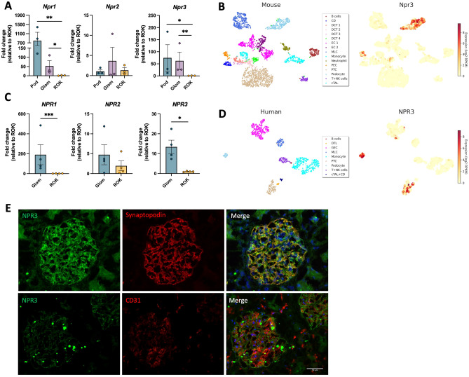
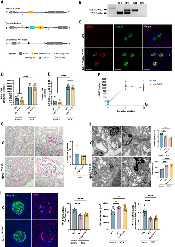
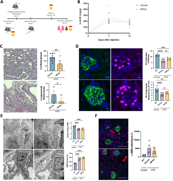
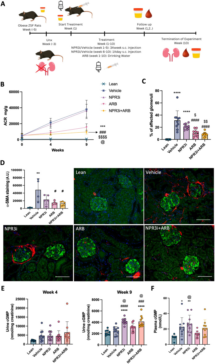
Natriuretic peptides (NPs) are cardio-derived hormones that have a crucial role in maintaining cardiovascular homeostasis. Physiological effects of NPs are mediated by binding to natriuretic peptide receptors 1 and 2 (NPR1/2), whereas natriuretic peptide receptor 3 (NPR3) acts as a clearance receptor that removes NPs from the circulation. Mouse studies have shown that local NP-signaling in the kidney glomerulus is important for the maintenance of renal homeostasis. In this study we examined the expression of NPR3 in kidney tissue and explored its involvement in renal physiology and disease by generating podocyte-specific knockout mice (NPR3podKO) as well as by using an NPR3 inhibitor (NPR3i) in rodent models of kidney disease. NPR3 was highly expressed by podocytes. NPR3podKO animals showed no renal abnormalities under healthy conditions and responded similarly to nephrotoxic serum (NTS) induced glomerular injury. However, NPR3i showed reno-protective effects in the NTS-induced model evidenced by decreased glomerulosclerosis and reduced podocyte loss. In a ZSF1 rat model of diabetic kidney injury, therapy alone with NPR3i did not have beneficial effects on renal function/histology, but when combined with losartan (angiotensin receptor blocker), NPR3i potentiated its ameliorative effects on albuminuria. In conclusion, these results suggest that NPR3 may contribute to kidney disease progression.


Conflict of interest statement

EC, PT, BR, ABG and ML are/were employed by AstraZeneca. JP’s research lab is financially supported by AstraZeneca. JP is employed by Unilabs. All other authors have no competing interest.

Figures

Figure 1
Figure 1
Expression of NPR3 in normal human and mouse kidneys. (A) RT-qPCR for NPR3 in FAC-sorted mouse podocyte (Pod), glomerular (Glom) and rest of kidney fractions (ROK). GAPDH was used as a house keeping gene. (B) Mouse single-cell RNA-sequencing data extracted from Kidney glomerular single cell atlas (data available at https://patrakkalab.se/kidney/). (C) RT-qPCR for NPR3 in human glomerular (Glom) and rest of kidney fractions (ROK). 28S was used as a housekeeping gene. (D) Human single-cell RNA-sequencing data extracted from Kidney glomerular single cell atlas (data available at https://patrakkalab.se/kidney/). The left panels show a t-SNE plot of cell clusters based on the specific cell markers. The right panels show t-SNE plot of NPR3 expression in the different clusters. CD collecting ducts, DCT distal convoluted tubules, EC endothelial cells, MLC mesangial-like cells, PEC glomerular parietal epithelial cells, PTC proximal tubular cells, T + NK T + natural killer lymphocytes, cTAL cortical thick ascending limb of Henle’s loop, DTL descending thin limb of Henle’s loop, GEC glomerular endothelial cells, cTAL + CD cortical thick ascending limb of Henle’s loop + collecting duct. (E) Immunofluorescence labelling in human kidney glomeruli for NPR3 (green) with podocyte marker Synaptopodin (red) and endothelial cell marker CD31 (red). Values are expressed as mean ± SD (n = 3). *p ≤ 0.05; **p ≤ 0.01; ***p ≤ 0.001.
Figure 2
Figure 2
Characterization of NPR3 Pod-KO at baseline and after NTS-induced injury. (A) Generation of conditional knockout mice in which NPR3 is specifically ablated in podocytes using Cre–LoxP recombination system. Exon 3 is deleted upon NPHS2-Cre-mediated recombination (L/R = left/right genotyping primer). (B) Genotyping by ear preparation and PCR at 4 weeks of age. (C) Expression of Tomato-reporter gene (red) and Podocyte marker Nephrin (green), scale bar; 100 μm. (D) Urinary cGMP levels in WT and NPR3Pod-KO mice, before and after NTS challenge (urine cGMP WT; n = 6, urine cGMP NPR3Pod-KO; n = 8, urine cGMP NTS WT; n = 5, urine cGMP NTS NPR3Pod-KO; n = 10). (E) Serum cGMP levels in WT and NPR3Pod-KO mice, before and after NTS challenge (serum cGMP WT; n = 4, serum cGMP NPR3Pod-KO; n = 5, serum cGMP NTS WT; n = 9, serum cGMP NTS NPR3Pod-KO; n = 13). (F) Urinary albumin/ creatinine ratios (U-ACR) in WT and NPR3Pod-KO after NTS-induced glomerulonephritis (WT; n = 5, NPR3Pod-KO; n = 6). (G) Histology and quantification of affected glomeruli in NTS-challenged mice. 30 randomly selected glomeruli were evaluated using PAS staining (WT; n = 4, NPR3Pod-KO; n = 5). (H) Electron microscopic findings and quantification of mean glomerular basement membrane (GBM) thickness and the number of slits per μm GBM in control and NTS challenged mice. Quantification was performed from transmission electron microscopy images (control; n = 5, WT; n = 4, NPR3Pod-KO; n = 4). (I) WT1 staining and quantification of positive cells in glomeruli, glomerular area and WT1 positive podocytes/glomerular area control and NTS challenged mice. Nephrin staining (green) was used for the quantification of glomerular area and WT1 (red) for number of podocytes. Quantification was performed from immunofluorescent images (control; n = 7, WT; n = 9, NPR3Pod-KO; n = 13). *p ≤ 0.05; **p ≤ 0.01; ***p ≤ 0.001, ****p ≤ 0.0001.
Figure 3
Figure 3
Treatment with NPR3 inhibitor via subcutaneous osmotic-pumps in mice with NTS-induced glomerular injury. (A) Schematic representation of the experimental treatment setup. (B) Urinary albumin/ creatinine ratios (U-ACR) in vehicle and NPR3i-treated mice after induction of glomerulonephritis (n = 5 for both). (C) Histology and quantification of affected glomeruli in treated mice. 30 randomly selected glomeruli were evaluated using PAS and Masson Trichrome staining (vehicle; n = 8, NPR3i; n = 9). (D) WT1 staining and quantification of positive cells in glomeruli, glomerular area and WT-1 positive podocytes/glomerular area in control and treated mice. Nephrin staining (green) was used for the quantification of glomerular area and WT1 (red) for number of podocytes. Quantification was performed from immunofluorescent images; 20 randomly selected glomeruli were evaluated (WT; n = 6, vehicle; n = 9, NPR3i; n = 8). (E) Electron microscopic findings and quantification of mean glomerular basement membrane (GBM) thickness and the number of slits per μm GBM in control and NTS challenged mice. Quantification was performed from transmission electron microscopy images (control; n = 5, WT; n = 4, NPR3Pod-KO; n = 4). (F) Staining and quantification of fibrosis marker ⍺-sma positive area in kidney cortex of control and treated mice. Quantification was performed from immunofluorescent images; an average of 10–12 randomly selected cortical cross sections with visible glomeruli were evaluated (WT; n = 2, vehicle; n = 8, NPR3i; n = 9). Scale bars: 100 μm. *p ≤ 0.05; **p ≤ 0.01; ***p ≤ 0.001, ****p ≤ 0.0001.
Figure 4
Figure 4
Treatment with selective NPR3 inhibitor monotherapy or combination therapy with ARB in diabetic nephropathy rat model. (A) Schematic representation of the experimental and treatment setup. (B) Urinary albumin/ creatinine ratios (U-ACR) in different treatment groups of diabetic nephropathy rat model (lean; n = 8, vehicle; n = 10, NPR3i; n = 10, ARB; n = 10, NPR3i + ARB; n = 10). (C) Quantification of affected glomeruli in treated rats. 30 randomly selected glomeruli were evaluated using PAS staining (lean; n = 8, vehicle; n = 10, NPR3i; n = 10, ARB; n = 10, NPR3i + ARB; n = 10). (D) Immunofluorescence and quantification of fibrosis marker ⍺-sma positive area in kidney cortex of control and treated mice (lean; n = 3, vehicle; n = 4, NPR3i; n = 5, ARB; n = 4, NPR3i + ARB; n = 5). Quantification was performed from immunofluorescent images; an average of 10–12 randomly selected cortical cross sections with visible glomeruli were evaluated. Nephrin staining (green) was used to locate the glomeruli and cortex area. ⍺-sma positive area (red) was used for fibrosis quantification. (E) Urinary cGMP levels at week 4 and 9 respectively corrected to creatinine levels in different treatment groups (lean; n = 8, vehicle; n = 10, NPR3i; n = 10, ARB; n = 10, NPR3i + ARB; n = 10). (F) Plasma cGMP levels in different treatment groups (lean; n = 8, vehicle; n = 10, NPR3i; n = 10, ARB; n = 10, NPR3i + ARB; n = 10). Treatment groups: lean = unchallenged lean rats, vehicle = ZSF1, UNx rats treated with subcutaneous injection of a vehicle (1 ml/kg), NPR3i = ZSF1, UNx rats treated with subcutaneous injection with NPR3 selective inhibitor (15 mg/kg, 1 ml/kg), ARB = ZSF1, UNX rats treated with Losartan in drinking water (20 mg/kg/day), NPR3i + ARB = ZSF1, UNx rates treated with subcutaneous injection with NPR3 selective inhibitor (15 mg/kg, 1 ml/kg) and Losartan in drinking water (20 mg/kg/day). Plasma cGMP levels in different treatment groups (lean; n = 8, vehicle; n = 10, NPR3i; n = 10, ARB; n = 10, NPR3i + ARB; n = 10). *p ≤ 0.05; **p ≤ 0.01; ***p ≤ 0.001, ****p ≤ 0.0001. *Compared to lean, #compared to vehicle, @compared to ARB, $compared to NPR3i. Scale bars: 100 μm.

References

    1. Eckardt K-U, et al. Evolving importance of kidney disease: From subspecialty to global health burden. Lancet. 2013;382:158–169. doi: 10.1016/S0140-6736(13)60439-0. - DOI - PubMed
    1. Kitching AR, Hutton HL. The players: Cells involved in glomerular disease. Clin. J. Am. Soc. Nephrol. 2016;11:1664–1674. doi: 10.2215/CJN.13791215. - DOI - PMC - PubMed
    1. Potter LR, Yoder AR, Flora DR, Antos LK, Dickey DM. Natriuretic peptides: Their structures, receptors, physiologic functions and therapeutic applications. Handb. Exp. Pharmacol. 2009 doi: 10.1007/978-3-540-68964-5_15. - DOI - PMC - PubMed
    1. Staffel J, et al. Natriuretic peptide receptor guanylyl cyclase-A in podocytes is renoprotective but dispensable for physiologic renal function. J. Am. Soc. Nephrol. JASN. 2017;28:260–277. doi: 10.1681/ASN.2015070731. - DOI - PMC - PubMed
    1. Woroniecka KI, et al. Transcriptome analysis of human diabetic kidney disease. Diabetes. 2011;60:2354–2369. doi: 10.2337/db10-1181. - DOI - PMC - PubMed

Substances