. 2024 Sep;19(9):1409-1417.
doi: 10.1038/s41565-024-01680-8. Epub 2024 May 23.

Bone-marrow-homing lipid nanoparticles for genome editing in diseased and malignant haematopoietic stem cells

Affiliations

Bone-marrow-homing lipid nanoparticles for genome editing in diseased and malignant haematopoietic stem cells

Xizhen Lian et al. Nat Nanotechnol. 2024 Sep.

Abstract

Therapeutic genome editing of haematopoietic stem cells (HSCs) would provide long-lasting treatments for multiple diseases. However, the in vivo delivery of genetic medicines to HSCs remains challenging, especially in diseased and malignant settings. Here we report on a series of bone-marrow-homing lipid nanoparticles that deliver mRNA to a broad group of at least 14 unique cell types in the bone marrow, including healthy and diseased HSCs, leukaemic stem cells, B cells, T cells, macrophages and leukaemia cells. CRISPR/Cas and base editing is achieved in a mouse model expressing human sickle cell disease phenotypes for potential foetal haemoglobin reactivation and conversion from sickle to non-sickle alleles. Bone-marrow-homing lipid nanoparticles were also able to achieve Cre-recombinase-mediated genetic deletion in bone-marrow-engrafted leukaemic stem cells and leukaemia cells. We show evidence that diverse cell types in the bone marrow niche can be edited using bone-marrow-homing lipid nanoparticles.


Conflict of interest statement

Competing interests

UT Southwestern has filed patent applications on the technologies described in this manuscript with X. Lian and D.J.S. listed as inventors. D.J.S. discloses the following competing interests: ReCode Therapeutics, Signify Bio, Tome Biosciences, Jumble Therapeutics and Pfizer Inc. D.R.L. is a consultant and equity holder of Beam Therapeutics, Prime Medicine, Pairwise Plants, Chroma Medicine and Nvelop Therapeutics, companies that use or deliver gene-editing or epigenome-modulating agents. M.J.W. is a consultant for GlaxoSmithKline, Cellarity, Novartis and Dyne Therapeutics. J.S.Y. is an equity owner of Beam Therapeutics. The remaining authors declare no competing interests.

Figures

Fig. 1 |
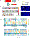
Fig. 1 |. Discovery and development of BM-homing LNPs.
a, Schematic of LNP preparation including covalent lipid species (covalent lipids and crosslinkers). b, Addition of a covalent lipid or crosslinker to the base-4-lipid LNP formulation leads to BM mRNA delivery and genome editing in a great breadth of unique BM cell types. c,d, Bioluminescence images of dissected femurs and summary of the average bioluminescence signal intensity on dissected femurs represented for covalent lipid (c) and crosslinker (d) molecular structures. The femurs were harvested from mice 6 h after the injection of BM-homing LNPs. Data are presented as mean ± standard deviation (s.d.) (n = 3 biologically independent samples).
Fig. 2 |
Fig. 2 |. tdTom expression in BM cells was activated by Cre mRNA BM-homing LNP delivery.
a, Schematic showing how the delivery of Cre mRNA activates tdTom expression in tdTom transgenic mice via Cre-mediated genetic deletion of the stop cassette. b, BM tdTom fluorescence was detected/quantified 72 h after the IV injection of LNPs loaded with Cre mRNA. c, In vivo evaluation of 23 BM-homing formulations in tdTom reporter mice showing the fluorescence images of dissected femur bones. d, Confocal microscopy imaging on BM slices was used to confirm the tdTom activation in BM. Scale bar, 100 μm. e, Flow cytometry was used to quantify Cre mRNA delivery efficacy in various BM cell types. Gating strategy for determining each cell type, full name of the cell sub-populations and cell surface markers are listed in detail in Supplementary Figs. 5–8 and Supplementary Table 1.
Fig. 3 |
Fig. 3 |. Factors that do and do not contribute to BM delivery tropism.
a,b, Hydrodynamic diameter and polydispersity index (PDI) (a) and ζ-potential (b) of selected BM-homing LNPs determined by dynamic light scattering (DLS). Data are presented as mean ± s.d. (n = 3 biologically independent samples). c, Protein composition of LNP-surface-adsorbed protein corona determined by unbiased mass spectrometry proteomics. d, Comparison of the luminescence signal intensity of selected BM-homing LNPs injected into wild-type (wt) C57BL/6 mice and ApoE knockout (ApoE−/−) mice. Data are presented as mean ± s.d. (n = 3 biologically independent samples). *P < 0.05, **P < 0.01, ***P < 0.001 determined by a two-tailed t-test.
Fig. 4 |
Fig. 4 |. In vivo genome and base editing of β-globin-disorder-relevant genes in HBBS/S Townes mice.
a, Extended β-globin locus, showing the target BCL11A binding motif in the promoters of the genes encoding γ-globin. BCL11A binding motif is represented by a red line and sgG34 is represented by a green line. b, HBBS/S Townes mice received two weekly IV injections of BM-homing LNPs encapsulating Cas9 mRNA and sgG34 or ABE8e_NRCH mRNA and sgHBB (n = 3; the total RNA dose per injection was 3 mg kg−1). BM samples were harvested and analysed seven days after the final injection. c, Insertions and deletions (indels) detected in CD117+ cells isolated from the BM of HBBS/S Townes mice. Data are presented as mean ± s.d. (n = 3 biologically independent samples). ***P < 0.001 determined by a two-tailed t-test. d, Representative NGS reads of a sample treated with LNPs (encapsulating Cas9 mRNA and sgG34). e, Edited region of HBB with target A at protospacer position 7 shown in orange and bystander edit in blue (silent). sgHBB is represented by a purple line. f, A–G conversion detected in CD117+ cells isolated from the BM of HBBS/S Townes mice. Data are presented as mean ± s.d. (n = 3 biologically independent samples). **P < 0.01 determined by a two-tailed t-test. g, Representative NGS reads of a sample treated with LNPs (encapsulating ABE8e_NRCH mRNA and sgHBB).
Fig. 5 |
Fig. 5 |. tdTom expression in MLL-AF9-driven AML model was activated by BM-homing LNP-mediated editing.
a, The MLL-AF9-IRES-YFP gene was installed into the genome of Lin cells extracted from the foetal liver of tdTom reporter mice and the cells were incubated with LNPs containing Cre mRNA in a 24-well plate for 24 h. b, Delivery of Cre mRNA activates tdTom expression in tdTom transgenic mice via the Cre-mediated genetic deletion of the stop cassette. c, Summary of the percentage of tdTom+ cells measured from the confocal microscopy images of the edited MLL-AF9-IRES-YFP Lin cells. Data are presented as mean ± s.d. (n = 3 biologically independent samples). d, Representative confocal microscopy images of the control and LNP-treated cells. Scale bar, 100 μm. e, DNA agarose gel of the PCR amplicon performed from the genomic DNA extracted from the control and LNP-treated cells with primers flanking the Ai14 locus (n = 3 biologically independent samples). f, C57BL/6 recipient mice were lethally irradiated and received IV transplantation of MLL-AF9-transfected Lin cells. Three weeks after the transplantation, the BM and spleen were extracted from the primary transplant recipient animal and the isolated cells were transplanted into a secondary transplant recipient to establish the model for LNP study. Three weeks after the secondary transplantation, Cre mRNA BM-homing LNPs were injected via IV administration. Leukaemic cells were harvested and analysed 72 h after LNP injection. g, Percentage of tdTom+ cells after LNP and BM-homing LNP injection on bone-engrafted leukaemia animals. BM-isolated leukaemic cells, leukaemic stem cells residing in BM (BM LSCs) and spleen-isolated leukaemic cells are characterized by flow cytometry. Data are presented as mean ± s.d. (n = 3 biologically independent samples).

References

    1. Laurenti E & Gottgens B From haematopoietic stem cells to complex differentiation landscapes. Nature 553, 418–426 (2018). - PMC - PubMed
    1. Bauer TR Jr. et al. Correction of the disease phenotype in canine leukocyte adhesion deficiency using ex vivo hematopoietic stem cell gene therapy. Blood 108, 3313–3320 (2006). - PMC - PubMed
    1. Blaese RM et al. T lymphocyte-directed gene therapy for ADA-SCID: initial trial results after 4 years. Science 270, 475–480 (1995). - PubMed
    1. Boztug K et al. Stem-cell gene therapy for the Wiskott-Aldrich syndrome. N. Engl. J. Med. 363, 1918–1927 (2010). - PMC - PubMed
    1. Cowan MJ et al. Early outcome of a phase I/II clinical trial (NCT03538899) of gene-corrected autologous CD34+ hematopoietic cells and low-exposure busulfan in newly diagnosed patients with Artemis-deficient severe combined immunodeficiency (ART-SCID). Biol. Blood Marrow Transpl. 26, S88–S89 (2020).

LinkOut - more resources