

MOG antibody-associated optic neuritis
- PMID: 38783085
- PMCID: PMC11306565
- DOI: 10.1038/s41433-024-03108-y
MOG antibody-associated optic neuritis
Abstract
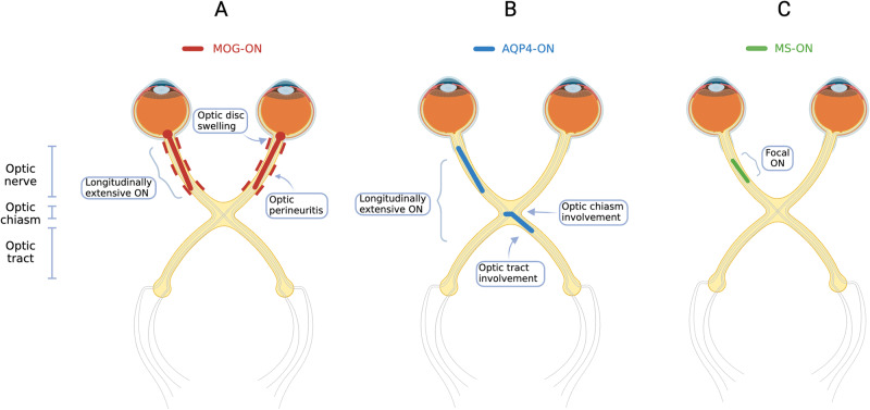
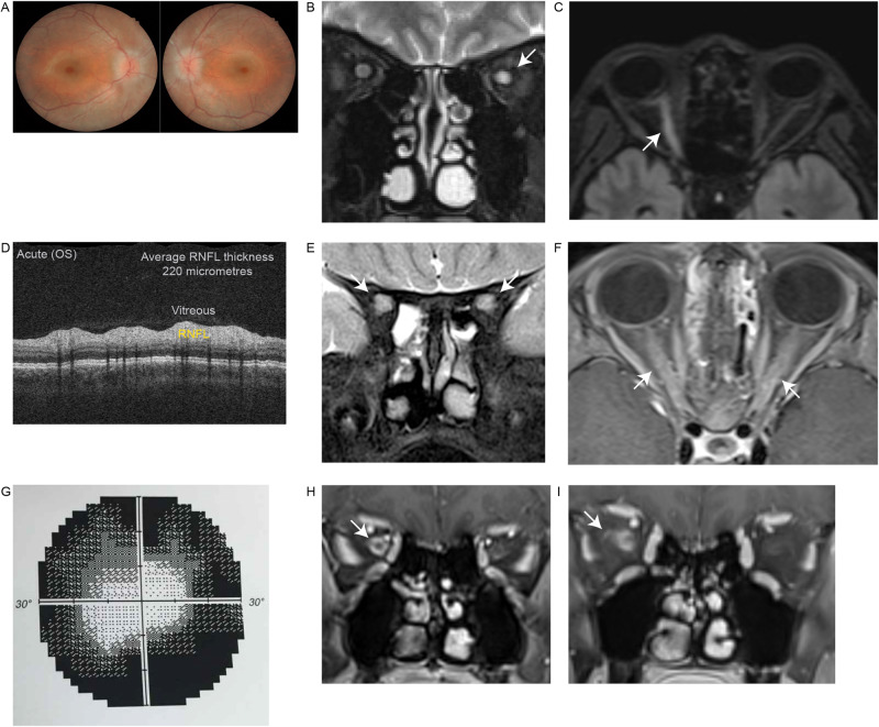
Myelin oligodendrocyte glycoprotein (MOG) antibody-associated disease (MOGAD) is a demyelinating disorder, distinct from multiple sclerosis (MS) and neuromyelitis optica spectrum disorder (NMOSD). MOGAD most frequently presents with optic neuritis (MOG-ON), often with characteristic clinical and radiological features. Bilateral involvement, disc swelling clinically and radiologically, and longitudinally extensive optic nerve hyperintensity with associated optic perineuritis on MRI are key characteristics that can help distinguish MOG-ON from optic neuritis due to other aetiologies. The detection of serum MOG immunoglobulin G utilising a live cell-based assay in a patient with a compatible clinical phenotype is highly specific for the diagnosis of MOGAD. This review will highlight the key clinical and radiological features which expedite diagnosis, as well as ancillary investigations such as visual fields, visual evoked potentials and cerebrospinal fluid analysis, which may be less discriminatory. Optical coherence tomography can identify optic nerve swelling acutely, and atrophy chronically, and may transpire to have utility as a diagnostic and prognostic biomarker. MOG-ON appears to be largely responsive to corticosteroids, which are often the mainstay of acute management. However, relapses are common in patients in whom follow-up is prolonged, often in the context of early or rapid corticosteroid tapering. Establishing optimal acute therapy, the role of maintenance steroid-sparing immunotherapy for long-term relapse prevention, and identifying predictors of relapsing disease remain key research priorities in MOG-ON.
摘要: 髓鞘少突胶质细胞糖蛋白 (MOG)抗体相关疾病(MOGAD)是一种脱髓鞘疾病, 有别于多发性硬化症 (MS) 和视神经脊髓炎视神经疾病 (NMOSD) 。MOGAD最常表现为视神经炎 (MOG-ON), 通常具有特征性的临床和影像学特征。MOGAD双侧受累、临床和影像学表现为视盘肿胀以及纵向MRI上广泛视神经高信号伴相关视神经束膜炎是有助于区分MOG-ON与其他病因所致视神经炎的关键特征。在具有相同临床表型的患者中, 利用基于活细胞的测定法检测血清MOG免疫球蛋白G对于MOGAD的诊断具有高度特异性。本综述将重点介绍加快诊断的关键临床和影像学特征及辅助检查, 如视野, 视觉诱发电位和脑脊液分析。这些辅助检查的诊断价值相对较弱, OCT可以识别急性视神经肿胀和慢性视神经萎缩, 并可能作为诊断和预后的生物标志物。MOG-ON似乎对皮质类固醇较敏感, 皮质类固醇通常是急性期治疗的一线药物。然而, 在随访时间延长的患者中很容易复发, 通常是在早期或快速减少皮质类固醇的情况下。最佳策略的的急性期治疗, 维持类固醇保留免疫疗法在长期预防复发中的作用, 以及确定疾病复发的预测因子仍然是MOG-ON的关键研究重点。.
© 2024. The Author(s).
Conflict of interest statement
NJ and ML have no financial disclosures to report. RD has received research funding from the Star Scientific Foundation, The Trish Multiple Sclerosis Research Foundation, Multiple Sclerosis Research Australia, the Petre Foundation and the NHMRC (Australia; Investigator Grant). He has also received honoraria from Biogen Idec as an invited speaker, and is on the IDMC for a Roche RCT in paediatric MS. He is on the medical advisory board (non-remunerated position) of The MOG Project. SR has received research funding from the National Health and Medical Research Council (NHMRC, Australia), the Petre Foundation, the Brain Foundation, the Royal Australasian College of Physicians, and the University of Sydney. She is supported by an NHMRC Investigator Grant (GNT2008339). She serves as a consultant on an advisory board for UCB and Limbic Neurology, and has been an invited speaker for educational/research sessions coordinated by Biogen, Alexion, Novartis, Excemed and Limbic Neurology. She is on the medical advisory boards (non-remunerated positions) of The MOG Project and the Sumaira Foundation.
Figures
References
-
- O’Connell K, Hamilton-Shield A, Woodhall M, Messina S, Mariano R, Waters P, et al. Prevalence and incidence of neuromyelitis optica spectrum disorder, aquaporin-4 antibody-positive NMOSD and MOG antibody-positive disease in Oxfordshire, UK. J Neurol Neurosurg Psychiatry. 2020;91:1126–8. 10.1136/jnnp-2020-323158 - DOI - PubMed
Publication types
MeSH terms
Substances
LinkOut - more resources
Full Text Sources
Miscellaneous
