. 2024 May 23;25(1):85.
doi: 10.1186/s10194-024-01791-6.

MERTK in the rat trigeminal system: a potential novel target for cluster headache?

Affiliations

MERTK in the rat trigeminal system: a potential novel target for cluster headache?

Jacob C A Edvinsson et al. J Headache Pain. .

Abstract

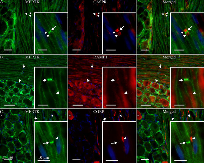
The trigeminal system is key to the pathophysiology of migraine and cluster headache, two primary headache disorders that share many features. Recently, MER proto-oncogene tyrosine kinase (MERTK), a cell surface receptor, was strongly associated with cluster headache through genetic studies. Further, the MERTK ligand galectin-3 has been found to be elevated in serum of migraine patients. In this study, MERTK and MERTK ligands were investigated in key tissue to better understand their potential implication in the pathophysiology of primary headache disorders. Immunohistochemistry was used to map MERTK and galectin-3 expression in rat trigeminal ganglia. RT-qPCR was used to assess MERTK gene expression in blood, and ELISA immunoassays were used for MERTK ligand quantification in serum from study participants with and without cluster headache. MERTK gene expression was elevated in blood samples from study participants with cluster headache compared to controls. In addition, MERTK ligand galectin-3 was found at increased concentration in the serum of study participants with cluster headache, whereas the levels of MERTK ligands growth arrest specific 6 and protein S unaffected. MERTK and galectin-3 were both expressed in rat trigeminal ganglia. Galectin-3 was primarily localized in smaller neurons and to a lesser extent in C-fibres, while MERTK was found in satellite glia cells and in the outer membrane of Schwann cells. Interestingly, a strong MERTK signal was found specifically in the region proximal to the nodes of Ranvier. The overexpression of MERTK and galectin-3 in tissue from study participants with cluster headache, as well as the presence of MERTK in rat peripheral satellite glia cells and Schwann cells in the trigeminal ganglia, further highlights MERTK signalling as an interesting potential future therapeutic target in primary headache.

Keywords: GWAS; Galectin-3; MER proto-oncogene tyrosine kinase; Migraine; Trigeminal system.


Conflict of interest statement

The authors report no competing interests.

Figures

Fig. 1
Fig. 1
MERTK expression in relation to RAMP1, CGRP and CASPR. A: MER proto-oncogene tyrosine kinase (MERTK) immunoreactivity was observed mainly in satellite glial cells (SGCs) and, to a lesser extent, in Schwann cells. Interestingly, MERTK was observed to have a striking expression in the region of the Schwann cell overhanging the nodes of Ranvier of Aδ-fibres (Arrow). This was confirmed by double staining with contactin associated protein 1 (CASPR), which is explicitly expressed in the para- and juxtaparanodal regions of myelinated fibres (Arrowheads). Insert-A: Close-up on an Aδ-fibre with para- and juxtaparanodal regions stained by CASPR (Arrowheads). Notably, a weak expression of MERTK can be seen in the outer layer of the Schwann cell, which is strikingly pronounced where the Schwann cells border the node of Ranvier (Arrow). B: To confirm the expression of MERTK in Schwann cells and SGCs, a double-staining was performed with a well-established antibody, RAMP1. MERTK can be seen expressed in SGCs enveloping a RAMP1 positive neuron (Arrowhead), and a striking immunoreactivity of MERTK can be seen in Schwann cell outer membranes proximal to the nodes of Ranvier (Arrow). Insert-B: Close-up on an Aδ-fibre immunoreactive for RAMP1 (Arrowhead). Flanking the axon, its associated Schwann cell indicates immunoreactivity for MERTK with a strong expression near the node of Ranvier (Arrow). C: The expression of MERTK in relation to calcitonin gene-related peptide (CGRP). CGRP was observed in C-fibre boutons (Arrowhead) and associated neuron cell bodies (Asterix). MERTK did not co-localize with CGRP but could be observed in SGCs surrounding CGRP-positive neurons, and in Schwann cell membranes (Arrow). Insert-C: A C-fibre displaying immunoreactivity for CGRP with a bouton (Arrowhead) in close proximation to the MERTK-positive Schwann cell membranes flanking the node of Ranvier of an Aδ-fibre (Arrow). Inserts are separate images from the lower magnification images A, B and C
Fig. 2
Fig. 2
MERTK and galectin-3 expression in rat TG. A: To evaluate the expression of MERTK in Schwann cells enveloping Aδ-fibres, a double-staining with myelin basic protein (MBP) was performed. MBP was observed flanking the axon of A-fibres (Arrowhead) but did not co-localize with MERTK in the outer layer of Schwann cells, or the region proximal to the nodes of Ranvier (Arrow). No immunoreactivity for MBP was observed in neuron cell bodies. Insert-A: Close-up displaying an Aδ-fibre axon (Asterix), its associated myelin sheath stained with MBP (Arrowhead) and a prominent MERTK expression near the node of Ranvier (Arrow). B: MERTK expression in relation to one of its ligands, galectin-3 (Gal-3). Interestingly, Gal-3 immunoreactivity was mainly expressed in a population of smaller neurons (Arrowhead) while larger neurons often were negative (Asterix). MERTK expression was again observed in satellite glial cells (SGCs) and Schwann cells (Arrow). Insert-B: A Gal-3 positive neuron (Arrowhead), enveloped by MERTK positive SGCs. In comparison, a negative neuron enveloped by MERTK immunoreactive SGCs can be observed at the bottom of the picture (Asterix). The arrow points out a nearby MERTK immunoreactive Schwann cell membranes flanking a node of Ranvier. C: Gal-3 was double stained with CGRP to determine co-localization in smaller neurons and C-fibres. CGRP was observed in a population of neurons and associated C-fibres (Arrowhead). Co-localization with Gal-3 and CGRP was observed in some, but not all, neuron cell bodies (Arrow). Insert-C: A weak expression of Gal-3 co-expressed with CGRP was observed in a C-fibre bouton (Arrowhead). Similarly, a weak expression of both Gal-3 and CGRP can be observed in a nearby neuron cell body (Arrow). Inserts are separate images from the lower magnification images A, B and C
Fig. 3
Fig. 3
MERTK gene expression analysis in blood from cluster headache patients and controls. mRNA levels are expressed as relative quantity, levels were normalized to TBP and IPO8 and to a control sample and log2 transformed. Boxplots represent the interquartile range and median value (horizontal line), whiskers correspond to 1.5 times the interquartile range in both directions, scattered symbols show the individual values; triangles = controls, circles = patients in active bout, squares = patients in remission. Patients n = 16 and controls n = 21. Group comparison by two-tailed t-test, p-value = 0.031, *; p-value < 0.05. TBP; TATA-Box Binding Protein, IPO8; Importin 8
Fig. 4
Fig. 4
Quantification of MERTK ligands Gal-3, GAS6 and PROS1 in serum from patients and controls. Concentrations of MERTK ligands in serum expressed as ng/ml serum. Left panel: Gal-3, patients n = 11 and controls n = 8. Middle panel: GAS6, patients n = 11 and controls n = 9. PROS1, patients n = 11 and controls n = 9. Boxplots represent the interquartile range and median value (horizontal line), whiskers correspond to 1.5 times the interquartile range in both directions, scattered symbols show the individual values; triangles = controls, circles = patients in active bout, squares = patients in remission. Group comparison by Wilcoxon test or two-tailed t-test, **; p-value < 0.01
Fig. 5
Fig. 5
Schematic of proposed mechanism of action at the trigeminal tripartite synapse. Top: Pseudo-unipolar trigeminal neuron projecting a myelinated Aδ-fibre which aligns with a C-fibre bouton. MERTK receptor expression can be observed in satellite glial cells (SGCs) and Schwann cells. Bottom-left: Depolarization of C-fibre axons causes the local release of headache relevant neuropeptides, e.g. calcitonin gene-related peptide (CGRP), substance P (SP) and neurokinin A (NKA) [43, 44, 59], these neuropeptides could traverse the nodal gap and activate their respective receptors, expressed in the Aδ-fibre axon at the node of Ranvier, leading to modulation and/or sensitization of the nociceptive signal projecting to higher order neurons. This is suggested to occur via activation of the cAMP dependent pathway, activating protein kinase A (PKA) and C (PKC), which leads to an increase in intracellular Ca2+ [60]. Similarly, Gal-3 could be co-released from the C-fibre bouton and activate MERTK receptors expressed in the Schwann cell membranes flanking the node of Ranvier. Bottom-right: The hypothesized local release of Gal-3 from C-fibre boutons likely activates MERTK receptors in the Schwann cell membrane. We propose that this activation could lead to the activation of the MAPK/ERK1/2 pathway in the Schwann cell which produces hypersensitivity which can be abolished by MEK1/2 inhibitor intervention, as shown in dorsal root ganglia [46]. Finally, the release of Gal-3 can also lead to the externalization of phosphatidylserine (Ptd-L-Ser), which potentially could be a driving factor for the mechanism of synaptic pruning

References

    1. Headache Classification Committee of the International Headache Society (IHS) The International classification of Headache disorders, 3rd edition. Cephalalgia. 2018;38(1):1–211. doi: 10.1177/0333102417738202. - DOI - PubMed
    1. Fischera M, Marziniak M, Gralow I, Evers S. The incidence and prevalence of cluster headache: a meta-analysis of population-based studies. Cephalalgia. 2008;28(6):614–618. doi: 10.1111/j.1468-2982.2008.01592.x. - DOI - PubMed
    1. Carmine Belin A, Ran C, Edvinsson L. Calcitonin gene-related peptide (CGRP) and cluster headache. Brain Sci. 2020;10(1):30. doi: 10.3390/brainsci10010030. - DOI - PMC - PubMed
    1. Hoffmann J, May A. Diagnosis, pathophysiology, and management of cluster headache. Lancet Neurol. 2018;17(1):75–83. doi: 10.1016/S1474-4422(17)30405-2. - DOI - PubMed
    1. Nesbitt AD, Goadsby PJ (2012) Cluster headache. BMJ. 344 - PubMed

LinkOut - more resources