

Breaking barriers: The potential of nanosystems in antituberculosis therapy
- PMID: 38783925
- PMCID: PMC11112550
- DOI: 10.1016/j.bioactmat.2024.05.013
Breaking barriers: The potential of nanosystems in antituberculosis therapy
Abstract
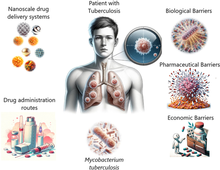
Tuberculosis (TB), caused by Mycobacterium tuberculosis, continues to pose a significant threat to global health. The resilience of TB is amplified by a myriad of physical, biological, and biopharmaceutical barriers that challenge conventional therapeutic approaches. This review navigates the intricate landscape of TB treatment, from the stealth of latent infections and the strength of granuloma formations to the daunting specters of drug resistance and altered gene expression. Amidst these challenges, traditional therapies often fail, contending with inconsistent bioavailability, prolonged treatment regimens, and socioeconomic burdens. Nanoscale Drug Delivery Systems (NDDSs) emerge as a promising beacon, ready to overcome these barriers, offering better drug targeting and improved patient adherence. Through a critical approach, we evaluate a spectrum of nanosystems and their efficacy against MTB both in vitro and in vivo. This review advocates for the intensification of research in NDDSs, heralding their potential to reshape the contours of global TB treatment strategies.
Keywords: Barriers; Drug resistance; Mycobacterium tuberculosis; Nanoscale drug delivery systems; Tuberculosis.
© 2024 The Authors.
Conflict of interest statement
All contributing authors declare no conflict of interest.
Figures
Similar articles
-
[Development of antituberculous drugs: current status and future prospects].Kekkaku. 2006 Dec;81(12):753-74. Kekkaku. 2006. PMID: 17240921 Review. Japanese.
-
Potential and development of inhaled RNAi therapeutics for the treatment of pulmonary tuberculosis.Adv Drug Deliv Rev. 2016 Jul 1;102:21-32. doi: 10.1016/j.addr.2016.04.013. Epub 2016 Apr 22. Adv Drug Deliv Rev. 2016. PMID: 27108702 Review.
-
Advanced drug delivery and therapeutic strategies for tuberculosis treatment.J Nanobiotechnology. 2023 Nov 9;21(1):414. doi: 10.1186/s12951-023-02156-y. J Nanobiotechnology. 2023. PMID: 37946240 Free PMC article. Review.
-
Lactate Metabolism and Signaling in Tuberculosis and Cancer: A Comparative Review.Front Cell Infect Microbiol. 2021 Feb 26;11:624607. doi: 10.3389/fcimb.2021.624607. eCollection 2021. Front Cell Infect Microbiol. 2021. PMID: 33718271 Free PMC article. Review.
-
Tuberculosis: An Update on Pathophysiology, Molecular Mechanisms of Drug Resistance, Newer Anti-TB Drugs, Treatment Regimens and Host- Directed Therapies.Curr Top Med Chem. 2021;21(6):547-570. doi: 10.2174/1568026621999201211200447. Curr Top Med Chem. 2021. PMID: 33319660 Review.
Cited by
-
Screening of novel narrow-spectrum benzofuroxan derivatives for the treatment of multidrug-resistant tuberculosis through in silico, in vitro, and in vivo approaches.Front Microbiol. 2024 Oct 11;15:1487829. doi: 10.3389/fmicb.2024.1487829. eCollection 2024. Front Microbiol. 2024. PMID: 39464394 Free PMC article.
-
Forging a New Frontier: Antimicrobial Peptides and Nanotechnology Converging to Conquer Gastrointestinal Pathogens.Small. 2025 Jul;21(26):e2501431. doi: 10.1002/smll.202501431. Epub 2025 May 19. Small. 2025. PMID: 40384187 Free PMC article. Review.
-
Advancing Nanotechnology: Targeting Biofilm-Forming Bacteria with Antimicrobial Peptides.BME Front. 2025 Mar 4;6:0104. doi: 10.34133/bmef.0104. eCollection 2025. BME Front. 2025. PMID: 40041091 Free PMC article. Review.
-
New Perspectives of Hydrogels in Chronic Wound Management.Molecules. 2025 Feb 4;30(3):686. doi: 10.3390/molecules30030686. Molecules. 2025. PMID: 39942790 Free PMC article. Review.
References
-
- Sankineni S., Chauhan S., Shegokar R., Pathak Y. Tubercular Drug Delivery Systems. Springer International Publishing; Cham: 2023. pp. 1–13.
-
- Vale N., Correia A., Figueiredo P., Santos H.A. J. Mol. Struct. 2016;1105:286.
-
- World Health Organization Global tuberculosis report. 2023 Geneva, 2023.
-
- Donnellan S., Giardiello M. vol. 4. 2019. p. 76. (J Interdiscip Nanomed).
Publication types
LinkOut - more resources
Full Text Sources
