

Are the class 18 myosins Myo18A and Myo18B specialist sarcomeric proteins?
- PMID: 38784114
- PMCID: PMC11112018
- DOI: 10.3389/fphys.2024.1401717
Are the class 18 myosins Myo18A and Myo18B specialist sarcomeric proteins?
Abstract
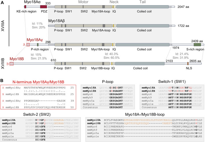
Initially, the two members of class 18 myosins, Myo18A and Myo18B, appeared to exhibit highly divergent functions, complicating the assignment of class-specific functions. However, the identification of a striated muscle-specific isoform of Myo18A, Myo18Aγ, suggests that class 18 myosins may have evolved to complement the functions of conventional class 2 myosins in sarcomeres. Indeed, both genes, Myo18a and Myo18b, are predominantly expressed in the heart and somites, precursors of skeletal muscle, of developing mouse embryos. Genetic deletion of either gene in mice is embryonic lethal and is associated with the disorganization of cardiac sarcomeres. Moreover, Myo18Aγ and Myo18B localize to sarcomeric A-bands, albeit the motor (head) domains of these unconventional myosins have been both deduced and biochemically demonstrated to exhibit negligible ATPase activity, a hallmark of motor proteins. Instead, Myo18Aγ and Myo18B presumably coassemble with thick filaments and provide structural integrity and/or internal resistance through interactions with F-actin and/or other proteins. In addition, Myo18Aγ and Myo18B may play distinct roles in the assembly of myofibrils, which may arise from actin stress fibers containing the α-isoform of Myo18A, Myo18Aα. The β-isoform of Myo18A, Myo18Aβ, is similar to Myo18Aα, except that it lacks the N-terminal extension, and may serve as a negative regulator through heterodimerization with either Myo18Aα or Myo18Aγ. In this review, we contend that Myo18Aγ and Myo18B are essential for myofibril structure and function in striated muscle cells, while α- and β-isoforms of Myo18A play diverse roles in nonmuscle cells.
Keywords: MYO18A; MYO18B; knockout (KO) mice; sarcomere; stress fibers; unconventional myosins.
Copyright © 2024 Horsthemke, Arnaud and Hanley.
Conflict of interest statement
The authors declare that the research was conducted in the absence of any commercial or financial relationships that could be construed as a potential conflict of interest.
Figures
Similar articles
-
A novel isoform of myosin 18A (Myo18Aγ) is an essential sarcomeric protein in mouse heart.J Biol Chem. 2019 May 3;294(18):7202-7218. doi: 10.1074/jbc.RA118.004560. Epub 2019 Feb 8. J Biol Chem. 2019. PMID: 30737279 Free PMC article.
-
Emerging concepts of myosin 18A isoform mechanobiology in organismal and immune system physiology, development, and function.FASEB J. 2024 May 31;38(10):e23649. doi: 10.1096/fj.202400350R. FASEB J. 2024. PMID: 38776246 Review.
-
Deficiency of Myo18B in mice results in embryonic lethality with cardiac myofibrillar aberrations.Genes Cells. 2008 Oct;13(10):987-99. doi: 10.1111/j.1365-2443.2008.01226.x. Epub 2008 Aug 29. Genes Cells. 2008. PMID: 18761673
-
Myo18b is essential for sarcomere assembly in fast skeletal muscle.Hum Mol Genet. 2017 Mar 15;26(6):1146-1156. doi: 10.1093/hmg/ddx025. Hum Mol Genet. 2017. PMID: 28104788
-
Myosin XVIII.Adv Exp Med Biol. 2020;1239:421-438. doi: 10.1007/978-3-030-38062-5_19. Adv Exp Med Biol. 2020. PMID: 32451870 Review.
Cited by
-
The myoblast methylome: multiple types of associations with chromatin and transcription.Epigenetics. 2025 Dec;20(1):2508251. doi: 10.1080/15592294.2025.2508251. Epub 2025 Jun 11. Epigenetics. 2025. PMID: 40497496 Free PMC article.
References
-
- Alexander C. J., Barzik M., Fujiwara I., Remmert K., Wang Y. X., Petralia R. S., et al. (2021). Myosin 18Aα targets the guanine nucleotide exchange factor β-Pix to the dendritic spines of cerebellar Purkinje neurons and promotes spine maturation. Faseb J. 35, e21092. 10.1096/fj.202001449R - DOI - PMC - PubMed
Publication types
LinkOut - more resources
Full Text Sources
Research Materials