. 2024 May 9:15:1400284.
doi: 10.3389/fmicb.2024.1400284. eCollection 2024.

Identification of a dCache-type chemoreceptor in Campylobacter jejuni that specifically mediates chemotaxis towards methyl pyruvate

Affiliations

Identification of a dCache-type chemoreceptor in Campylobacter jejuni that specifically mediates chemotaxis towards methyl pyruvate

Qi Zhao et al. Front Microbiol. .

Abstract

The foodborne pathogenic bacterium Campylobacter jejuni utilizes chemotaxis to assist in the colonization of host niches. A key to revealing the relationship among chemotaxis and pathogenicity is the discovery of signaling molecules perceived by the chemoreceptors. The C. jejuni chemoreceptor Tlp11 is encoded by the highly infective C. jejuni strains. In the present study, we report that the dCache-type ligand-binding domain (LBD) of C. jejuni ATCC 33560 Tlp11 binds directly to novel ligands methyl pyruvate, toluene, and quinoline using the same pocket. Methyl pyruvate elicits a strong chemoattractant response, while toluene and quinoline function as the antagonists without triggering chemotaxis. The sensory LBD was used to control heterologous proteins by constructing chimeras, indicating that the signal induced by methyl pyruvate is transmitted across the membrane. In addition, bioinformatics and experiments revealed that the dCache domains with methyl pyruvate-binding sites and ability are widely distributed in the order Campylobacterales. This is the first report to identify the class of dCache chemoreceptors that bind to attractant methyl pyruvate and antagonists toluene and quinoline. Our research provides a foundation for understanding the chemotaxis and virulence of C. jejuni and lays a basis for the control of this foodborne pathogen.

Keywords: Campylobacter jejuni; chemoreceptor Tlp11; chemotaxis; chimeras; ligands.


Conflict of interest statement

The authors declare that the research was conducted in the absence of any commercial or financial relationships that could be construed as a potential conflict of interest.

Figures

Figure 1
Figure 1
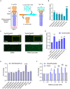
Identification of methyl pyruvate as the direct-binding ligand of Tlp11-LBD. (A) The effect of different concentrations of methyl pyruvate on the Tm of Tlp11-LBD. Each concentration of methyl pyruvate indicated in (A) was the working concentration in the system. The right panel shows the molecular structure of methyl pyruvate. Error bars represent the standard errors of three independent replicates, shown as mean ± SD. The p-values were calculated using the paired t-test; **p < 0.01, compared to buffer. (B) The thermal unfolding curves and calculated ΔTm of thermal shift assay measurements for Tlp11-LBD, in the absence and presence of 10 mM methyl pyruvate. (C) Microscale thermophoresis of Tlp11-LBD with methyl pyruvate. The blue and black lines indicate the thermophoresis of Tlp11-LBD labeled with fluorescent dyes, at different concentrations of methyl pyruvate and in the buffer, respectively. The working concentration of the proteins used for MST detection was 250 nM. The maximum working concentration of the ligand was 2.5 mM, and it was gradually diluted. Upper panel: raw thermophoretic data; lower panel: dose-response curves with fitting results. Error bars represent the standard errors of three independent replicates, shown as mean ± SD.
Figure 2
Figure 2
The effect of methyl pyruvate on chemotaxis and growth of C. jejuni. (A,C,D) The chemotactic responses of the C. jejuni NCTC 11168 strain expressing Tlp11 of ATCC 33560 (11168ΩTlp11) (A), WT NCTC 11168 strain (C), and non-chemotactic mutant 11168ΩTlp11/ΔCheY strain (D) towards different concentrations of methyl pyruvate, shown as relative chemotactic index. Error bars represent the standard errors of three independent replicates, shown as mean ± SD. The p-values were calculated using the paired t-test; *p < 0.05, **p < 0.01, and ***p < 0.001, compared to buffer. (B) Examples of the distributions of the C. jejuni NCTC 11168 WT or ΩTlp11 cells in the observation channel of the microfluidic device, obtained before addition of ligand and 30 min after response to 10 mM methyl pyruvate. The black arrow indicates the direction up the concentration gradient of methyl pyruvate. The response is characterized by measurement of the cell number (~200–600 cells) in the analysis region of the observation channel, as the view in (B). (E,F) The growth curves of the C. jejuni ATCC 33560 strain in Minimal Essential Medium supplemented with fetal bovine serum and different concentrations of methyl pyruvate (E) and pyruvate (F). Error bars represent the standard errors of 4 independent replicates, shown as mean ± SD.
Figure 3
Figure 3
Response of chimeras Tlp11-Tar and Tlp11-PhoQ to methyl pyruvate. (A) Design and construction of the Tlp11-Tar hybrid receptor. On the left is the schematic diagram of the E. coli chemoreceptor Tar, with the cytoplasmic region shown in red. C. jejuni Tlp11-LBD is connected to the cytoplasmic region of Tar to form the hybrid receptor Tlp11-Tar. The fusion site is located in the TM2. (B) The relative chemotactic index (CI) of the E. coli VS188 cells expressing Tlp342Tar200, Tlp343Tar200, Tlp346Tar204, Tlp347Tar204, Tlp348Tar204, or pin-head Tar as the sole receptor, in response to 10 mM glucose for 50 min. (C) Examples of the distribution of the E. coli cells expressing Tlp342Tar200 in the observation channel of the microfluidic device, acquired before addition of the ligand and 50 min after the response to 20 mM methyl pyruvate (scale bar: 100 μm). The x-component (black arrow) indicates the direction up the concentration gradient of methyl pyruvate. The response is characterized by measurements of the total fluorescence intensity (cell density) in the analysis region (225 × 150 μm) of the observation channel, indicated by a yellow rectangle. (D) The relative CI of E. coli VS188 cells expressing Tlp342Tar200 as the sole receptor, in response to the indicated concentrations of methyl pyruvate or buffer at 50 min. In (B,D), the corresponding values of the fluorescence intensities in the analysis regions were normalized to the fluorescence intensity of cells before adding the compound, to obtain CI. The CI of cells in response to compound gradient was then normalized to that of cells in the buffer, to get the relative CI. (E) The fold-change in fluorescence intensity of E. coli MG1655/ΔPhoQ expressing each hybrid kinase, PhoQ, or only containing empty vector pKG116, after stimulation with 20 mM methyl pyruvate for 40 min. (F) The responses of E. coli MG1655/ΔPhoQ expressing Tlp343PhoQ202 as the single chimera or PhoQ towards indicated concentrations of methyl pyruvate. Error bars represent the standard errors of three independent replicates, shown as mean ± SD. The p-values were calculated using the paired t-test; *p < 0.05, **p < 0.01, and ***p < 0.001.
Figure 4
Figure 4
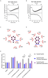
Binding interaction analysis using molecular docking and Tlp11-LBD mutant proteins. (A) Molecular docking analysis of the interaction of Tlp11-LBD with methyl pyruvate using Autodock. The conformation with the lowest binding free energy is shown with PyMOL. Methyl pyruvate is predicted to bind to the membrane-proximal pocket of Tlp11-LBD. The key residues in the ligand-binding pocket involved in methyl pyruvate binding are shown as sticks. The hydrogen bonds are shown as yellow dashed lines. (B–D) MST measurements for the interactions of Tlp11-LBD mutants N268A, Y291A, and T320A with methyl pyruvate. The upper panel indicates the representative curves for thermophoresis of mutant proteins with different concentrations of methyl pyruvate, while the lower panel indicates the dose–response curve with the fitting result. Error bars represent the standard errors of three independent replicates, shown as mean ± SD. The concentration for the mutant proteins was 250 nM, and the maximum concentration for the ligand was 2.5 mM, which was diluted gradually.
Figure 5
Figure 5
Detection of the antagonistic effects of toluene and quinoline. (A,B) MST measurements for the interactions of Tlp11-LBD with toluene (A) and quinoline (B). The upper panel indicates the representative curves for thermophoresis of Tlp11-LBD with different concentrations of toluene or quinoline, while the lower panel indicates the dose–response curve with the fitting result. Error bars represent the standard errors of three independent replicates, shown as mean ± SD. The concentration for Tlp11-LBD was 250 nM, and the maximum concentration for the ligand was 5 mM, which was gradually diluted. (C,D) Binding interaction analysis between toluene (C) or quinoline (D) and Tlp11-LBD. Molecular docking was performed using AutoDock, and the interactions were illustrated using LigPlus. The hydrogen bond is represented by orange dashed lines. (E) The effects of toluene and quinoline on the chemotaxis of E. coli expressing the hybrid chemoreceptor Tlp342Tar200 to methyl pyruvate. E. coli VS188 cells expressing Tlp342Tar200 were adapted in 10 mM toluene or quinoline, and their chemotaxis to methyl pyruvate was measured. Significant differences, as compared to the Tlp342Tar200 (toluene or quinoline saturation), were calculated using a paired t-test; *p < 0.05 and ***p < 0.001.
Figure 6
Figure 6
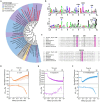
Identification of Tlp11-LBD homologues that bind to methyl pyruvate. (A) The phylogenetic tree showing the biological distribution of proteins with dCache domains that potentially bind to methyl pyruvate. The homologues selected for experimental verification are highlighted in red. (B) The conservation pattern found in Tlp11-LBD homologues. The sequence region corresponds to the membrane-proximal pocket of Tlp11-LBD (residues 239-322). The five crucial residues in Tlp11 that are present at the highest frequencies, L264, N268, I276, Y291, and T320, are indicated by blue arrows, while V318 is indicated by a grey arrow. (C) The sequence alignment of Tlp11-LBD homologues in C. jejuni, C. coli, H. equorum, H. himalayensis, H. mesocricetorum, H. ganmani, and C. upsaliensis. The red and grey areas indicate the key residues involved in the formation of hydrogen bonds and hydrophobic interactions, respectively, with methyl pyruvate. (D–F) The binding of Tlp11-LBD homologues in C. coli (D), H. equorum (E), and H. himalayensis (F) to methyl pyruvate, as measured using MST. Upper panel: thermophoresis raw data; lower panel: dose-response curve with the fitting result. Error bars represent the standard errors of three replicates. The concentration for the mutant protein was 250 nM, and the maximum concentration for the ligand was 2.5 mM, which was gradually diluted.

Similar articles

Cited by

References

    1. Barupal D. K., Fiehn O. (2019). Generating the blood exposome database using a comprehensive text mining and database fusion approach. Environ. Health Perspect. 127:97008. doi: 10.1289/ehp4713, PMID: - DOI - PMC - PubMed
    1. Bi S., Pollard A. M., Yang Y., Jin F., Sourjik V. (2016). Engineering hybrid chemotaxis receptors in bacteria. ACS Synth. Biol. 5, 989–1001. doi: 10.1021/acssynbio.6b00053, PMID: - DOI - PubMed
    1. Bi S., Sourjik V. (2018). Stimulus sensing and signal processing in bacterial chemotaxis. Curr. Opin. Microbiol. 45, 22–29. doi: 10.1016/j.mib.2018.02.002, PMID: - DOI - PubMed
    1. Bi S., Yu D., Si G., Luo C., Li T., Ouyang Q., et al. . (2013). Discovery of novel chemoeffectors and rational design of Escherichia coli chemoreceptor specificity. Proc. Natl. Acad. Sci. USA 110, 16814–16819. doi: 10.1073/pnas.1306811110, PMID: - DOI - PMC - PubMed
    1. Borrok M. J., Zhu Y., Forest K. T., Kiessling L. L. (2009). Structure-based design of a periplasmic binding protein antagonist that prevents domain closure. ACS Chem. Biol. 4, 447–456. doi: 10.1021/cb900021q, PMID: - DOI - PMC - PubMed

LinkOut - more resources