. 2024 Mar 12;27(4):109484.
doi: 10.1016/j.isci.2024.109484. eCollection 2024 Apr 19.

Caenorhabditis elegans neuropeptide NLP-27 enhances neurodegeneration and paralysis in an opioid-like manner during fungal infection

Affiliations

Caenorhabditis elegans neuropeptide NLP-27 enhances neurodegeneration and paralysis in an opioid-like manner during fungal infection

Maria Pop et al. iScience. .

Abstract

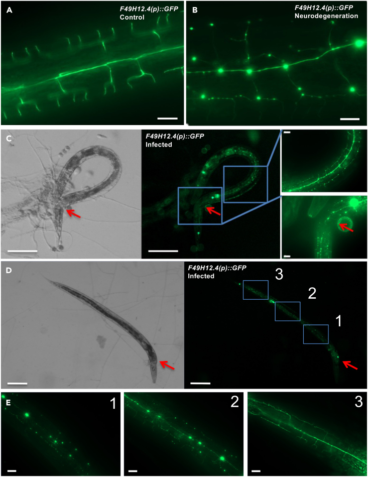
The nervous system of metazoans is involved in host-pathogen interactions to control immune activation. In Caenorhabditis elegans, this includes sleep induction, mediated by neuropeptide-like proteins (NLPs), which increases the chance of survival after wounding. Here we analyzed the role of NLP-27 in the infection of C. elegans with the nematode-trapping fungus Arthrobotrys flagrans. Early responses of C. elegans were the upregulation of nlp-27, the induction of paralysis (sleep), and neurodegeneration of the mechanosensing PVD (Posterior Ventral Process D) neurons. Deletion of nlp-27 reduced neurodegeneration during fungal attack. Induction of nlp-27 was independent of the MAP kinase PMK-1, and expression of nlp-27 in the hypodermis was sufficient to induce paralysis, although NLP-27 was also upregulated in head neurons. NLP-27 contains the pentapeptide YGGYG sequence known to bind the human μ- and κ-type opioid receptors suggesting NLP-27 or peptides thereof act on opioid receptors. The opioid receptor antagonist naloxone shortened the paralysis time like overexpression of NLP-27.

Keywords: Immunology; Molecular neuroscience; Neuroscience.


Conflict of interest statement

The authors declare no competing interests.

Figures

None
Graphical abstract
Figure 1
Figure 1
Analysis of the expression changes of the nlp-29 gene cluster in C. elegans after infection with A. flagrans (A) Alignment of the peptides encoded by the nlp-29 gene cluster without NLP-34. (B) Relative expression of the nlp-29 gene cluster genes during infection with A. flagrans determined by qRT PCR with actin for normalization. Young adult nematodes were co-incubated with A. flagrans for 24 h and total RNA was extracted using the TRIzol method, ∗∗p < 0.01. (C–F) Expression analyses using promoter-reporter assays with fluorescence microscopy. Promoters of nlp-29, nlp-31, and nlp-27 were fused to a DNA fragment encoding an NLS and to the wrmScarlet or GFP-coding gene. (C) Expression of nlp-29 and nlp-31 in nlp-29(rfl5) in uninfected control nematodes and (D) during infection with A. flagrans. (E) Expression of nlp-27 in nlp-27(rfl2) in uninfected control nematodes and (F) during infection with A. flagrans. The fluorescence signals of Figures 1C–1F were quantified (Figure S1). The arrows indicate the site of infection. The scale bar represents 100 μm.
Figure 2
Figure 2
nlp-27 is induced in the head area of C. elegans after infection with A. flagrans and after wounding Induction of nlp-27 in nlp-27(rfl2) after trapping of the nematode in the (A) head and (B) tail area. The arrows indicate the site of infection. The scale bar represents 100 μm. (C) Comparison of the fluorescence of the reporter construct in untrapped and in trapped nematodes. The above panels show the marked area of the pharynx, while the graph shows the total cell fluorescence measured in ImageJ. Unpaired Student’s t test was calculated for all experiments. Ns p > 0.05, ∗p < 0.05, ∗∗p < 0.01, ∗∗∗p < 0.001. (D) Visualization of the fluorescence of the reporter construct after wounding in nlp-27(rfl8). The scale bar represents 100 μm. (E) Expression of nlp-27 in ASI and ASH head neurons nlp-27(rfl2). Yellow triangle and green star point at ASI and ASH neurons. The graph represents the GFP, and wrmScarlet (RFP) intensity measured with ImageJ along the line shown in the close-up. The scale bar represents 20 μm.
Figure 3
Figure 3
NLP-27 is required for fast paralysis during fungal infection (A) Scheme of the nlp-27-deletion mutant nlp-27(rfl1). Dark gray boxes represent the promoter region and the 3′ UTR, the light gray box represents the signal peptide, the white box represents the first exon and the dashed box the intron region. (B) nlp-27 relative expression in wild type C. elegans (N2) and the nlp-27-deletion strain nlp-27(rfl1), ∗∗p < 0.01. (C) Infection assay of N2 and the nlp-27 deletion strain nlp-27(rfl1) with A. flagrans. ∗∗∗∗p < 0.0001. (D) Lifespan assays of N2 and the nlp-27-deletion strain nlp-27(rfl1). (E) NLP-27 overexpression in N2. nlp-27 mRNA fold change in nlp-27(rfl13) overexpressing nlp-27 under the native promoter relative to N2. ∗∗p < 0.01. (F) Time to paralysis of N2 and the nlp-27(rfl13) strain. ∗p < 0.05. (G) Lifespan assays of N2 and the nlp-27(rfl13) strain overexpressing nlp-27 under nlp-27 promoter in N2. Lifespans were performed in triplicates with at least 50 nematodes per replicate. Relative expression (2-ΔCt) is presented for each condition. act-1 was used to normalize the Ct value of the genes. The mRNA fold change was calculated as a ratio of the relative expression of the condition tested to the control. Unpaired Student’s t test was calculated for all experiments. ns p > 0.05, ∗p < 0.05, ∗∗p < 0.01, ∗∗∗p < 0.001. Error bars represent SD.
Figure 4
Figure 4
Expression analysis of nlp-27 (A) nlp-27 relative expression in the pmk-1(km25) mutant and sta-2(ok1860) mutant. RNA was extracted from mutant strains after 4 h of co-incubation with induced A. flagrans, ns p > 0.05. (B) nlp-27 expression in a nipi-3(fr4) mutant background, ∗p < 0.05. (C) Scheme of the PMK-1/p38 MAPK cascade activated by wounding or infection with D. coniospora. (D) Time to paralysis of the dcar-1(tm2484), ∗∗∗p < 0.001. (E) nlp-27 complementation with nlp-27 expressed under the native promoter nlp-27(rfl6) and under the hypodermal promoter col-12 nlp-27(rfl7), ∗∗∗p < 0.001. (F) Time needed to paralyze the ASI(−) and ASH(−) ablated strains, ns p > 0.05. (G) nlp-27 mRNA fold change in infection with A. flagrans in the ASI(−) and ASH(−) ablated strains, ns p > 0.05. (H) nlp-27 mRNA fold change after exposure to 5 mM DHCA in the ASI(−) and ASH(−) ablated strains. For the virulence assays unpaired Student’s t test was calculated for all experiments. ns p > 0.05, ∗p < 0.05, ∗∗p < 0.01, ∗∗∗p < 0.001. Fold change was calculated as the relative expression (2-ΔCt) ratio of the infected condition to the control condition for each strain in biological triplicates. Error bars represent sd. act-1 was used to normalize the Ct value of the genes.
Figure 5
Figure 5
Neurodegeneration during A. flagrans infection (A) Control PVD neurons in NC1686 expressing F49H12.4(p)::GFP. The scale bar represents 10 μm. (B) Neurodegeneration of PVD neurons in NC1686 strain expressing F49H12.4(p)::GFP. The scale bar represents 10 μm. (C) C. elegans NC1686 infected with A. flagrans. The arrows indicate the site of infection. The scale bar represents 100 μm. (D) C. elegans NC1686 infected with A. flagrans. The arrows indicate the site of infection. The scale bar represents 100 μm. (E) 1–3 are close ups from the regions indicated in (D). The scale bar represents 10 μm.
Figure 6
Figure 6
NLP-27 processing (A) Percentage of NC1686 nematodes with PVD dendrite degeneration expressing empty-vector control (black), nlp-27 (red), nlp-27(rfl3), or nlp-29 (blue) nlp-29(rfl4). ∗∗∗∗p < 0.0001. One-way ANOVA test was calculated. (B) Scheme of NLP-27 with a cleavage site predicted with the NeuroCS prediction tool and Phobius., The gray box represents the signal peptide, the red box peptide A and the blue box peptide B. Peptide A and peptide B were each fused to the SP for expression of the individual peptides in (C). (C) Percentage of NC1686 nematodes with PVD dendrite degeneration expressing empty-vector control (black), nlp-27A (red) (strain nlp-27(rfl10)), nlp-27B (blue) (strain nlp-27(rfl11)). ns p > 0.5, ∗∗∗p < 0.001, ∗∗∗∗p < 0.0001. A one-way ANOVA test was applied. (D) Time to paralysis of the nep-1(by159) mutant, ns p > 0.05. (E) COBALT alignment of NLP-24 and NLP-27 with the YGGYG motif marked. (F) Time to paralysis of wild type C. elegans exposed to 10 mM naloxone for 4 h before the virulence assay. ∗p < 0.05. The unpaired Student’s t test was applied.

Similar articles

Cited by

References

    1. Procaccini C., Pucino V., De Rosa V., Marone G., Matarese G. Neuro-endocrine networks controlling immune system in health and disease. Front. Immunol. 2014;5:143. doi: 10.3389/fimmu.2014.00143. - DOI - PMC - PubMed
    1. McKay F.M., McCoy C.J., Crooks B., Marks N.J., Maule A.G., Atkinson L.E., Mousley A. In silico analyses of neuropeptide-like protein (NLP) profiles in parasitic nematodes. Int. J. Parasitol. 2022;52:77–85. doi: 10.1016/j.ijpara.2021.07.002. - DOI - PMC - PubMed
    1. Nathoo A.N., Moeller R.A., Westlund B.A., Hart A.C. Identification of neuropeptide-like protein gene families in Caenorhabditis elegans and other species. Proc. Natl. Acad. Sci. USA. 2001;98:14000–14005. doi: 10.1073/pnas.241231298. - DOI - PMC - PubMed
    1. Li C., Kim K. WormBook. 2008. Neuropeptides; pp. 1–36. - DOI
    1. Couillault C., Pujol N., Reboul J., Sabatier L., Guichou J.F., Kohara Y., Ewbank J.J. TLR-independent control of innate immunity in Caenorhabditis elegans by the TIR domain adaptor protein TIR-1, an ortholog of human SARM. Nat. Immunol. 2004;5:488–494. doi: 10.1038/ni1060. - DOI - PubMed