. 2024 Jul 2;12(7):e0025324.
doi: 10.1128/spectrum.00253-24. Epub 2024 May 24.

A novel multiplex fluorescent-labeling method for the visualization of mixed-species biofilms in vitro

Affiliations

A novel multiplex fluorescent-labeling method for the visualization of mixed-species biofilms in vitro

Olivia Aherne et al. Microbiol Spectr. .

Abstract

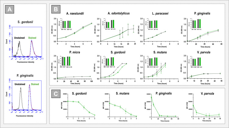
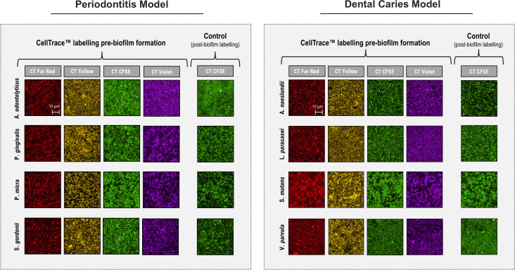
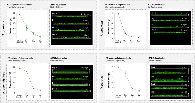
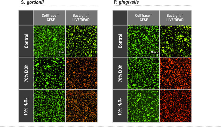
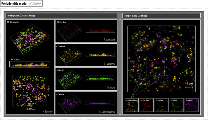
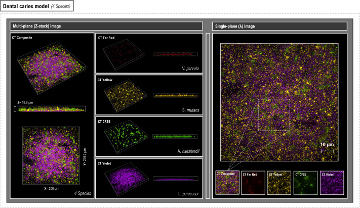
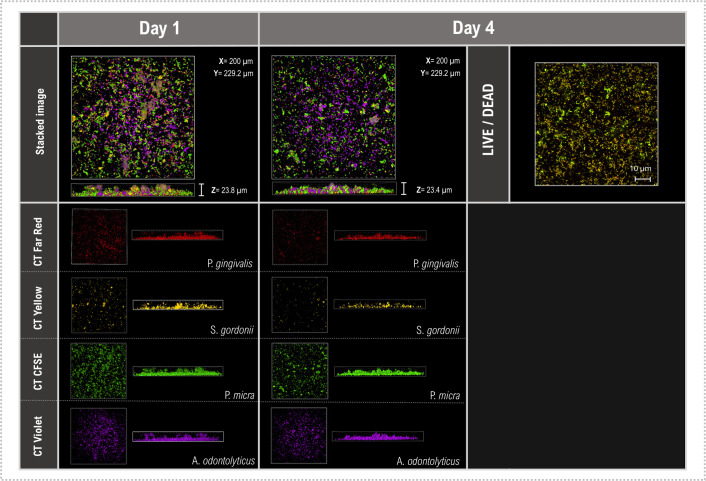
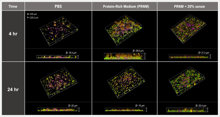
In nature, bacteria usually exist as mixed-species biofilms, where they engage in a range of synergistic and antagonistic interactions that increase their resistance to environmental challenges. Biofilms are a major cause of persistent infections, and dispersal from initial foci can cause new infections at distal sites thus warranting further investigation. Studies of development and spatial interactions in mixed-species biofilms can be challenging due to difficulties in identifying the different bacterial species in situ. Here, we apply CellTrace dyes to studies of biofilm bacteria and present a novel application for multiplex labeling, allowing identification of different bacteria in mixed-species, in vitro biofilm models. Oral bacteria labeled with CellTrace dyes (far red, yellow, violet, and CFSE [green]) were used to create single- and mixed-species biofilms, which were analyzed with confocal spinning disk microscopy (CSDM). Biofilm supernatants were studied with flow cytometry (FC). Both Gram-positive and Gram-negative bacteria were well labeled and CSDM revealed biofilms with clear morphology and stable staining for up to 4 days. Analysis of CellTrace labeled cells in supernatants using FC showed differences in the biofilm dispersal between bacterial species. Multiplexing with different colored dyes allowed visualization of spatial relationships between bacteria in mixed-species biofilms and relative coverage by the different species was revealed through segmentation of the CSDM images. This novel application, thus, offers a powerful tool for studying structure and composition of mixed-species biofilms in vitro.IMPORTANCEAlthough most chronic infections are caused by mixed-species biofilms, much of our knowledge still comes from planktonic cultures of single bacterial species. Studies of formation and development of mixed-species biofilms are, therefore, required. This work describes a method applicable to labeling of bacteria for in vitro studies of biofilm structure and dispersal. Critically, labeled bacteria can be multiplexed for identification of different species in mixed-species biofilms using confocal spinning disk microscopy, facilitating investigation of biofilm development and spatial interactions under different environmental conditions. The study is an important step in increasing the tools available for such complex and challenging studies.

Keywords: CellTrace; biofilm detachment; biofilm growth; confocal spinning disc microscopy; flow cytometry; live imaging; microscopy; oral bacteria; oral disease; staining.


Conflict of interest statement

The authors declare no conflict of interest.

Figures

Fig 1
Fig 1
Effects of CellTrace labeling on bacteria. (A) FC analysis of bacteria labeled with CellTrace. Gram-positive S. gordonii and Gram-negative P. gingivalis were labeled for 1 hour with CellTrace™ CFSE (green) and analyzed using FC. Graphs show CFSE staining expressed as the relative fluorescence intensity (X-axis) and the relative number of events (counts, Y-axis). The data show three experiments carried out using independent biological replicates and a single population of unstained cells was used as a control. (B) Growth curve analysis of CellTrace CFSE-labeled planktonic cultures. Cells were pre-labeled for 1 hour, and growth was measured as optical density. Unlabeled cells were used for comparison. Graphs show mean ± SD of three independent biological replicates (except for P. gingivalis and P. micra where n = 2). Bar charts (inserts) show mean ± SD of CFU counts at experiment start and a later stage of growth for each bacterial species. Labeled and unlabeled cells are shown in green and black, respectively. (C) Fluorescence intensity of CellTrace™ CFSE-labeled planktonic cultures. Graphs showing mean ± SD of MFI values over time measured using FC. Two representative Gram positive (S. gordonii and S. mutans) and Gram negative (P. gingivalis and V. parvula) bacteria are shown.
Fig 2
Fig 2
CDSM visualization of SS biofilms labeled with CellTrace. Representative CDSM images of SS biofilms related to oral disease models. Biofilms were formed overnight using unlabeled or CellTrace (far red, yellow, CFSE, or violet)-labeled cells. Unlabeled biofilms were post-stained with CellTrace CFSE for comparison. The periodontitis model is represented by S. gordonii, A. odontolyticus, P. gingivalis, and P. micra, whereas the dental caries model is comprised of S. mutans, V. parvula, L. paracasei, and A. naeslundii. The scale bar (10 µm) applies to all panels.
Fig 3
Fig 3
Examination of biofilm structure and cell detachment from SS biofilms over time. Gram-positive (S. gordonii and A. odontolyticus) and Gram-negative (P. gingivalis and V. parvula) bacteria labeled with CellTrace CFSE (green) were used to create SS biofilms. Graphs (left panels) show percentages of CFSE-stained cells present in the starting inocula and biofilm supernatants. Supernatants were extracted and analyzed with FC on days 1, 2, and 3. The right panels show representative Z-plane CDSM images of SS biofilms after 4 hours (day 0) and 1, 2, and 3 days.
Fig 4
Fig 4
Assessment of CellTrace staining as a marker of bacterial viability in biofilms representative CDSM and CLSM images of overnight SS biofilms stained with CellTrace CFSE (green) or BacLight LIVE/DEAD stain. Biofilms of S. gordonii and P. gingivalis were maintained in PRNM (control) or treated with 70% ethanol or H2O2 for 1 hour. The scale bar (10 µm) applies to all panels.
Fig 5
Fig 5
Visualization of MS biofilms representing a periodontitis model labeled by multiplexing CellTrace dyes. Representative CDSM Z-stack (left) and single-plane (right) images of overnight MS biofilms stained with CellTrace dyes. A unique CellTrace color was used to label four species (P. gingivalis [red], S. gordonii [yellow], P. micra [green], and A. odontolyticus [violet]). Both multiplex (4-color compilation) and the color-segmented images are shown. The percentage coverage for each species within the biofilm models was calculated using a region of interest (ROI) within the delineated area in the single-plane image. Z-stack images were used to provide information regarding bacterial spatial location and relationships within the overall biofilm.
Fig 6
Fig 6
Visualization of MS biofilms representing a dental caries model labeled by multiplexing CellTrace dyes. Representative CDSM Z-stack (left) and single-plane (right) images of overnight MS biofilms stained with CellTrace dyes. A unique CellTrace color was used to label four species (V. parvula [red], S. mutans [yellow], A. naeslundii [green], and L. paracasei [violet]). Both multiplex (4-color compilation) and the color segmented images are shown. The percentage coverage for each species within the biofilm models was calculated using a region of interest (ROI) within the delineated area in the single-plane image. Z-stack images were used to provide information regarding bacterial spatial location and relationships within the overall biofilm.
Fig 7
Fig 7
Assessment of CellTrace durability in MS biofilms. Representative CSDM and CLSM images of MS biofilms. Biofilms were created using the periodontitis model (P. gingivalis [red], S. gordonii [yellow], P. micra [green], and A. odontolyticus [violet]) and visualized following overnight growth (day 1) and after 4 days through CDSM Z-stack images. Parallel, non-labeled biofilms were also grown, stained with BacLight LIVE/DEAD stain on day 4 and images using CLSM to assess viability. The scale bar (10 µm) applies to all panels.
Fig 8
Fig 8
Investigation of the influence of environment on early biofilm formation. Representative CDSM Z-stack images of overnight MS biofilms. Biofilms were created using the periodontitis model (P. gingivalis [red], S. gordonii [yellow], P. micra [green], and A. odontolyticus [violet]). They were then grown in either PBS, PRNM, or PRNM + serum, and imaged after 4 or 24 hours.

Similar articles

Cited by

  • MUC5B modulation of early oral biofilm glucose metabolism.
    Robertsson C, Davies J, Svensäter G, Nord AB, Norrström N, Wickström C. Robertsson C, et al. Front Oral Health. 2025 Feb 11;6:1516025. doi: 10.3389/froh.2025.1516025. eCollection 2025. Front Oral Health. 2025. PMID: 40008185 Free PMC article.

References

    1. Stoodley P, Sauer K, Davies DG, Costerton JW. 2002. Biofilms as complex differentiated communities. Annu Rev Microbiol 56:187–209. doi:10.1146/annurev.micro.56.012302.160705 - DOI - PubMed
    1. Jakubovics NS. 2015. Intermicrobial interactions as a driver for community composition and stratification of oral biofilms. J Mol Biol 427:3662–3675. doi:10.1016/j.jmb.2015.09.022 - DOI - PubMed
    1. Luo A, Wang F, Sun D, Liu X, Xin B. 2021. Formation, development, and cross-species interactions in biofilms. Front Microbiol 12:757327. doi:10.3389/fmicb.2021.757327 - DOI - PMC - PubMed
    1. Kreth J, Zhang Y, Herzberg MC. 2008. Streptococcal antagonism in oral biofilms: Streptococcus sanguinis and Streptococcus gordonii interference with Streptococcus mutans. J Bacteriol 190:4632–4640. doi:10.1128/JB.00276-08 - DOI - PMC - PubMed
    1. Abisado RG, Benomar S, Klaus JR, Dandekar AA, Chandler JR. 2018. Bacterial quorum sensing and microbial community interactions. mBio 9:e02331–17. doi:10.1128/mBio.02331-17 - DOI - PMC - PubMed

MeSH terms

Substances

LinkOut - more resources