

A Tissue Engineered 3D Model of Cancer Cell Invasion for Human Head and Neck Squamous-Cell Carcinoma
- PMID: 38785518
- PMCID: PMC11119844
- DOI: 10.3390/cimb46050250
A Tissue Engineered 3D Model of Cancer Cell Invasion for Human Head and Neck Squamous-Cell Carcinoma
Abstract
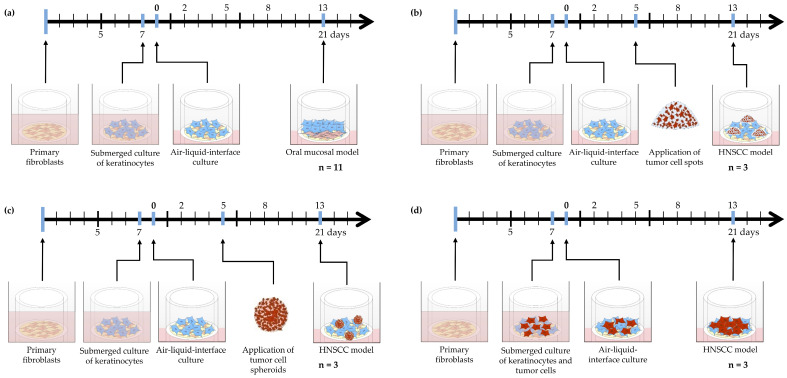
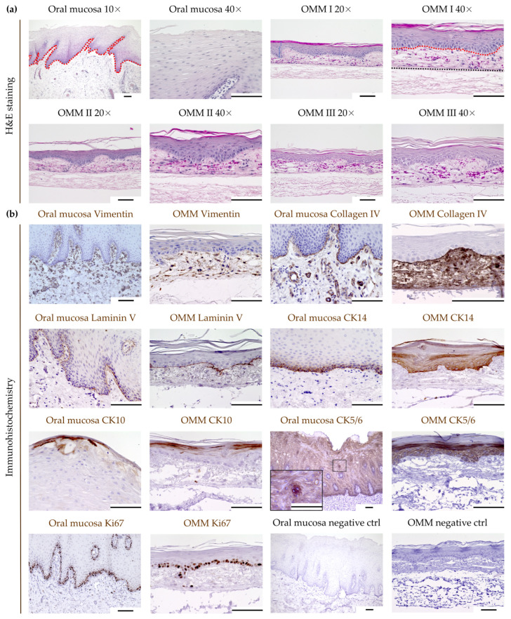
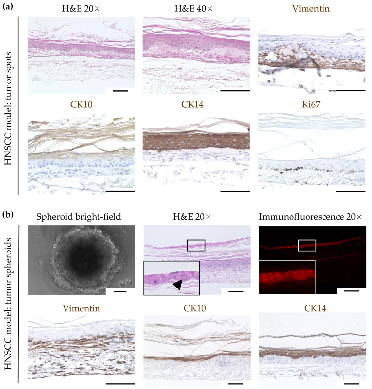
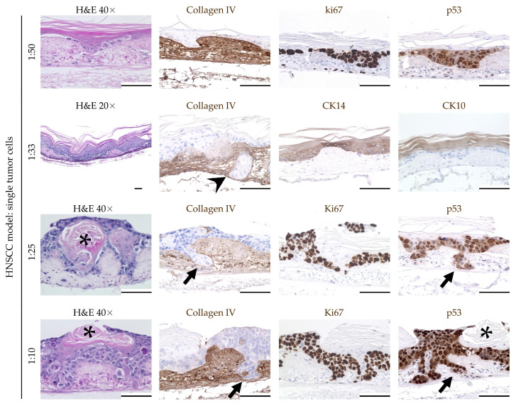
Head and neck squamous-cell carcinoma (HNSCC) is associated with aggressive local invasiveness, being a main reason for its poor prognosis. The exact mechanisms underlying the strong invasive abilities of HNSCC remain to be elucidated. Therefore, there is a need for in vitro models to study the interplay between cancer cells and normal adjacent tissue at the invasive tumor front. To generate oral mucosa tissue models (OMM), primary keratinocytes and fibroblasts from human oral mucosa were isolated and seeded onto a biological scaffold derived from porcine small intestinal submucosa with preserved mucosa. Thereafter, we tested different methods (single tumor cells, tumor cell spots, spheroids) to integrate the human cancer cell line FaDu to generate an invasive three-dimensional model of HNSCC. All models were subjected to morphological analysis by histology and immunohistochemistry. We successfully built OMM tissue models with high in vivo-in vitro correlation. The integration of FaDu cell spots and spheroids into the OMM failed. However, with the integration of single FaDu cells into the OMM, invasive tumor cell clusters developed. Between segments of regular epithelial differentiation of the OMM, these clusters showed a basal membrane penetration and lamina propria infiltration. Primary human fibroblasts and keratinocytes seeded onto a porcine carrier structure are suitable to build an OMM. The HNSCC model with integrated FaDu cells could enable subsequent investigations into cancer cell invasiveness.
Keywords: 3D tissue model; head and neck squamous-cell carcinoma; oral mucosa; tissue engineering.
Conflict of interest statement
All authors have no conflicts of interest to declare that are relevant to the content of this article.
Figures
Similar articles
-
Differential Angiogenic Potential of 3-Dimension Spheroid of HNSCC Cells in Mouse Xenograft.Int J Mol Sci. 2021 Jul 31;22(15):8245. doi: 10.3390/ijms22158245. Int J Mol Sci. 2021. PMID: 34361027 Free PMC article.
-
Early stage mechanical remodeling of collagen surrounding head and neck squamous cell carcinoma spheroids correlates strongly with their invasion capability.Acta Biomater. 2019 Jan 15;84:280-292. doi: 10.1016/j.actbio.2018.11.046. Epub 2018 Nov 27. Acta Biomater. 2019. PMID: 30500449
-
Development of tissue-engineered models of oral dysplasia and early invasive oral squamous cell carcinoma.Br J Cancer. 2011 Nov 8;105(10):1582-92. doi: 10.1038/bjc.2011.403. Epub 2011 Oct 11. Br J Cancer. 2011. PMID: 21989184 Free PMC article.
-
Animal models of head and neck squamous cell carcinoma.Vet J. 2016 Apr;210:7-16. doi: 10.1016/j.tvjl.2015.11.006. Epub 2015 Nov 24. Vet J. 2016. PMID: 26965084 Review.
-
Role of Vitamin D in Head and Neck Cancer-Immune Function, Anti-Tumour Effect, and Its Impact on Patient Prognosis.Nutrients. 2023 May 31;15(11):2592. doi: 10.3390/nu15112592. Nutrients. 2023. PMID: 37299554 Free PMC article. Review.
References
-
- Hashim D., Sartori S., Brennan P., Curado M.P., Wunsch-Filho V., Divaris K., Olshan A.F., Zevallos J.P., Winn D.M., Franceschi S., et al. The role of oral hygiene in head and neck cancer: Results from International Head and Neck Cancer Epidemiology (INHANCE) consortium. Ann. Oncol. 2016;27:1619–1625. doi: 10.1093/annonc/mdw224. - DOI - PMC - PubMed
LinkOut - more resources
Full Text Sources
