. 2024 May 19;13(5):357.
doi: 10.3390/biology13050357.

α-Catenin and Piezo1 Mediate Cell Mechanical Communication via Cell Adhesions

Affiliations

α-Catenin and Piezo1 Mediate Cell Mechanical Communication via Cell Adhesions

Mingxing Ouyang et al. Biology (Basel). .

Abstract

Cell-to-cell distant mechanical communication has been demonstrated using in vitro and in vivo models. However, the molecular mechanisms underlying long-range cell mechanoresponsive interactions remain to be fully elucidated. This study further examined the roles of α-Catenin and Piezo1 in traction force-induced rapid branch assembly of airway smooth muscle (ASM) cells on a Matrigel hydrogel containing type I collagen. Our findings demonstrated that siRNA-mediated downregulation of α-Catenin or Piezo1 expression or chemical inhibition of Piezo1 activity significantly reduced both directional cell movement and branch assembly. Regarding the role of N-cadherin in regulating branch assembly but not directional migration, our results further confirmed that siRNA-mediated downregulation of α-Catenin expression caused a marked reduction in focal adhesion formation, as assessed by focal Paxillin and Integrin α5 localization. These observations imply that mechanosensitive α-Catenin is involved in both cell-cell and cell-matrix adhesions. Additionally, Piezo1 partially localized in focal adhesions, which was inhibited by siRNA-mediated downregulation of α-Catenin expression. This result provides insights into the Piezo1-mediated mechanosensing of traction force on a hydrogel. Collectively, our findings highlight the significance of α-Catenin in the regulation of cell-matrix interactions and provide a possible interpretation of Piezo1-mediated mechanosensing activity at focal adhesions during cell-cell mechanical communication.

Keywords: Piezo1; cell mechanical communication; directional migration; focal adhesion; mechanotransduction; α-Catenin.


Conflict of interest statement

The authors declare no conflicts of interest.

Figures

Figure 1
Figure 1
α-Catenin regulates both branch formation and directed migration. ASM cells were transfected with α-Catenin (ctnna1) siRNA or control siRNA. After incubating for 52 h, the cells were seeded onto a hydrogel for time-lapse imaging. (A) Experimental setup for cell culture on a hydrogel. (B,C) Branch assembly of ASM cells transfected with control siRNA (NC) on a hydrogel and migration analysis of trajectory plots (left) and displacement maps (right) (B) and of cells transfected with ctnna1 siRNA (C). (D) Statistical quantification of the ASM cell movement rate and velocity (mean ± S.D., n = 34 and 38, respectively) and branch length (n = 54 and 118, respectively) under conditions (B,C). (E) Statistical quantification of the movement rate and velocity of ASM cells transfected with control or N-CAD siRNA (mean ± S.D., n = 50, 47, and 48, respectively) and branch length (n = 45, 38, and 49, respectively). Cell images and migration trajectories are shown in Figure S2. *, **, ***, and **** indicate p < 0.05, 0.01, 0.001, and 0.0001, respectively, for significant differences in statistical comparisons; “ns” refers to no significant difference.
Figure 2
Figure 2
Role of mechanosensitive Piezo1 channels during directed cell migration and self-assembly. Normal ASM cells were inoculated onto a hydrogel with or without an inhibitor in the culture media; DMSO was used for control cells. The cells were allowed to incubate for 6 h, after which time-lapse imaging was performed every 0.5 h for 15.5 h. (AD) Cell images at 6 h and 21.5 h. Trajectory analysis plots and displacement maps are shown for the normal state (A), control conditions (DMSO) (B), treatment with GsMTx4 (3 μM) (C), and treatment with GdCl3 (100 μM) (D). (E) Statistical quantification of the ASM cell movement rate and velocity (n = 46, 45, 58, and 58, respectively) and branch length (n = 25, 17, 49, and 55, respectively) under different conditions (AD). (F,G) Branch assembly and migration analysis of ASM cells transfected with control (NC) or Piezo1 siRNA on the hydrogel. (H) Statistical quantification of the cell movement rate and velocity, and branch length under conditions (F,G). *, **, ***, and **** indicate p < 0.05, 0.01, 0.001, and 0.0001, respectively in statistical comparisons; “ns” refers to no significant difference.
Figure 3
Figure 3
The effect of α-Catenin on the distribution of paxillin or integrin at focal adhesions in ASM cells. (A,D) Images showing paxillin (A) or integrin (D) fluorescence in normal ASM cells and in those transfected with control siRNA or ctnna1 siRNA. Fluorescence clusters showing paxillin or integrin α5 expression at focal adhesions. (B,E) Sample demonstrations for the quantification of the numbers and average fluorescence intensity of focal adhesions based on fluorescent paxillin (B) and integrin α5 expression (E). The details are described in the Methods section. (C,F) Statistical analysis of the number of Paxillin-labeled focal adhesions per cell (n = 17, 12, and 11, respectively). (C) The number of integrin-labeled focal adhesions per cell (n = 14, 10, and 10, respectively). (F) The average Paxillin or Integrin α5 fluorescence intensity in focal adhesions and the total area of focal adhesions per cell. *, **, and **** indicate p < 0.05, 0.01, and 0.0001 in statistical comparisons, respectively; “ns” refers to no significant difference.
Figure 4
Figure 4
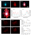
Paxillin and Piezo1 colocalize at cellular focal adhesions. (A) Representative images showing colocalization of fluorescent Paxillin and Piezo1 at focal adhesions in ASM cells spread on fibronectin-coated glass for 4 h. Fluorescence images showing Piezo1 expression (left) and Paxillin expression (middle), and merged images showing both Piezo1 and Paxillin expression (right). (B) Fluorescence intensities of Piezo1 (blue) and paxillin (red) in regions of cellular focal adhesions in the selected lines. (C) Fluorescent paxillin at the focal adhesions of ASM cells in the control group or in the group treated with the inhibitor GsMTx4 or GdCl3. (D) Statistical analysis of the number of focal adhesions and Paxillin fluorescence intensity (n = 9, 8, 9, and 9, respectively) under the conditions described in (C). “ns” indicates no significant difference.
Figure 5
Figure 5
The effect of α-Catenin on Piezo1 focal localization and a hypothetical model of molecular mechanosensation. (A,B) α-Catenin-mediated regulation of Piezo1 focal targeting. After transfection with control or α-Catenin siRNA, ASM cells were further transfected with fluorescent Piezo1 and paxillin. Images of focal adhesions were acquired (A), followed by the counting of Piezo1 focal loci per cell (B). Only the cells showing visible focal adhesions were counted. (C) Hypothetical model of the roles of α-Catenin and Piezo1 along with actomyosin contraction, Integrin, and calcium channels in cell-to-cell mechanical communication on a hydrogel matrix. Cellular contraction generates traction force, which is transmitted through the hydrogel. α-Catenin regulates both traction force-induced cell–cell adherens junctions and cell-matrix adhesions. Piezo1 partially localizing at focal adhesions together mechanosense the stretch in the matrix for cell–cell distant communication. The arrows indicate the force-pulling directions or targets. **** indicates p < 0.0001 in statistical comparison.

Similar articles

References

    1. Wang H., Abhilash A.S., Chen C.S., Wells R.G., Shenoy V.B. Long-range force transmission in fibrous matrices enabled by tension-driven alignment of fibers. Biophys. J. 2014;107:2592–2603. doi: 10.1016/j.bpj.2014.09.044. - DOI - PMC - PubMed
    1. Alisafaei F., Chen X., Leahy T., Janmey P.A., Shenoy V.B. Long-range mechanical signaling in biological systems. Soft Matter. 2021;17:241–253. doi: 10.1039/D0SM01442G. - DOI - PMC - PubMed
    1. Yang C., Wang X., Xie R., Zhang Y., Xia T., Lu Y., Ye F., Zhang P., Cao T.M., Xu Y., et al. Dynamically Reconstructed Collagen Fibers for Transmitting Mechanical Signals to Assist Macrophages Tracing Breast Cancer Cells. Adv. Funct. Mater. 2023;33:2211807. doi: 10.1002/adfm.202211807. - DOI
    1. Postma M., van Haastert P.J. Mathematics of Experimentally Generated Chemoattractant Gradients. Methods Mol. Biol. 2016;1407:381–396. - PubMed
    1. Hardin C.C., Chattoraj J., Manomohan G., Colombo J., Nguyen T., Tambe D., Fredberg J.J., Birukov K., Butler J.P., Del Gado E., et al. Long-range stress transmission guides endothelial gap formation. Biochem. Biophys. Res. Commun. 2018;495:749–754. doi: 10.1016/j.bbrc.2017.11.066. - DOI - PMC - PubMed