. 2024 May 11;13(10):822.
doi: 10.3390/cells13100822.

Effects of Garlic on Breast Tumor Cells with a Triple Negative Phenotype: Peculiar Subtype-Dependent Down-Modulation of Akt Signaling

Affiliations

Effects of Garlic on Breast Tumor Cells with a Triple Negative Phenotype: Peculiar Subtype-Dependent Down-Modulation of Akt Signaling

Federica Brugnoli et al. Cells. .

Abstract

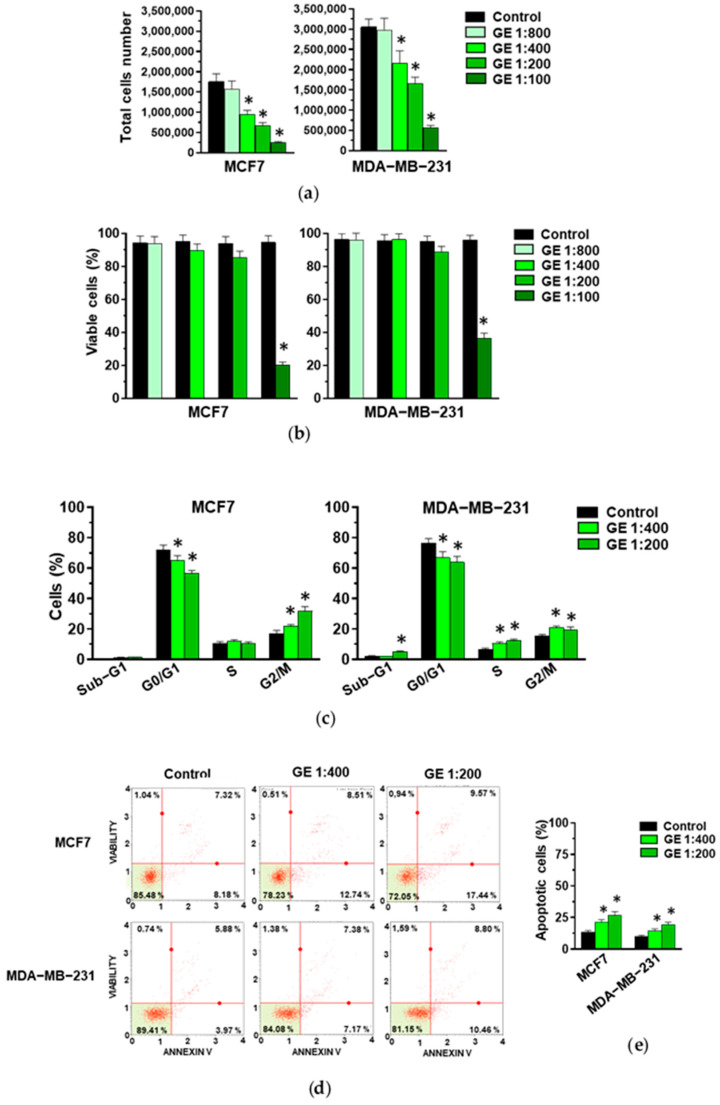
Breast cancer includes tumor subgroups with morphological, molecular, and clinical differences. Intrinsic heterogeneity especially characterizes breast tumors with a triple negative phenotype, often leading to the failure of even the most advanced therapeutic strategies. To improve breast cancer treatment, the use of natural agents to integrate conventional therapies is the subject of ever-increasing attention. In this context, garlic (Allium sativum) shows anti-cancerous potential, interfering with the proliferation, motility, and malignant progression of both non-invasive and invasive breast tumor cells. As heterogeneity could be at the basis of variable effects, the main objective of our study was to evaluate the anti-tumoral activity of a garlic extract in breast cancer cells with a triple negative phenotype. Established triple negative breast cancer (TNBC) cell lines from patient-derived xenografts (PDXs) were used, revealing subtype-dependent effects on morphology, cell cycle, and invasive potential, correlated with the peculiar down-modulation of Akt signaling, a crucial regulator in solid tumors. Our results first demonstrate that the effects of garlic on TNBC breast cancer are not unique and suggest that only more precise knowledge of the mechanisms activated by this natural compound in each tumor will allow for the inclusion of garlic in personalized therapeutic approaches to breast cancer.

Keywords: Akt; PDX-derived cells.; TNBC subtypes; breast cancer; garlic extract.


Conflict of interest statement

The authors declare no conflicts of interest.

Figures

Figure 1
Figure 1
Effect of GE on growth of MCF7 and MDA-MB-231 cells. After 72 hours of culture in control conditions (Control) or in the presence of the reported dilutions of garlic extract (GE), cells were subjected to an evaluation of proliferation (a) and the percentage of viable cells (b), and to a cytofluorimetric analysis of cell cycle distribution (c). (d) A representative apoptosis profile obtained after an Annexin V assay on MCF7 and MDA-MB-231 cells under the indicated conditions. (e) Percentage of total (early and late) apoptotic cells. Results represent the mean of three experiments ± SD. * p < 0.05 compared to control.
Figure 2
Figure 2
Effect of GE on migration and invasion of breast cancer derived cells. (a) Representative xCELLigence-driven dynamic monitoring of migration and invasion of MCF7 and MDA-MB-231 cells after 72 hours in control conditions or in the presence of the indicated GE dilutions. Curves represent mean ± SD of Cell Index (CI). (b,c) Slope analysis of migration and invasion CI curves, respectively. Data represent the mean of three experiments ± SD. * p < 0.05 compared to control.
Figure 3
Figure 3
Effect of garlic extracts on growth of HBCX-17, T174, HBCX-9, and HBCX-39 cells. (a) Proliferation of the indicated PDX-derived cell lines after 72 hours of culture in control conditions (Control) or in the presence of the reported dilutions of garlic extract (GE). (b) Percentage of viable cells under the above-reported conditions. Results represent the mean of three independent experiments ± SD. * p < 0.05 compared to untreated cells (Control).
Figure 4
Figure 4
Effect of garlic extracts on cell cycle and apoptosis of PDX-derived cells. (a) Cytofluorimetric analysis of cell cycle distribution of HBCX-17, T174, HBCX-9, and HBCX-39 cell lines treated with 1:400 GE for 72 h. (b) Muse Cell Analyzer apoptosis profile of the same cells treated as indicated above. (c) Percentage of total apoptotic cells. Results represent the mean of three independent experiments ±SD. * p < 0.05 compared to control cells.
Figure 5
Figure 5
Effect of garlic extract on invasive capabilities of PDX-derived cell lines. (a) xCELLigence-driven dynamic monitoring of invasion of HBCX-17, T174, HBCX-9, and HBCX-39 cells treated or not with GE for 72 h. Curves represent mean ± SD of CI. (b) Correspondent slope analysis, indicating steepness, inclination, gradient, and changing rate of CI curve over time. Results represent the mean of three independent experiments ± SD. * p < 0.05 compared to untreated cells (Control).
Figure 6
Figure 6
Morphological analysis of MDA-MB-231 and PDX-derived TNBC cell lines after GE treatment. (a) Representative phase-contrast image of MDA-MB-231 cells growing on glass dishes for 72 h in control conditions or in the presence of 1:400 diluted GE. (b) Representative fluorescence images after immunocytochemical analysis with anti-α-tubulin antibody of MDA-MB-231 cells in the same conditions. (c) Representative florescence images of PDX-derived cell lines grown on glass dishes for 72 h in the presence or not of 1:400 diluted GE and then subjected to immunocytochemical analysis of α-tubulin antibody. Scale bar: 50 μm.
Figure 7
Figure 7
Effect of garlic extract on EMT markers in MDA-MB-231 and PDX-derived TNBC cell lines. (a) Representative Western blot analysis with the indicated antibodies of total lysates from the reported cell lines treated with GE for 72 h. β-Tubulin was used as internal control for equivalence of loaded proteins. (b) Histograms showing protein levels, normalized using β-Tubulin, as deduced from band densitometry obtained through chemiluminescence signal. Asterisks indicate statistically significant differences with respect to corresponding controls taken as 1. Results represent the mean of three independent experiments ± SD. * p < 0.05 compared to control cells.
Figure 8
Figure 8
Effect of garlic extract on Akt status in MDA-MB-231 and in PDX-derived cell lines. (a) Representative immunoblot analysis with the indicated antibodies of total lysates from MDA-MB-231 and PDX-derived cells after 72 hours of culture in control conditions (Control) or in the presence of 1:400 diluted GE. (b) Histograms, as deduced from the densitometry of Western blot bands, reporting the levels of the indicated proteins normalized to β-Tubulin. Results represent the mean of three independent experiments ± SD. * p < 0.05 compared to control cells taken as 1.
Figure 9
Figure 9
Effect of garlic extract on Akt targets in MDA-MB-231 and in PDX-derived cell lines. (a), representative immunoblot analysis with the indicated antibodies of lysates from MDA-MB-231 and PDX-derived cells after 72 hours of culture in control conditions (Control) or in the presence of garlic extract (GE). (b) Levels of proteins normalized to β-Tubulin, as deduced from the densitometry of Western blot bands. Results represent the mean of three independent experiments ± SD. * p < 0.05 compared to untreated cells (Control) taken as 1.
Figure 10
Figure 10
Effect of Akt inhibition on MDA-MB-231 cells. Proliferation (a), percentage of viable cells (b), cytofluorimetric analysis of cell cycle distribution (c), and invasive potential (d,e) of MDA-MB-231 cells after 24 hours of culture in control conditions (Vehicle) or in the presence of the reported concentrations of MK-2206. Results represent the mean of three independent experiments ± SD. * p < 0.05 compared to control cells (Vehicle).

Similar articles

Cited by

References

    1. Wilkinson L., Gathani T. Understanding Breast Cancer as a Global Health Concern. Br. J. Radiol. 2022;95:20211033. doi: 10.1259/bjr.20211033. - DOI - PMC - PubMed
    1. Iacopetta D., Ceramella J., Baldino N., Sinicropi M.S., Catalano A. Targeting Breast Cancer: An Overlook on Current Strategies. Int. J. Mol. Sci. 2023;24:3643. doi: 10.3390/ijms24043643. - DOI - PMC - PubMed
    1. Ye F., Dewanjee S., Li Y., Jha N.K., Chen Z.-S., Kumar A., Vishakha, Behl T., Jha S.K., Tang H. Advancements in Clinical Aspects of Targeted Therapy and Immunotherapy in Breast Cancer. Mol. Cancer. 2023;22:105. doi: 10.1186/s12943-023-01805-y. - DOI - PMC - PubMed
    1. Barrett S.V. Breast Cancer. J. R. Coll. Physicians Edinb. 2010;40:335–338; quiz 339. doi: 10.4997/JRCPE.2010.418. - DOI - PubMed
    1. Perou C.M., Sørlie T., Eisen M.B., van de Rijn M., Jeffrey S.S., Rees C.A., Pollack J.R., Ross D.T., Johnsen H., Akslen L.A., et al. Molecular Portraits of Human Breast Tumours. Nature. 2000;406:747–752. doi: 10.1038/35021093. - DOI - PubMed

Publication types

MeSH terms

Substances