Review
. 2024 May 16;13(10):844.
doi: 10.3390/cells13100844.

Immune Cell Migration to Cancer

Affiliations
Review

Immune Cell Migration to Cancer

Allison T Ryan et al. Cells. .

Abstract

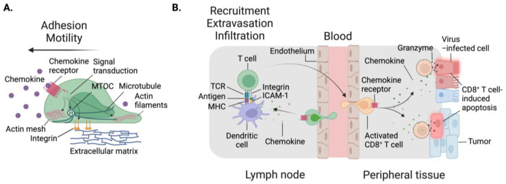
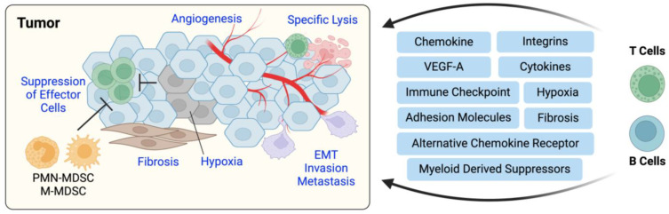
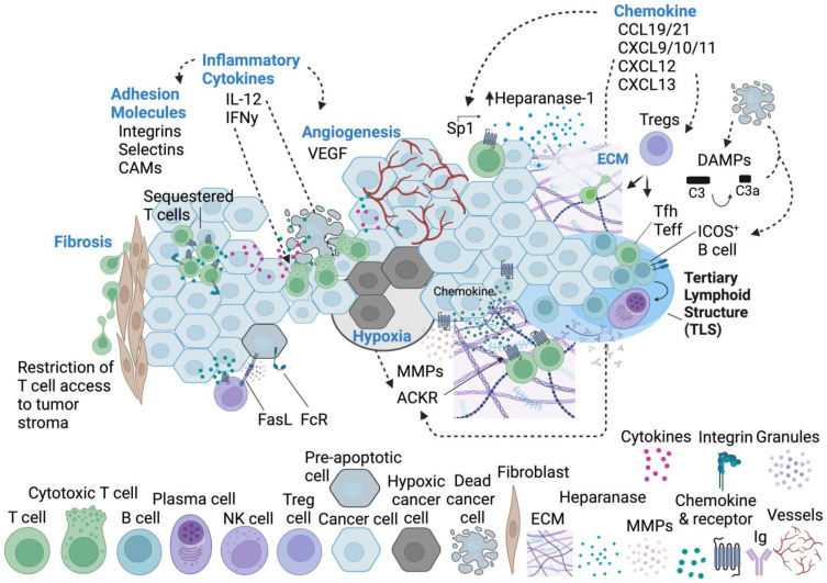
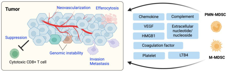
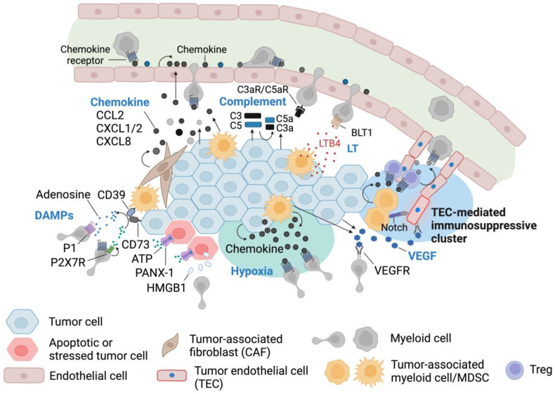
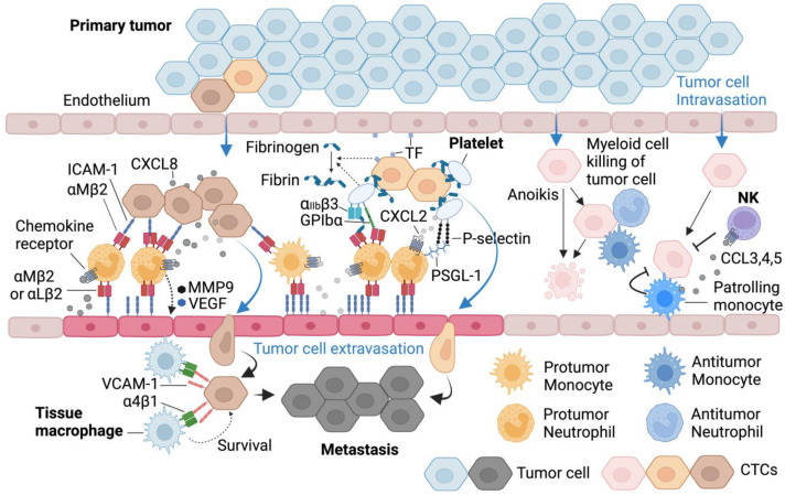
Immune cell migration is required for the development of an effective and robust immune response. This elegant process is regulated by both cellular and environmental factors, with variables such as immune cell state, anatomical location, and disease state that govern differences in migration patterns. In all cases, a major factor is the expression of cell surface receptors and their cognate ligands. Rapid adaptation to environmental conditions partly depends on intrinsic cellular immune factors that affect a cell's ability to adjust to new environment. In this review, we discuss both myeloid and lymphoid cells and outline key determinants that govern immune cell migration, including molecules required for immune cell adhesion, modes of migration, chemotaxis, and specific chemokine signaling. Furthermore, we summarize tumor-specific elements that contribute to immune cell trafficking to cancer, while also exploring microenvironment factors that can alter these cellular dynamics within the tumor in both a pro and antitumor fashion. Specifically, we highlight the importance of the secretome in these later aspects. This review considers a myriad of factors that impact immune cell trajectory in cancer. We aim to highlight the immunotherapeutic targets that can be harnessed to achieve controlled immune trafficking to and within tumors.

Keywords: MDSC; T cell; cancer; chemokine; chemotaxis; immune cell; leukocyte; migration; myeloid-derived suppressor cell; trafficking; tumor; tumor microenvironment.


Conflict of interest statement

The authors declare no conflicts of interest.

Figures

Figure 1
Figure 1
Cellular migration is dependent on cell intrinsic factors, as well as multiple signal inputs from the environment. (A) External signals such as chemokines are sensed by membrane-bound receptors on immune cells. The receptor-induced intracellular signaling reorganizes the actin cytoskeleton and the microtubule organizing center (MTOC) and activates integrins, resulting in adhesion to extracellular matrix or counterpart cells, followed by directional migration. (B) CD8+ T cells migrate to lymph nodes where they are primed by tumor- or virus-antigens presented by dendritic cells. These activated T cells are then recruited to cancerous or infected tissues and eliminate transformed or infected cells by secreting cytotoxic molecules including Granzymes. Immune cells have remarkable capabilities to adapt their mode of adhesion and migration to their surroundings. TCR, T cell receptor; MHC, Major histocompatibility complex.
Figure 2
Figure 2
Tumor recruitment of adaptive immune cells can have positive and negative effects to the host. The main effectors, T cells and B cells, are attracted via chemokines, cytokines, and growth factors, which continue to modulate their activity within the tumor microenvironment. Chemokines, adhesion molecules, checkpoint molecules contribute to specific lysis or immune evasion contextually. Recruited immune cells or tumor cells can secrete VEGF-A to further modulate immune cell recruitment. Cancer-associated fibroblasts can modulate the extracellular matrix to further augment effector cell migration within the microenvironment. Tumors become hypoxic, limiting effector capability. All of these factors may contribute to tumor invasion and metastasis. EMT, Epithelial-mesenchymal transition; PMN-MDSC, neutrophil-derived myeloid-derived suppressor cell. M-MDSC, monocyte/macrophage-derived myeloid-derived suppressor cell.
Figure 3
Figure 3
Lymphoid cell recruitment to tumors by multiple mechanisms. Immune cell migration through ECM, tissue, blood, and lymphatics is highly dependent on expression of integrins, selectins, and cell adhesion molecules such as ICAM family ligands. Chemoattractants produced within the TME recruit and retain lymphocytes, which play a significant role in tumor elimination. Chemokine receptor expression on immune cells and tumor cells is highly dynamic. Tumors may alter chemokine receptor and cell adhesion factor expression to sequester or restrict T cell access within tumor stroma, limiting their effector capability. Inflammatory cytokines from immune cells or tumor/tumor associated cells can induce additional alterations to these cell surface molecules. Furthermore, inflammatory cytokines can promote T cell cytotoxicity, causing tumor cell apoptosis. These cytokines can also promote angiogenic programs, increasing flow of nutrients to the tumor, as well as myeloid derived suppressor cells (not pictured). Angiogenic programs can result in aberrant vasculature patterns, resulting in hypoxic tumor regions with limited effector cell presence. TME factors can also induce the expression of atypical chemokine receptors, which scavenge free ligands, disrupting intratumoral immune cell signaling and activation, as there is less bioavailable chemokine for typical receptors. Chemokine and cytokine expression patterns can also cause alternations to the ECM through induction of MMPs or heparanase-1, which can both promote or disrupt immune cell migration depending on the context. Chemokines also recruit Treg cells to the tumor which can induce immunosuppression. DAMPs from apoptotic cancer cells can activate complement signaling cascades and recruit B cells. Interactions between Tfh and B cells promote tertiary lymphoid structure formation, which supports the antitumor response. Furthermore, within tertiary lymphoid structures, interactions with antigen, B cells, and Tfh give rise to plasma cells, which produce large amounts of Igs. NK cells also contribute to immunosurveillance within the TME through recognition of Fas Ligand (FasL), Fc receptors, or TRAIL, though tumor cells have evolved mechanisms to downregulate those receptors to evade antitumor immune surveillance. Fibroblast remodeling contributes to tumor fibrosis, and may disrupt immune cell migration patterns or tumor access. Tfh, T follicular helper cell; Teff, effector T cell; Tregs, regulatory T cells; MMPs, matrix metalloproteases; DAMPs, damage-associated molecular patterns; Ig, Immunoglobulin.
Figure 4
Figure 4
Tumor recruitment of innate immune cells is essential for tumor development, invasion and metastasis. Tumor cells or tumor-associated cells attract and retain protumoral innate immune cells by secreting chemokines, complement, leukotrienes (e.g., LTB4), and damage-associated molecular patterns (e.g., adenosine and HMGB1) or via platelets as a bridge. Recruited myeloid cells support tumorigenesis by suppressing antitumoral T cell response, promoting angiogenesis and genomic instability, clearing dying cells via efferocytosis, and by facilitating tumor invasion and metastasis to neighboring and remote tissue sites. PMN-MDSC, neutrophil-derived myeloid-derived suppressor cell; M-MDSC, monocyte/macrophage-derived myeloid derived suppressor cell.
Figure 5
Figure 5
Myeloid cell recruitment to tumors by multiple mechanisms. Chemoattractants produced within the TME recruit and retain myeloid cells, which are indispensable for tumor growth and development. Tumor cells, tumor-associated macrophages (TAMs), and cancer-associated fibroblasts (CAFs) secrete myeloid cell-attracting chemokines such as CCL2, CXCL1/2, and CXCL8. Recruited myeloid cells are reprogramed to support the entirety of tumorigenesis, from tumor initiation and growth to invasion and metastasis. Endothelial cells also produce chemokines and/or display them on glycocalyx that they have on the luminal side. Some tumor types express complement C3 and C5. Their cleaved forms, C3a and C5a, are potent chemoattractants of myeloid cells which express receptors for C3a and C5a (C3aR and C5aR). Leukotrienes (LTs) are critical signaling mediators in mammalian biology. They also play significant roles in the immune system, including cancer immunity. While leukotriene-metabolism is present in a range of cell types, myeloid cells are the major producer of leukotrienes in the TME. Some cancer cells can also generate a large amount of LTs. LT receptors such as BLT1 are expressed broadly in myeloid immune cells and LT binding to their receptors induces adhesion and chemotactic migration of neutrophils, monocytes and macrophages. Hypoxic conditions are inevitable in solid tumors and are an obstacle for growing tumors. However, successful tumors adapt themselves to hypoxia, inducing neovascularization and using it as a tool for immune evasion. Tumor cells and TAMs in hypoxic conditions attract and retain myeloid immune cells by enhanced expression of chemokines, then those recruited myeloid cells facilitate angiogenesis by providing proangiogenic growth factors and MMPs. Myeloid cells under hypoxia are reprogramed toward an immunosuppressive phenotype through anti-inflammatory cytokines including IL-10. VEGF, a critical factor for angiogenesis which is elevated significantly under hypoxia, is also a myeloid cell attractant. TME also has high levels of damage-associated molecular patterns (DAMPs) including High mobility group box 1 (HMGB1) and extracellular nucleotides/nucleosides. DAMPs are secreted passively upon cell death or actively released under stress conditions. Secreted HMGB1 has multiple inflammatory functions including immune cell recruitment. Tissues under inflammatory and cancerous conditions are rich in ATP, which is actively released from cells through specialized plasma membrane channels such as PANX-1. ATP is a strong stimulator of both fast cell migration (mediated by PANX-1/P2X7R complex) and direct chemotaxis of immune cells. ATP can be rapidly metabolized to adenosine by ectonucleotidases including CD39 and CD73, then adenosine induces chemotactic migration and immunosuppression of innate immune cells. Tumor endothelial cells (TECs) are unique structurally and functionally. In hepatocellular carcinoma, VEGF from the TME generates TECs and the TECs are an immune-signaling hub that recruits MDSCs and Tregs.
Figure 6
Figure 6
Myeloid cell recruitment to circulating tumor cells (CTCs). CTCs are cancer cells that escape their primary site into the bloodstream, becoming seeds of metastasis. CTCs utilize immune cells, especially myeloid cells and platelets, to survive and disseminate into distant tissues. CTCs can express ICAM-1, and thus recruit and retain MAC-1 (αMβ2 integrin)-expressing neutrophils. Those neutrophils adhere to ICAM-1-expressing endothelium at the same time, thus guiding CTCs to extravasate into distant tissue sites. CXCL8 from CTCs help retain and activate neutrophils in CTC-neutrophil microaggregates while the neutrophils precondition endothelium for tumor cell extravasation by secreting VEGF and MMPs. Monocytes might have a similar process for CTCs to adhere to and extravasate endothelium. CTCs can interact with myeloid cells via platelets. CTC-platelet complex binds to neutrophils via the interaction between molecules on platelets and neutrophils, such as P-Selectin/PSGL-1, GPIbα/MAC-1, αIIbβ3/fibrinogen/MAC-1. Platelets further sustain the ternary complex by producing chemokines to recruit, retain, and activate neutrophils, and by facilitating coagulation in the complex. Tissue factor (TF) derived from CTCs or primary tumor sites drives the CTC-platelet-myeloid cell complex formation. Metastatic tumor cells can express VCAM-1 and tissue-resident α4β1+ macrophages promote survival of the VCAM-1+ tumor cells in the metastatic sites. Physical association of these two cells via α4β1–VCAM-1 interaction underlies the tumor cell survival. However, humans and mice have antitumoral innate immune mechanisms in blood and in the premetastatic sites to prevent tumor cell spread. Neutrophils, eosinophils, and monocytes block tumor cells in premetastatic sites by directly killing them or via recruiting cytotoxic lymphocytes and NK cells.

Similar articles

Cited by

References

    1. Guak H., Krawczyk C.M. Implications of cellular metabolism for immune cell migration. Immunology. 2020;161:200–208. doi: 10.1111/imm.13260. - DOI - PMC - PubMed
    1. Simula L., Fumagalli M., Vimeux L., Rajnpreht I., Icard P., Birsen G., An D., Pendino F., Rouault A., Bercovici N., et al. Mitochondrial metabolism sustains CD8(+) T cell migration for an efficient infiltration into solid tumors. Nat. Commun. 2024;15:2203. doi: 10.1038/s41467-024-46377-7. - DOI - PMC - PubMed
    1. SenGupta S., Parent C.A., Bear J.E. The principles of directed cell migration. Nat. Rev. Mol. Cell. Biol. 2021;22:529–547. doi: 10.1038/s41580-021-00366-6. - DOI - PMC - PubMed
    1. Yamada K.M., Sixt M. Mechanisms of 3D cell migration. Nat. Rev. Mol. Cell. Biol. 2019;20:738–752. doi: 10.1038/s41580-019-0172-9. - DOI - PubMed
    1. Moreau H.D., Piel M., Voituriez R., Lennon-Dumenil A.M. Integrating Physical and Molecular Insights on Immune Cell Migration. Trends Immunol. 2018;39:632–643. doi: 10.1016/j.it.2018.04.007. - DOI - PubMed

Publication types