. 2024 May 17;13(10):854.
doi: 10.3390/cells13100854.

The Role of TLR-2 in Lethal COVID-19 Disease Involving Medullary and Resident Lung Megakaryocyte Up-Regulation in the Microthrombosis Mechanism

Affiliations

The Role of TLR-2 in Lethal COVID-19 Disease Involving Medullary and Resident Lung Megakaryocyte Up-Regulation in the Microthrombosis Mechanism

Giuseppe Pannone et al. Cells. .

Abstract

Patients with COVID-19 have coagulation and platelet disorders, with platelet alterations and thrombocytopenia representing negative prognostic parameters associated with severe forms of the disease and increased lethality.

Methods: The aim of this study was to study the expression of platelet glycoprotein IIIa (CD61), playing a critical role in platelet aggregation, together with TRL-2 as a marker of innate immune activation.

Results: A total of 25 patients were investigated, with the majority (24/25, 96%) having co-morbidities and dying from a fatal form of SARS-CoV-2(+) infection (COVID-19+), with 13 men and 12 females ranging in age from 45 to 80 years. When compared to a control group of SARS-CoV-2 (-) negative lungs (COVID-19-), TLR-2 expression was up-regulated in a subset of patients with deadly COVID-19 fatal lung illness. The proportion of Spike-1 (+) patients found by PCR and ISH correlates to the proportion of Spike-S1-positive cases as detected by digital pathology examination. Furthermore, CD61 expression was considerably higher in the lungs of deceased patients. In conclusion, we demonstrate that innate immune prolonged hyperactivation is related to platelet/megakaryocyte over-expression in the lung.

Conclusions: Microthrombosis in deadly COVID-19+ lung disease is associated with an increase in the number of CD61+ platelets and megakaryocytes in the pulmonary interstitium, as well as their functional activation; this phenomenon is associated with increased expression of innate immunity TLR2+ cells, which binds the SARS-CoV-2 E protein, and significantly with the persistence of the Spike-S1 viral sequence.

Keywords: ARDS; CD61; SARS-CoV-2; TRL-2; TRLs; lethal COVID-19; lung disease; megakaryocytes; micro-thrombosis.


Conflict of interest statement

The authors declare no conflict of interest.

Figures

Figure 1
Figure 1
CD-61 immunostaining with standard LSAB-HRP, nuclear counterstaining with Gill’s type-II Haematoxylin; (a) Further magnification of an X60 histological picture. Megakaryocyte lies on the basement membrane of the capillary vessel and its offshoot proplatelets are in contact with two adjacent endothelial cells. The staining shows the connections between the megakaryocytes and endothelium (full arrows) and between the endothelium and platelets (empty arrows). Note: the proplatelets (full arrowheads) and released platelets (stars); in the lower-caliber microvessels (<10 micron), red blood cells must stack in a narrow lumen, which in cross-section, is lined by two endothelial cells and one megakaryocyte (bottom of the figure). (b) Further magnification of a ×60 histological picture. CD61 in uninfected lung interstitial vessel. (c) Further magnification of a ×60 histological picture). Interstitial vessel knots stained with megakaryocytes/platelet CD61 marker in COVID-19 disease. (d) Further magnification of a ×20 histological picture. CD61+ platelets in lung micro-thrombus. (e) Further magnification of a ×20 histological picture. Monocyte (CD61−) and platelet (CD61+) interaction in vessels of a SARS-COV-2+ patient. This is responsible for hypercoagulation, thrombosis, and Disseminated Intravascular Coagulation (DIC), a late event associated with lethality.
Figure 2
Figure 2
Means of cell numbers CD61/TLR2 COVID-19-positive and COVID-19-negative, as displayed by a heat map.
Figure 3
Figure 3
TLR2 IHC expression in megakaryocytes and macrophages. (a) Megakaryocytes are highlighted with a green polygonal line, and macrophages with red circles (DAB, Original magnification ×20, NanoZoomer S60 C13210 series Hamamatsu Photonics K-K, Visiopharm software version 2021.02). TLR-2 up-regulation in lethal COVID-19 lung disease (b) as compared to uninfected lung and (c) further magnification of an x20 histological picture. (d,e) TLR2 (CD282) was strongly up-regulated in both Spike (+) and Spike (−) SARS-CoV-2-related autoptic lethal lungs when compared to SARS-CoV-2-negative controls; Mann–Whitney U-test statistical test (p < 0.001), with a further increase in the levels of expression in Spike (+) cases (p < 0.0001).
Figure 4
Figure 4
Representative rank plots of TLR2 distribution in the lungs of COVID-19 (+) cases, compared with COVID-19 (−) controls: The ranks plot reveals key differences between COVID-19 patients (red) and controls (blue). COVID-19 patients exhibit a bell-shaped distribution, whereas controls are predominantly distributed toward lower ranks. The trend for COVID-19 patients is slightly positive (increasing rank with value), whereas for controls, it is negative (decreasing rank).
Figure 4
Figure 4
Representative rank plots of TLR2 distribution in the lungs of COVID-19 (+) cases, compared with COVID-19 (−) controls: The ranks plot reveals key differences between COVID-19 patients (red) and controls (blue). COVID-19 patients exhibit a bell-shaped distribution, whereas controls are predominantly distributed toward lower ranks. The trend for COVID-19 patients is slightly positive (increasing rank with value), whereas for controls, it is negative (decreasing rank).
Figure 5
Figure 5
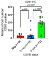
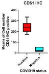
Platelet-megakaryocyte up-regulation is strongly associated with the persistence of Spike-1 in lethal COVID-19 lung disease. Expression of CD61 showed significant up-regulation in the lungs of patients with lethal COVID-19 when compared to the control group of SARS-CoV-2-negative lungs. The subgroup of patients with significantly high levels of CD61+ platelets and megakaryocytes, associated with a systemic pro-thrombotic state and with the formation of tissue micro-thrombosis, corresponds to the Spike-S1-positive cases demonstrated by PCR and ISH methods. Means and standard deviations were obtained by immunohistochemical (IHC) expression of CD61 (glycoprotein IIIa), as evaluated by digital pathology analysis. Spike-positive status was detected by PCR-based methods and in situ hybridization; Mann–Whitney U-test statistical test indicated the comparisons were statistically significant (p < 0.001).
Figure 6
Figure 6
Spike-1 (+) status was detected by ISH. Immunohistochemistry showed a mean count in SARS-CoV-2 (−) controls of 26.50 ± 7.41 SEM, versus mean count of 35.23 ± 17.46 SEM, and 180.43 ± 35.33 SEM in Spike-1 (−) and Spike-1 (+) COVID-19 deceased patients, respectively. Comparisons were statistically significant as evaluated by the Mann–Whitney U-test (p < 0.001).
Figure 7
Figure 7
Representative rank plots of CD61 distribution in the lungs of COVID-19 (+) cases, compared with COVID-19 (−) controls: The ranks plot displays two distinct distributions, one for COVID-19-positive cases (red points) and one for control cases (blue points). The positive data exhibits a bell-shaped curve, with the majority concentrated in the central part of the ranks and a slight tendency to increase in rank with value. Conversely, the negative data shows an asymmetric distribution, with the majority accumulated in the lower ranks and a negative slope, indicating a decrease in rank with increasing value. Overall, the negative data appears to be more variable than the positive data, as evidenced by the greater dispersion of the blue points compared to the red ones. Additionally, the plot indicates the presence of more data with positive ranks compared to negative ranks.
Figure 8
Figure 8
CD61 IHC expression evaluated by digital pathology analysis. (a) (Original magnification ×10, NanoZoomer S60 C13210 series Hamamatsu Photonics K-K, Visiopharm software version 2021.02). (b) Further amplification from the bottom of the figure (a) showing a detail of a megakaryocyte surrounded by platelets. (Original magnification ×60, NanoZoomer S60 C13210 series Hamamatsu Photonics K-K, Visiopharm software version 2021.02).

Similar articles

Cited by

References

    1. Menter D.G., Kopetz S., Hawk E., Sood A.K., Loree J.M., Gresle P., Honn V.K. Platelet “first responders” in wound response, cancer, and metastasis. Cancer Metastasis Rev. 2017;36:199–213. doi: 10.1007/s10555-017-9682-0. - DOI - PMC - PubMed
    1. Yin S., Huang M., Li D., Tang N. Difference of coagulation features between severe pneumonia induced by SARS-CoV2 and non-SARS-CoV2. J. Thromb. Thrombolysis. 2020;51:1107–1110. doi: 10.1007/s11239-020-02105-8. - DOI - PMC - PubMed
    1. Lippi G., Plebani M., Henry B.M. Thrombocytopenia is associated with severe coronavirus disease 2019 (COVID-19) infections: A meta-analysis. Clin. Chim. Acta. 2020;506:145–148. doi: 10.1016/j.cca.2020.03.022. - DOI - PMC - PubMed
    1. Yang X., Yang Q., Wang Y., Wu Y., Xu J., Yu Y., Shang Y. Thrombocytopenia and its Association with Mortality in Patients with COVID-19. J. Thromb. Haemost. 2020;18:1469–1472. doi: 10.1111/jth.14848. - DOI - PMC - PubMed
    1. D’Atri L.P., Schattner M. Platelet toll-like receptors in thromboinflammation. Front. Biosci. 2017;22:1867–1883. - PubMed

Publication types

MeSH terms