Review
. 2024 May 17;13(10):864.
doi: 10.3390/cells13100864.

Cross-Talks between Raf Kinase Inhibitor Protein and Programmed Cell Death Ligand 1 Expressions in Cancer: Role in Immune Evasion and Therapeutic Implications

Affiliations
Review

Cross-Talks between Raf Kinase Inhibitor Protein and Programmed Cell Death Ligand 1 Expressions in Cancer: Role in Immune Evasion and Therapeutic Implications

Mai Ho et al. Cells. .

Abstract

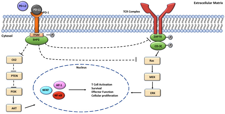
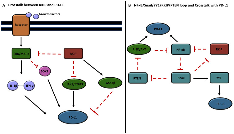
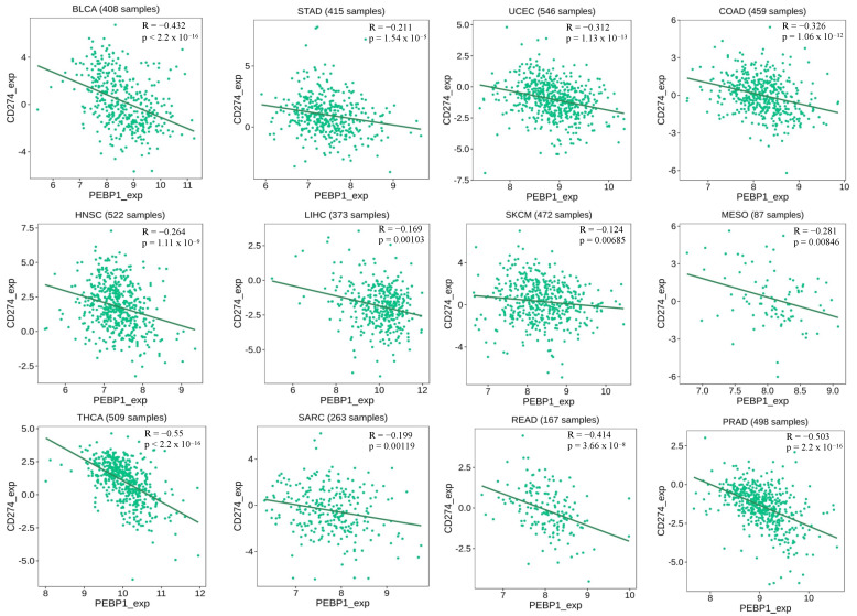
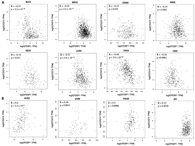
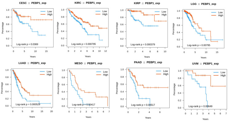
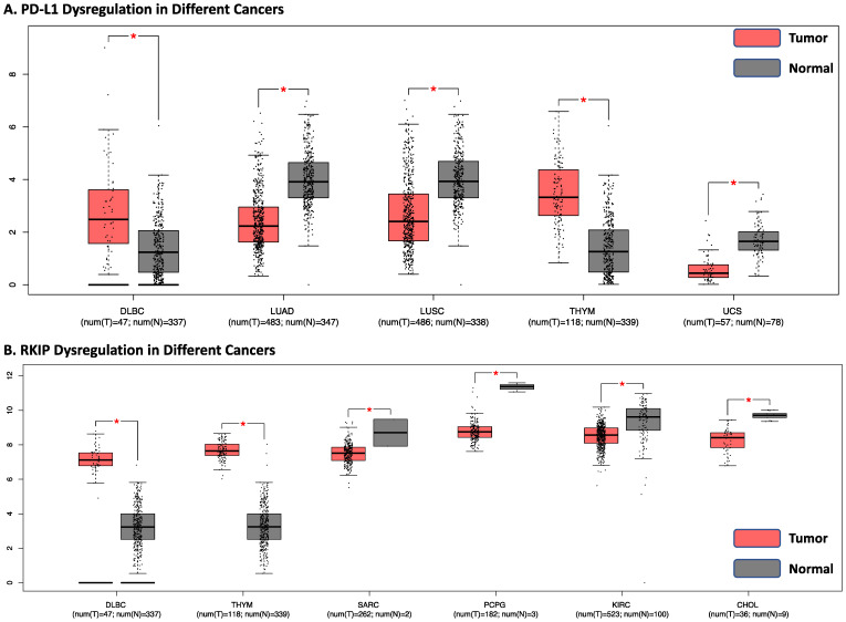
Innovations in cancer immunotherapy have resulted in the development of several novel immunotherapeutic strategies that can disrupt immunosuppression. One key advancement lies in immune checkpoint inhibitors (ICIs), which have shown significant clinical efficacy and increased survival rates in patients with various therapy-resistant cancers. This immune intervention consists of monoclonal antibodies directed against inhibitory receptors (e.g., PD-1) on cytotoxic CD8 T cells or against corresponding ligands (e.g., PD-L1/PD-L2) overexpressed on cancer cells and other cells in the tumor microenvironment (TME). However, not all cancer cells respond-there are still poor clinical responses, immune-related adverse effects, adaptive resistance, and vulnerability to ICIs in a subset of patients with cancer. This challenge showcases the heterogeneity of cancer, emphasizing the existence of additional immunoregulatory mechanisms in many patients. Therefore, it is essential to investigate PD-L1's interaction with other oncogenic genes and pathways to further advance targeted therapies and address resistance mechanisms. Accordingly, our aim was to investigate the mechanisms governing PD-L1 expression in tumor cells, given its correlation with immune evasion, to uncover novel mechanisms for decreasing PD-L1 expression and restoring anti-tumor immune responses. Numerous studies have demonstrated that the upregulation of Raf Kinase Inhibitor Protein (RKIP) in many cancers contributes to the suppression of key hyperactive pathways observed in malignant cells, alongside its broadening involvement in immune responses and the modulation of the TME. We, therefore, hypothesized that the role of PD-L1 in cancer immune surveillance may be inversely correlated with the low expression level of the tumor suppressor Raf Kinase Inhibitor Protein (RKIP) expression in cancer cells. This hypothesis was investigated and we found several signaling cross-talk pathways between the regulations of both RKIP and PD-L1 expressions. These pathways and regulatory factors include the MAPK and JAK/STAT pathways, GSK3β, cytokines IFN-γ and IL-1β, Sox2, and transcription factors YY1 and NFκB. The pathways that upregulated PD-L1 were inhibitory for RKIP expression and vice versa. Bioinformatic analyses in various human cancers demonstrated the inverse relationship between PD-L1 and RKIP expressions and their prognostic roles. Therefore, we suspect that the direct upregulation of RKIP and/or the use of targeted RKIP inducers in combination with ICIs could result in a more targeted anti-tumor immune response-addressing the therapeutic challenges related to PD-1/PD-L1 monotherapy alone.

Keywords: PD-L1; RKIP; cancer; cross-talk; immune evasion; targeted therapy.


Conflict of interest statement

The authors declare no conflicts of interest.

Figures

Figure 1
Figure 1
A pathway depicting the activation of the PD-1 pathway via PD-L1. Activation via PD-L1 will phosphorylate the ITSM region, triggering the recruitment of SHP2. The interactions with SHP2 and ITSM lead to the inhibition of positive signals (CD-3ζ and ZAP70) that occur through the T cell receptor (TCR). These inhibitory signals will lead to the suppression of the Ras/MEK/ERK/MAPK and the PI3K/AKT pathways. Eventually, the inhibition of those pathways will lead to the downstream suppression of transcription factors AP-1, NFAT, and NF-κB in the nucleus. These will affect T cell activation and survival, effector function, and cellular proliferation.
Figure 2
Figure 2
Cross-talks between RKIP and PD-L1 signaling pathways. (A) The pathway depicts various cross-talks between RKIP and PD-L1. The activation of the ERK/MAPK pathways lead to an increased production of cytokines IFN-γ and IL-1β, upregulating PD-L1 expression. RKIP inhibits this pathway, suppressing downstream signaling and PD-L1 expression. Moreover, RKIP inhibits SOX2 through ERK inhibition and also inhibits the JAK/STAT pathway, both of which contribute to PD-L1 upregulation. Additionally, RKIP activates GSK3β signaling, promoting PD-L1 degradation. (B) A pathway depicting the dysregulated loop NFκB/Snail/YY1/RKIP/PTEN. High RKIP expression inhibits NF-κB, leading to the downregulation of Snail and YY1 expressions, while promoting PTEN and downregulating the PI3K/AKT pathway, leading to reduced PD-L1 regulation. Downregulation is represented by the red blocking arrows, while upregulation is represented by the black arrows. Black arrows upregulating PD-L1 leads to tumor-inducing signaling, while the arrows mediated by RKIP seeks to show RKIP’s prevention of dysregulated signaling.
Figure 3
Figure 3
Negative correlation between RKIP expression and PD-L1 expression in different types of cancer. The graphs and data were derived and produced using TISIDE (Accessed January 2024). RKIP and PD-L1 alternative names (PEBP1/CD274) were used. The Spearman R (correlation coefficient), p-values, and number of samples for the individual correlations are indicated.
Figure 4
Figure 4
Negative and positive correlations between RKIP and PD-L1 expressions in various cancers. (A) (row 1–2) represents negative correlations between RKIP and PD-L1 in 8 different cancer types. (B) shows positive correlations between RKIP and PD-L1 in 4 different types of cancers. RKIP and PD-L1 alternative names (PEBP1/CD274) were used. Spearman correlation (R) and p-values are indicated in each graph. Graphs are produced using GEIPA with data from TCGA (Accessed January 2024).
Figure 5
Figure 5
Associations between RKIP (PEBP1) expression and overall survival across human cancers. Higher expression levels of RKIP showed longer survival rates for CESC, KIRC, KIRP, LGG, LUAD, MESO, PAAD, and UVM. Graphs and data were derived and produced using TISIDE (Accessed January 2024).
Figure 6
Figure 6
RKIP and PD-L1 gene expressions. RKIP and PD-L1 gene expression samples showing significant dysregulation (p < 0.01) compared to the controls. (A) PD-L1 dysregulation in DLBC, LUAD, LUSC, THYM, and UCS. (A) shows tumor expression is significantly different compared to normal tissues with PD-L1. (B) RKIP dysregulation in DLBC, THYM, SARC, PCPG, KIRH, and CHOL. (B) shows tumor expression is significantly different compared to normal tissues with RKIP. The box plots shown in pink represent tumor expression levels while the grey box plots represent expression levels in normal tissues. The relative expression levels were first log2(TPM+1)-transformed and the log2FC was defined as the median (tumor)–median (normal), where TPM is the transcript count per million. Graphs are produced using GEIPA with data from TCGA.

Similar articles

Cited by

References

    1. Abbott M., Ustoyev Y. Cancer and the Immune System: The History and Background of Immunotherapy. Semin. Oncol. Nurs. 2019;35:150923. doi: 10.1016/j.soncn.2019.08.002. - DOI - PubMed
    1. Olszanski A.J. Principles of Immunotherapy. J. Natl. Compr. Cancer Netw. 2015;13:670–672. doi: 10.6004/jnccn.2015.0199. - DOI - PubMed
    1. Woo S.-R., Corrales L., Gajewski T.F. Innate Immune Recognition of Cancer. Annu. Rev. Immunol. 2015;33:445–474. doi: 10.1146/annurev-immunol-032414-112043. - DOI - PubMed
    1. Lee M.Y., Jeon J.W., Sievers C., Allen C.T. Antigen Processing and Presentation in Cancer Immunotherapy. J. Immunother. Cancer. 2020;8:e001111. doi: 10.1136/jitc-2020-001111. - DOI - PMC - PubMed
    1. Vesely M.D., Kershaw M.H., Schreiber R., Smyth M. Natural Innate and Adaptive Immunity to Cancer. Annu. Rev. Immunol. 2011;29:235–271. doi: 10.1146/annurev-immunol-031210-101324. - DOI - PubMed

MeSH terms