. 2024 May 18;13(10):871.
doi: 10.3390/cells13100871.

Retinoid Synthesis Regulation by Retinal Cells in Health and Disease

Affiliations

Retinoid Synthesis Regulation by Retinal Cells in Health and Disease

Massimiliano Andreazzoli et al. Cells. .

Abstract

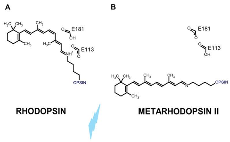
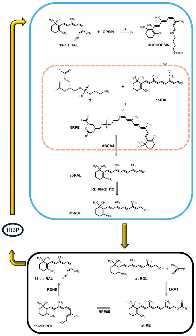
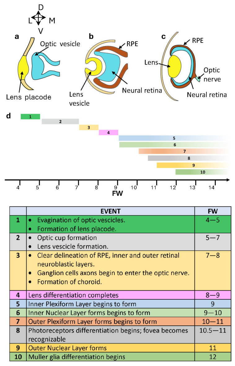
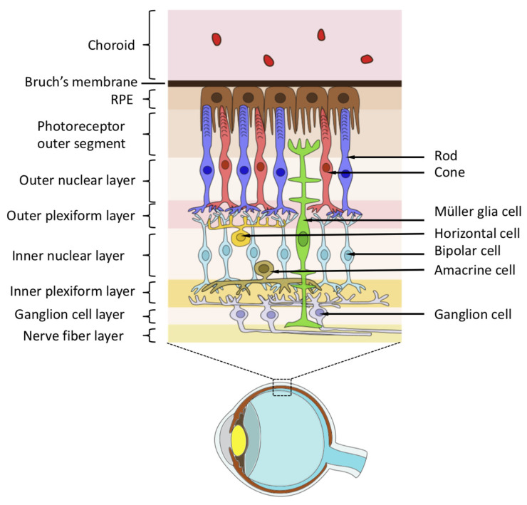
Vision starts in retinal photoreceptors when specialized proteins (opsins) sense photons via their covalently bonded vitamin A derivative 11cis retinaldehyde (11cis-RAL). The reaction of non-enzymatic aldehydes with amino groups lacks specificity, and the reaction products may trigger cell damage. However, the reduced synthesis of 11cis-RAL results in photoreceptor demise and suggests the need for careful control over 11cis-RAL handling by retinal cells. This perspective focuses on retinoid(s) synthesis, their control in the adult retina, and their role during retina development. It also explores the potential importance of 9cis vitamin A derivatives in regulating retinoid synthesis and their impact on photoreceptor development and survival. Additionally, recent advancements suggesting the pivotal nature of retinoid synthesis regulation for cone cell viability are discussed.

Keywords: 11cis retinaldehyde; 9cis retinaldehyde; Müller glial cells; RPE; RPE65; cone photoreceptors; retinal dystrophies; retinoic acid; rod photoreceptors; vision.


Conflict of interest statement

The authors declare no conflicts of interest.

Figures

Figure 1
Figure 1
(A) In ciliary photoreceptors expressing c-type opsin with bound 11cis-RAL, the negative charge provided by E113 stabilizes the positive charge of the protonated Schiff base. (B) Light (cyan) isomerizes 11cis-RAL to at-RAL. After several quick rearrangements, E133 may not stabilize the metarhodopsin II deprotonated Schiff base, which became protonated, preventing at-RAL photoreversal to 11cis isomers.
Figure 2
Figure 2
Outer (OS) and inner (IS) segments of cone (green) and rod (cyan) cells lie in the subretinal space (double-arrow line), which is limited by the junctional complexes (orange) of the retinal pigment epithelium (RPE) and those of the outer limiting membrane (OLM) between Müller glial cells (MGCs) (pink) and rod and cone IS.
Figure 3
Figure 3
The cyan box shows chemical events in the OS. Opsin and 11cis-RAL react, forming a protonated Schiff base between a lysin amino group of the opsin and the aldehyde. After light-induced (hv) 11cis-RAL isomerization, at-RAL is released inside the disk (orange dotted box). It reacts spontaneously with the phosphatidyl ethanolamine (PE) amino group, generating N-retinylidene ethanolamine (NRPE). NRPE is transferred to the OS cytoplasm by the flippase ABCA4. at-RAL dissociates from PE in the cytoplasm and is reduced to all-trans retinol (at-ROL or vitamin A) by retinol dehydrogenase 8 and 12 (RDH8 and RDH12). In the RPE (black box), retinol generated and released by the OS, is esterified by lecithin retinol acyl transferase (LRAT) into at-RE. RPE65 exhibits isomerohydrolase activity, converting at-RE into 11cis-ROL. Retinol dehydrogenase 5 (RDH5) converts 11cis-ROL into 11cis-RAL, which may be bound by cellular retinaldehyde-binding protein (CRALBP) (not displayed in the Figure) to prevent spontaneous isomerization. CRALBP eventually transfers 11cis-RAL to the interphotoreceptor-binding protein (IRBP), which conveys it to the OS by diffusion across the subretinal space (yellow arrows). * indicates a spontaneous reaction between amino groups and an aldehyde group.
Figure 4
Figure 4
Schematic representation of critical phases in human eye development. (a) Formation of the optic vesicle and the lens placode. (b) Progression in optic cup and lens vesicle development, with simultaneous specification of the neural retina and the RPE. (c) The advancements in organized eye structure involve neural retina stratification and optic nerve growth. (d) Timeline and table of the 10 main developmental steps of the human retina from FW4 to FW12. As the table below indicates, steps 1–4 occur within the timeline. Steps 5–10 start at the indicated time at the location of the presumptive foveal. They will spread in the same order to adjacent retinal areas, reaching the peripheral retina around FW 40.
Figure 5
Figure 5
Structure of the mature retina.
Figure 6
Figure 6
(a) A SOX binding site in the proximal region upstream of the 5′ transcription start site (TSS) of RPE65, RLBP1, RGR, LRAT, and RDH5. (b) Transcription factors SOX9 (blue oval) and OTX2 (red square) bind the promoter region of RPE65 and RLB1; SOX9 and LXH2 bind the RGR promoter region. (c) The black curve plots RPE65 isomerohydrolase activity as a function of all-trans-retinol palmitate (at-RP) concentration. The horizontal blue double-arrow segment indicates the substrate concentration range in the RPE, indicating the enzyme operates at its maximal velocity. (d) Translational control involves microRNA (miRNA) (purple half circle) binding at the 3′ untranslated region (3′ UTR) of RPE65 mRNA to prevent its translation (red cross superimposed on the green arrow) into PP.

Similar articles

Cited by

References

    1. Yau K.W., Hardie R.C. Phototransduction motifs and variations. Cell. 2009;139:246–264. doi: 10.1016/j.cell.2009.09.029. - DOI - PMC - PubMed
    1. Arendt D., Tessmar-Raible K., Snyman H., Dorresteijn A.W., Wittbrodt J. Ciliary photoreceptors with a vertebrate-type opsin in an invertebrate brain. Science. 2004;306:869–871. doi: 10.1126/science.1099955. - DOI - PubMed
    1. Lamb T.D. Evolution of vertebrate retinal photoreception. Philos. Trans. R. Soc. Lond. B Biol. Sci. 2009;364:2911–2924. doi: 10.1098/rstb.2009.0102. - DOI - PMC - PubMed
    1. Hardie R.C., Juusola M. Phototransduction in Drosophila. Curr. Opin. Neurobiol. 2015;34:37–45. doi: 10.1016/j.conb.2015.01.008. - DOI - PubMed
    1. Fain G.L., Hardie R., Laughlin S.B. Phototransduction and the evolution of photoreceptors. Curr. Biol. 2010;20:R114–R124. doi: 10.1016/j.cub.2009.12.006. - DOI - PMC - PubMed

LinkOut - more resources