. 2024 May 18;13(10):873.
doi: 10.3390/cells13100873.

Modification of Gas6 Protein in the Brain by a Functional Endogenous Tissue Vitamin K Cycle

Affiliations

Modification of Gas6 Protein in the Brain by a Functional Endogenous Tissue Vitamin K Cycle

Nadide Aydin et al. Cells. .

Abstract

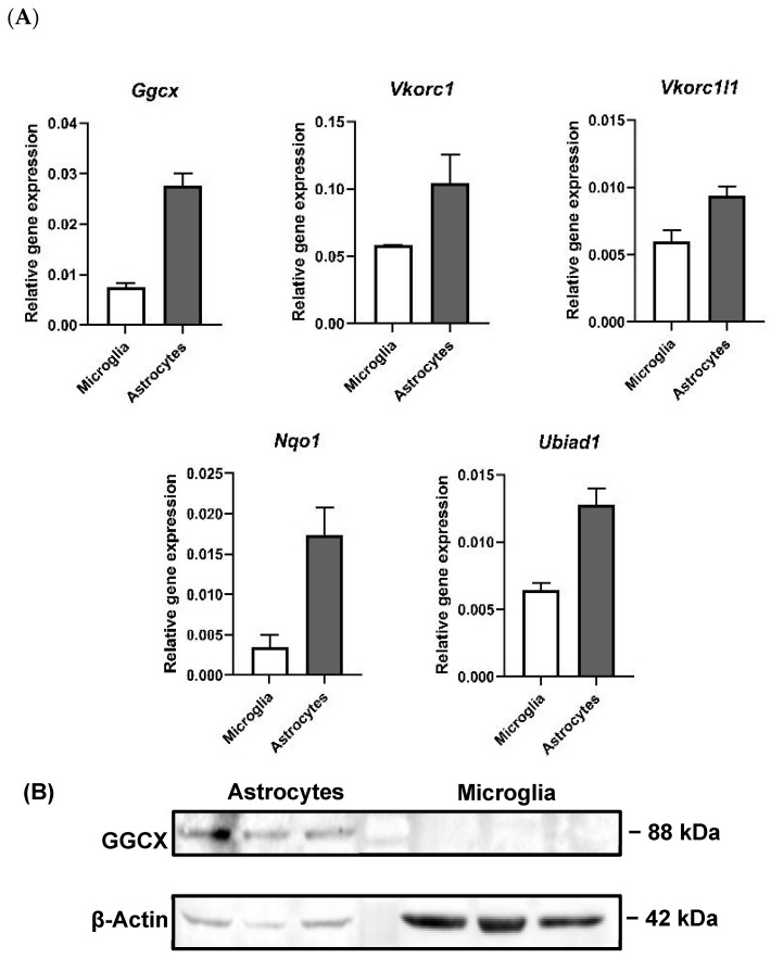
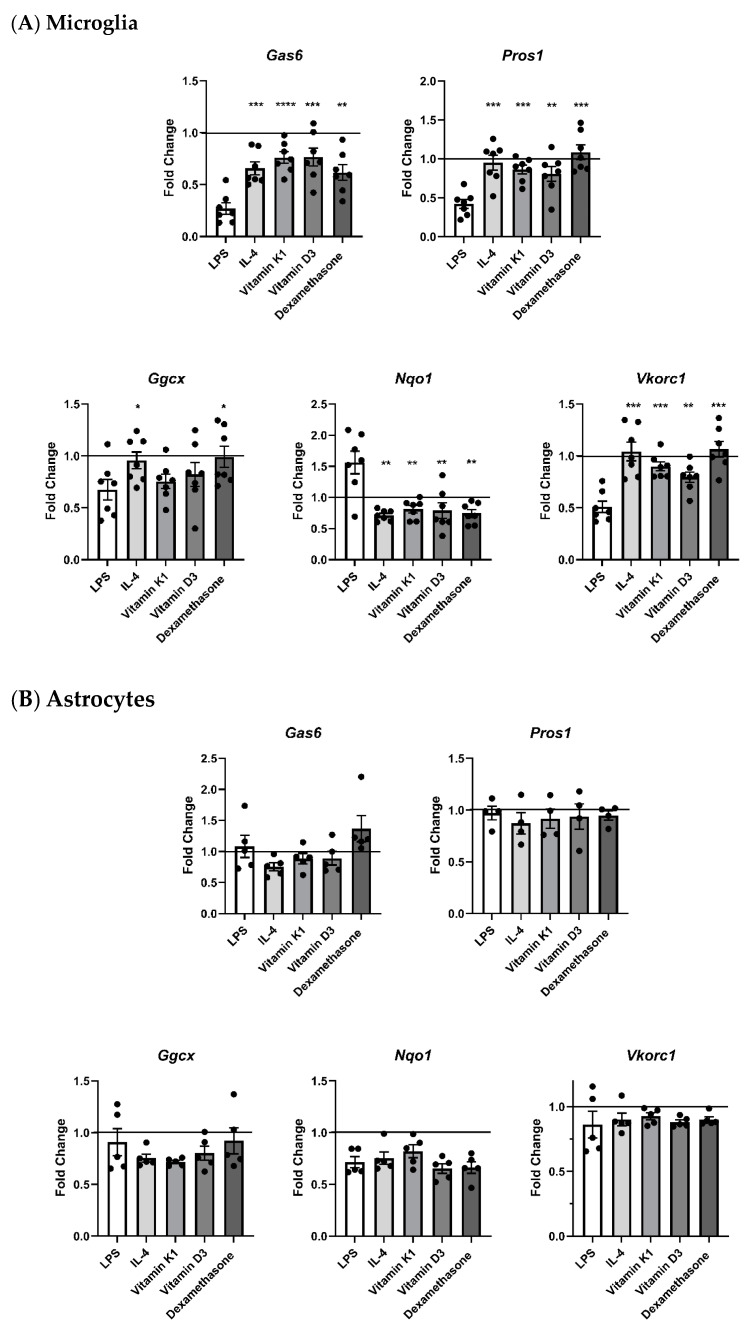
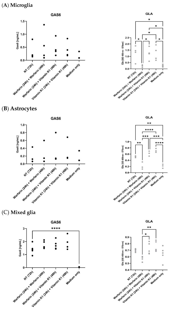
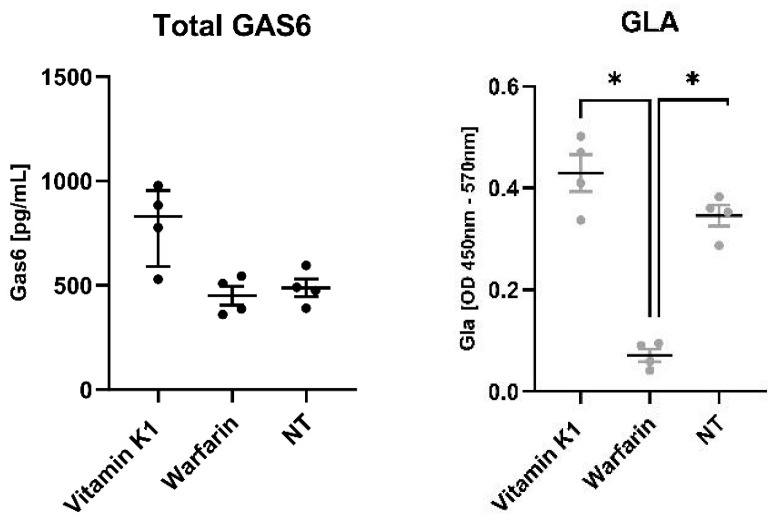
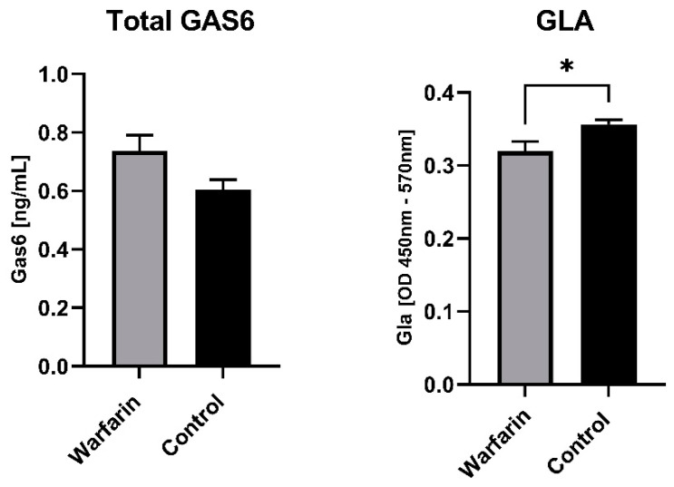
The TAM receptor ligand Gas6 is known for regulating inflammatory and immune pathways in various organs including the brain. Gas6 becomes fully functional through the post-translational modification of multiple glutamic acid residues into γ-carboxyglutamic in a vitamin K-dependent manner. However, the significance of this mechanism in the brain is not known. We report here the endogenous expression of multiple components of the vitamin K cycle within the mouse brain at various ages as well as in distinct brain glial cells. The brain expression of all genes was increased in the postnatal ages, mirroring their profiles in the liver. In microglia, the proinflammatory agent lipopolysaccharide caused the downregulation of all key vitamin K cycle genes. A secreted Gas6 protein was detected in the medium of both mouse cerebellar slices and brain glial cell cultures. Furthermore, the endogenous Gas6 γ-carboxylation level was abolished through incubation with the vitamin K antagonist warfarin and could be restored through co-incubation with vitamin K1. Finally, the γ-carboxylation level of the Gas6 protein within the brains of warfarin-treated rats was found to be significantly reduced ex vivo compared to the control brains. In conclusion, we demonstrated for the first time the existence of a functional vitamin K cycle within rodent brains, which regulates the functional modification of endogenous brain Gas6. These results indicate that vitamin K is an important nutrient for the brain. Furthermore, the measurement of vitamin K-dependent Gas6 functionality could be an indicator of homeostatic or disease mechanisms in the brain, such as in neurological disorders where Gas6/TAM signalling is impaired.

Keywords: Gas6; TAM receptor; glia; microglia; neurodegenerative disease; neuroinflammation; nutrition; vitamin K; warfarin; γ-carboxyglutamic acid; γ-carboxylation.


Conflict of interest statement

The authors declare no conflicts of interest. The funders had no role in the design of the study; in the collection, analyses, or interpretation of data; in the writing of the manuscript; or in the decision to publish the results.

Figures

Figure 1
Figure 1
Gene expression of TAM ligands and TAM receptors in adult mouse tissues. RT-qPCR analysis of relative gene expression in adult mouse liver and brain of TAM ligands Gas6 and Pros1 and TAM receptors Tyro3, Axl and Mertk. Relative gene expression was analysed by using 2−ΔCt method, using Gapdh as housekeeping gene (mean ± SEM, n = 3 tissues).
Figure 2
Figure 2
Gene expression of TAM ligands and TAM receptors in mouse brain glial cells. RT-qPCR analysis of gene expression of TAM ligands Gas6 and Pros1 and TAM receptors Tyro3, Axl and Mertk in primary cultures of astrocytes and microglia. Relative gene expression was analysed by using 2−ΔCt methods, using Gapdh as housekeeping gene (mean ± SEM; n = 3 cultures).
Figure 3
Figure 3
Analysis of gene and protein expression of vitamin K cycle enzymes in mouse liver and brain during postnatal development. (A) Relative gene expression was determined by RT-qPCR and data was analysed by 2−ΔCt method, using Gapdh as housekeeping gene (mean ± SEM; n = 4 tissues; brain embryo (E) n = 3). ANOVA with Tukey’s multiple expression multiple comparison post hoc analysis * p < 0.05, ** p < 0.01, *** p < 0.001, **** p < 0.0001. (B) Western blot detection of GGCX and VKORC1 proteins in extracts from liver (P0—adult (A); upper blot) and brain (E–A; lower blot). Accompanying graphs show protein quantification by band densitometry analysis. Data are mean ± SEM protein expression normalised against β-Actin as loading control protein. Data underwent unpaired t-test; * p < 0.05, ** p < 0.01 between samples as indicated (n = 3).
Figure 3
Figure 3
Analysis of gene and protein expression of vitamin K cycle enzymes in mouse liver and brain during postnatal development. (A) Relative gene expression was determined by RT-qPCR and data was analysed by 2−ΔCt method, using Gapdh as housekeeping gene (mean ± SEM; n = 4 tissues; brain embryo (E) n = 3). ANOVA with Tukey’s multiple expression multiple comparison post hoc analysis * p < 0.05, ** p < 0.01, *** p < 0.001, **** p < 0.0001. (B) Western blot detection of GGCX and VKORC1 proteins in extracts from liver (P0—adult (A); upper blot) and brain (E–A; lower blot). Accompanying graphs show protein quantification by band densitometry analysis. Data are mean ± SEM protein expression normalised against β-Actin as loading control protein. Data underwent unpaired t-test; * p < 0.05, ** p < 0.01 between samples as indicated (n = 3).
Figure 4
Figure 4
Gene and protein expression of vitamin K cycle enzymes in primary cultured mouse brain astrocytes and microglia. (A) Relative gene expression was analysed by using 2−ΔCt method, using Gapdh as housekeeping gene (mean ± SEM; n = 3 cultures). (B) Western blot showing GGCX protein expression in astrocytes. β-Actin was used as protein loading control. n = 3 separate culture extracts loaded.
Figure 5
Figure 5
RT-qPCR analysis of mRNA expression of TAM ligands and vitamin K cycle enzymes in glial cells undergoing treatment with different agents. Expression of K cycle enzyme genes (Ggcx, Nqo1, Vkorc1) and Gas6 and Pros1 in pure primary cultures of (A) microglia and (B) astrocytes after 8 h incubation with agents. Relative gene expression was analysed by 2−ΔΔCt method, using Gapdh as housekeeping gene (mean ± SEM, microglia n = 7 cultures, astrocytes n = 5 cultures (n = 4 for Pros1)). Horizontal line in each graph shows baseline expression in untreated cells, with fold changes displayed relative to that. Statistical significance was determined using Welch’s t-test with * p < 0.05, ** p < 0.01, *** p < 0.001, **** p < 0.0001 vs. LPS.
Figure 6
Figure 6
Mouse brain glial cells in culture release endogenous Gas6 protein that can be suppressed in its γ-carboxylation by warfarin and subsequently reversed by exogenous vitamin K1. Pure cultures of microglia (A), astrocytes (B) as well as mixed glia (C) were pre-treated with warfarin (3.24 μM) or vitamin K1 (11 μM) for 24 h, followed by replacement with 2% serum medium with added warfarin or vitamin K1 for a further 48 h. ‘NT’ is conditioned medium that has been incubated with cells but with nothing extra added, whereas ‘medium only’ is medium that has not been incubated with cells. Media were analysed by specific ELISA assays for total mouse Gas6 protein (left graph, black dots) and Gla-Gas6 (right graph, grey dots). Statistical significance was determined by one-way ANOVA with Tukey’s multiple comparisons test, * p < 0.05, ** p < 0.01, *** p < 0.001, **** p < 0.0001 for comparisons indicated by lines (microglia n = 4, astrocytes n = 3, mixed glia n = 5 on separate cultures).
Figure 7
Figure 7
Effects of vitamin K1 and warfarin on levels of total and γ-carboxylated Gas6 released by mouse cerebellar slice cultures. γ-carboxylated Gas6 (right graph, grey dots) is significantly decreased by warfarin in comparison to vitamin K1 and control. Statistical significance was determined using Mann– Whitney test; * p < 0.05 for comparisons indicated by lines (n = 4 separate cultures).
Figure 8
Figure 8
γ-carboxylated Gas6 protein is downregulated in the brains of warfarin-treated rats in vivo. Eight-week-old male Wistar rats were treated with 14 mg/kg per day with warfarin in the drinking water and subcutaneous vitamin K1 (85 mg/kg/day) injections three times per week for 10 weeks. Control animals were treated with normal water and injected with saline three times per week for 10 weeks in total. Control group were fed with AIN-93-based diet containing 750 μg vitamin K1/kg. The bar graphs show comparisons of control (black bars) vs. warfarin (grey bars) for levels of total Gas6 and Gla-Gas6. Statistical analysis was determined with unpaired t-test * p < 0.05 (n = 8 separate tissues).

Similar articles

Cited by

References

    1. Hafizi S., Dahlback B. Signalling and Functional Diversity within the Axl Subfamily of Receptor Tyrosine Kinases. Cytokine Growth Factor Rev. 2006;17:295–304. doi: 10.1016/j.cytogfr.2006.04.004. - DOI - PubMed
    1. Hafizi S., Dahlback B. Gas6 and Protein S. Vitamin K-Dependent Ligands for the Axl Receptor Tyrosine Kinase Subfamily. FEBS J. 2006;273:5231–5244. doi: 10.1111/j.1742-4658.2006.05529.x. - DOI - PubMed
    1. Nakano T., Higashino K., Kikuchi N., Kishino J., Nomura K., Fujita H., Ohara O., Arita H. Vascular Smooth Muscle Cell-Derived, Gla-Containing Growth-Potentiating Factor for Ca(2+)-Mobilizing Growth Factors. J. Biol. Chem. 1995;270:5702–5705. doi: 10.1074/JBC.270.11.5702. - DOI - PubMed
    1. Goruppi S., Ruaro E., Schneider C. Gas6, the Ligand of Axl Tyrosine Kinase Receptor, Has Mitogenic and Survival Activities for Serum Starved NIH3T3 Fibroblasts. Oncogene. 1996;12:471–480. - PubMed
    1. Angelillo-Scherrer A., Burnier L., Flores N., Savi P., DeMol M., Schaeffer P., Herbert J.-M., Lemke G., Goff S.P., Matsushima G.K., et al. Role of Gas6 Receptors in Platelet Signaling during Thrombus Stabilization and Implications for Antithrombotic Therapy. J. Clin. Investig. 2005;115:237–246. doi: 10.1172/JCI22079. - DOI - PMC - PubMed

Publication types

LinkOut - more resources