. 2024 May 20;13(10):881.
doi: 10.3390/cells13100881.

NADH Intraperitoneal Injection Prevents Lung Inflammation in a BALB/C Mice Model of Cigarette Smoke-Induced Chronic Obstructive Pulmonary Disease

Affiliations

NADH Intraperitoneal Injection Prevents Lung Inflammation in a BALB/C Mice Model of Cigarette Smoke-Induced Chronic Obstructive Pulmonary Disease

Nada Slama et al. Cells. .

Abstract

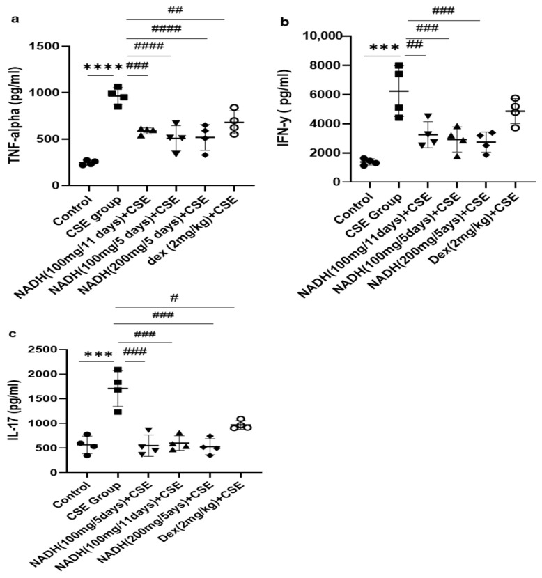
Cigarette smoke is one of the main factors in Chronic Obstructive Pulmonary Disease (COPD), a respiratory syndrome marked by persistent respiratory symptoms and increasing airway obstruction. Perturbed NAD+/NADH levels may play a role in various diseases, including lung disorders like COPD. In our study, we investigated the preventive effect of NADH supplementation in an experimental model of COPD induced by cigarette smoke extract (CSE). N = 64 mice randomly distributed in eight groups were injected with NADH (two doses of 100 mg/kg or 200 mg/kg) or dexamethasone (2 mg/kg) before being exposed to CSE for up to 9 weeks. Additionally, NADH supplementation preserved lung antioxidant defenses by preventing the functional loss of key enzymes such as superoxide dismutase (SOD), glutathione peroxidase (GPX), catalase, and the expression levels of glutathione (GSH) (n = 4, p < 0.001). It also reduced oxidative damage markers, such as malondialdehyde (MDA) and nitrites (n = 4, p < 0.001). A marked increase in tissue myeloperoxidase activity was assessed (MPO), confirming neutrophils implication in the inflammatory process. The latter was significantly ameliorated in the NADH-treated groups (p < 0.001). Finally, NADH prevented the CSE-induced secretion of cytokines such as Tumor Necrosis Factor alpha (TNF-α), IL-17, and IFN-y (n = 4, p < 0.001). Our study shows, for the first time, the clinical potential of NADH supplementation in preventing key features of COPD via its unique anti-inflammatory and antioxidant properties.

Keywords: NADH; animal model; cigarette smoke; inflammation; oxidative stress.


Conflict of interest statement

All authors (except for Birkmayer) declare no competing interests. The NADH used in the present study was a gift from the late Birkmayer, the founder of the Birkmayer NADH Company, who tragically died on 28 August 2022. The Birkmayer NADH Company was not involved in the study conception and design, data collection, analysis and interpretation of results, or manuscript preparation. None of the authors received any financial or project funding support from the Birkmayer NADH Company.

Figures

Figure 1
Figure 1
NADH effects on mice body weights. Mice were weighed every 11 days; ### p < 0.001 compared with CSE group (n = 8/per group).
Figure 2
Figure 2
NADH, irrespective to the dose and treatment regimen, prevented blood inflammatory changes in mice exposed to CSE. Blood was collected at the end of the experiment from the control groups (diluent, dexamethasone alone, and NADH alone) and the groups treated with CSE alone or with NADH in a5-day or 11-day regimen. The numbers of total white blood cells (a), lymphocytes (b), granulocytes (neutrophils, eosinophils, or basophils) (c), and MID (monocyte and all non-granulocyte or lymphocyte blood cells) (d) were then determined as described in the Section 2. ** p < 0.05, *** p < 0.001, and **** p < 0.0001 compared with the control group; # p < 0.05, ## p < 0.01, ### p < 0.001, and #### p < 0.0001 compared with CSE group (n = 4/per group).
Figure 3
Figure 3
NADH reduced markers of oxidative stress in the lungs of mice exposed to CSE. The levels of malondialdehyde (MDA) (a), nitrites (b), and MPO (c) were assessed as described in the Section 2 in lung homogenates from the control groups (diluent, dexamethasone alone, and NADH alone) or the groups treated with CSE alone or with NADH every 5 or 11 days. *** p < 0.001 and **** p < 0.0001 versus the control group; # p < 0.05, ## p < 0.01, ### p < 0.001, and #### p < 0.0001 compared with the CSE group (n = 4 animals/condition).
Figure 4
Figure 4
NADH prevented the reduced antioxidant response in the lungs of mice exposed to CSE. The activities of key detoxifying enzymes, including superoxide dismutase (SOD) (a), glutathione peroxidase (GPX) (b), glutathione (GSH) (c), and catalase (d), were assessed in lung homogenates from the control groups (diluent, dexamethasone alone, and NADH alone) or the groups treated with CSE alone or with NADH in the 5-day or 11-day regimen as described in the Section 2. * p < 0.05, ** p < 0.01 and *** p < 0.001 compared with the control group; # p < 0.05, ## p < 0.01, ### p < 0.001 and #### p < 0.0001 compared with the CSE group (n = 4/per group).
Figure 5
Figure 5
The effects of NADH on levels of TNF-alpha, IFN-γ, and IL-17 in the lungs of mice exposed to CSE. The levels of cytokines, including TNF-alpha (a), IFN-γ (b), and IL-17 (c), were assessed in lung homogenates from the control group or the groups treated with CSE alone or with NADH in the 5-day or 11-day regimen as described in the Section 2. *** p < 0.001 and **** p < 0.0001 compared with the control group; # p < 0.05 ## p < 0.01, ### p < 0.001, and #### p < 0.0001 compared with the CSE group (n = 4/per group).
Figure 6
Figure 6
NADH ameliorated CSE-induced air space enlargement. Representative lung tissue sections (airways and parenchyma) stained with hematoxylin–eosin (H&E), taken at 400× magnification. (A,B) Control group; (C,D) CSE group. (E,F) NADH (100 mg/11 days) + CSE group. (G,H) NADH (100 mg/5 days) + CSE group. (I,J) NADH (200 mg/5 days) + CSE group. (K,L) Dexamethasone (2 mg/kg) + CSE group. Images are representative of n = 4 animals per condition. AWS = airways; double headed arrow = alveolar space enlargement.
Figure 7
Figure 7
Lung morphometric analysis following CSE exposure in different treatment groups. Mean linear intercept (MLI) (a) and bronchial wall thickening (b) were calculated using Image J-v1.54j software. *** p < 0.001 versus control group, ## p < 0.01 and ### p < 0.001 vs. CSE group (n = 4/condition).
Figure 8
Figure 8
NADH protected against CSE-induced airway fibrosis. Representative lung tissue sections stained with Masson’s trichrome, taken at 400× magnification. Top panel: (A) control group, (B) CSE group, (C) NADH (200 mg/5 days) + CSE, (D) NADH (100 mg/5-day regimen) + CSE, (E) NADH (100 mg/11-day regimen) + CSE, (F) dexamethasone (2 mg/kg) + CSE, (G) assessment of collagen content. Bottom panel: comparison of extent of airway fibrosis (μm2) in lungs of different experimental groups compared to control group. **** p < 0.05 versus control group, ### p < 0.01 and #### p < 0.0001 versus CSE group, n = 4/per group.
Figure 9
Figure 9
An overview of the therapeutic effect of NADH in the murine COPD model induced by systemic CSE exposure. The NADH-treated groups presented with a significant reduction in COPD key features, such as emphysema and airway remodeling (fibrosis). The therapeutic benefit of NADH is believed to result from blunted inflammatory pathways, as evidenced by the marked reductions in systemic granulocytes and lung neutrophils (reflected by decreased MPO levels). A significant decrease in the key COPD inflammatory cytokines (TNF-a, IFN-g, and IL-17) in the lungs may, in part, explain NADH’s beneficial action. These cytokines and other growth factors (TGF-b and CTGF) are known to play central roles in the chemoattraction and activation of macrophages/monocytes and neutrophils, thereby contributing to the amplification/perpetuation of inflammation/oxidative stress/remodeling processes in lung tissues. NADH supplementation was also found to markedly reduce CSE-induced oxidative damage, as shown by the diminution of typical biomarkers (MDA and nitrites) and the prevention of CSE-associated depletion of multiple antioxidant defense mechanisms (GPX, SOD, GSH, and catalase). ↔: Indicates the impact of NADH on both airway inflammation and oxidative stress may explain its beneficial action in COPD. GPX: glutathione peroxidase, GSH: glutathione, MDA: malondialdehyde, MPO: myeloperoxidase, SOD: superoxide dismutase. The image of the lung is from Pixabay.

Similar articles

Cited by

References

    1. Dorababu A., Maraswami M. Recent Advances (2015–2020) in Drug Discovery for Attenuation of Pulmonary Fibrosis and COPD. Molecules. 2023;28:3674. doi: 10.3390/molecules28093674. - DOI - PMC - PubMed
    1. Gomes F., Cheng S.-L. Pathophysiology, Therapeutic Targets, and Future Therapeutic Alternatives in COPD: Focus on the Importance of the Cholinergic System. Biomolecules. 2023;13:476. doi: 10.3390/biom13030476. - DOI - PMC - PubMed
    1. Barnes P.J. Inflammatory Mechanisms in Patients with Chronic Obstructive Pulmonary Disease. J. Allergy Clin. Immunol. 2016;138:16–27. doi: 10.1016/j.jaci.2016.05.011. - DOI - PubMed
    1. Sidhaye V.K., Koval M. Lung Epithelial Biology in the Pathogenesis of Pulmonary Disease. Academic Press; Cambridge, MA, USA: 2017.
    1. Strzelak A., Ratajczak A., Adamiec A., Feleszko W. Tobacco Smoke Induces and Alters Immune Responses in the Lung Triggering Inflammation, Allergy, Asthma and Other Lung Diseases: A Mechanistic Review. Int. J. Environ. Res. Public Health. 2018;15:1033. doi: 10.3390/ijerph15051033. - DOI - PMC - PubMed

Publication types

MeSH terms