. 2024 May 4;13(5):421.
doi: 10.3390/antibiotics13050421.

The MraY Inhibitor Muraymycin D2 and Its Derivatives Induce Enlarged Cells in Obligate Intracellular Chlamydia and Wolbachia and Break the Persistence Phenotype in Chlamydia

Affiliations

The MraY Inhibitor Muraymycin D2 and Its Derivatives Induce Enlarged Cells in Obligate Intracellular Chlamydia and Wolbachia and Break the Persistence Phenotype in Chlamydia

Iris Löckener et al. Antibiotics (Basel). .

Abstract

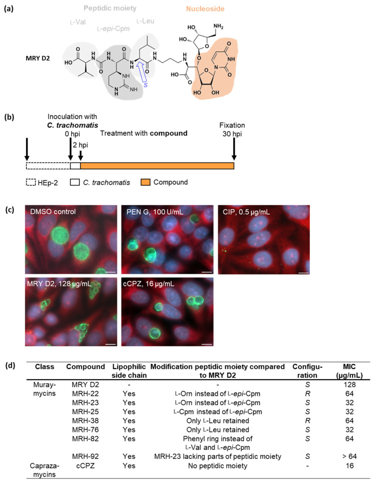
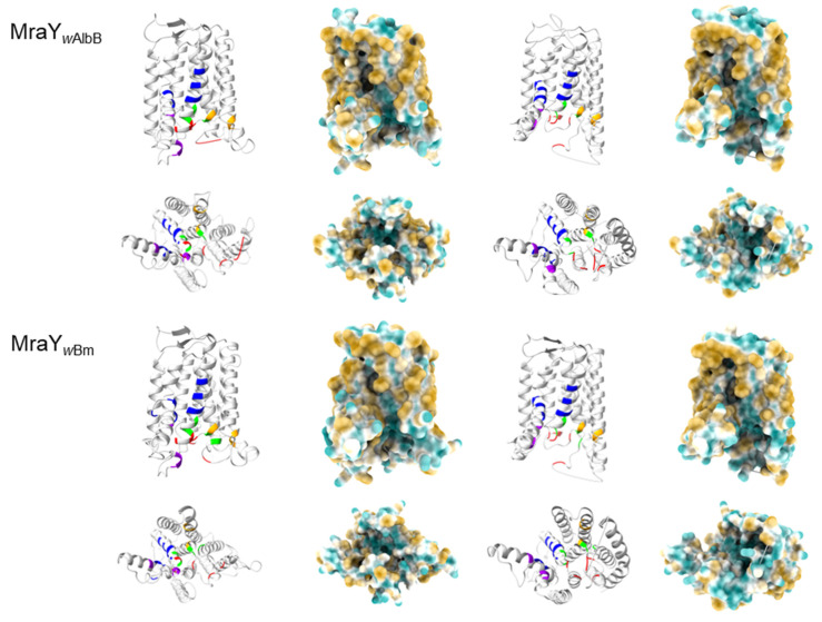
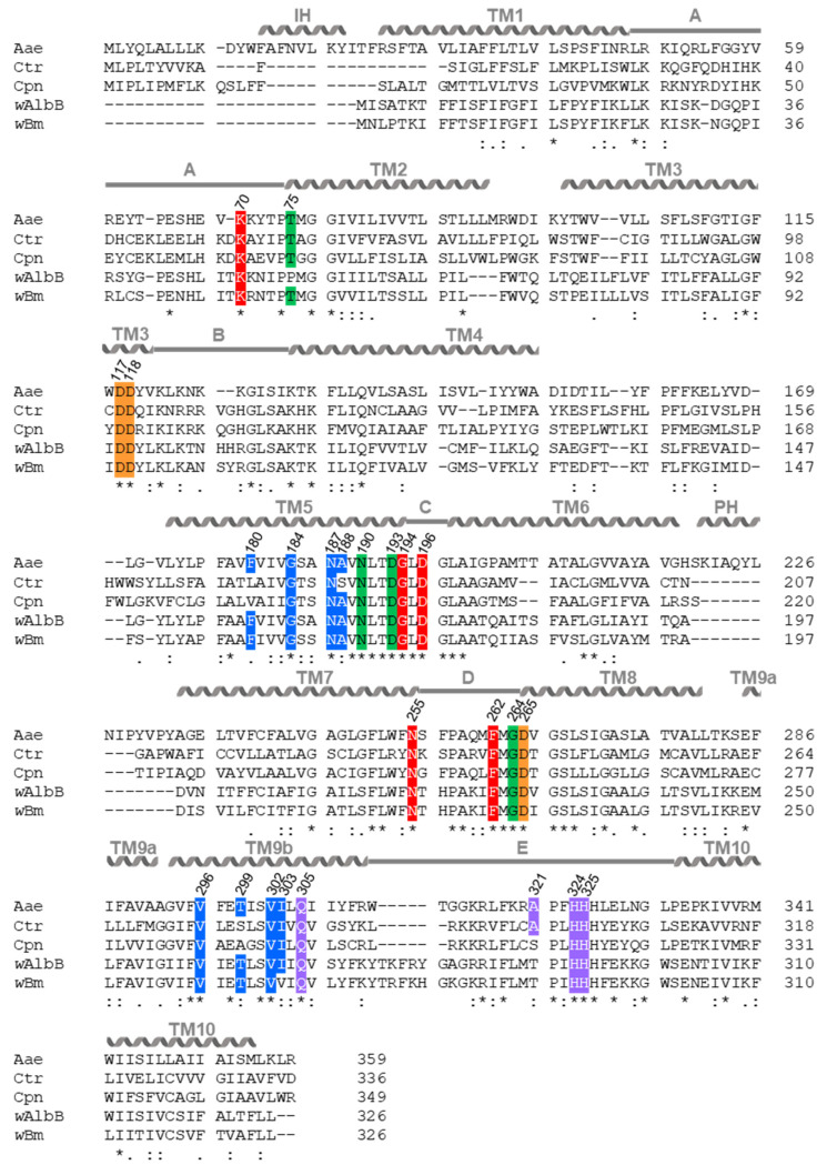
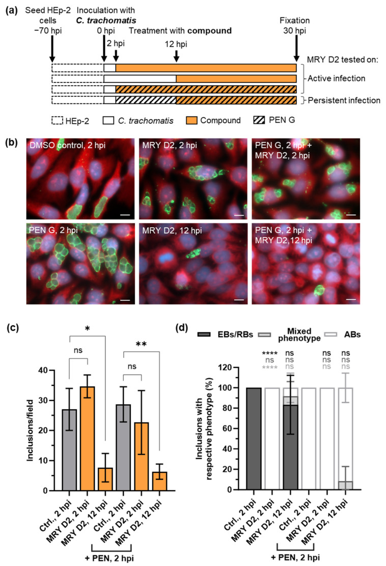
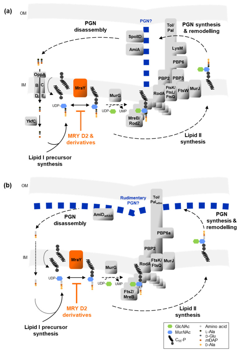
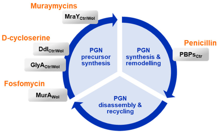
Chlamydial infections and diseases caused by filarial nematodes are global health concerns. However, treatment presents challenges due to treatment failures potentially caused by persisting Chlamydia and long regimens against filarial infections accompanied by low compliance. A new treatment strategy could be the targeting of the reduced peptidoglycan structures involved in cell division in the obligate intracellular bacteria Chlamydia and Wolbachia, the latter being obligate endosymbionts supporting filarial development, growth, and survival. Here, cell culture experiments with C. trachomatis and Wolbachia showed that the nucleoside antibiotics muraymycin and carbacaprazamycin interfere with bacterial cell division and induce enlarged, aberrant cells resembling the penicillin-induced persistence phenotype in Chlamydia. Enzymatic inhibition experiments with purified C. pneumoniae MraY revealed that muraymycin derivatives abolish the synthesis of the peptidoglycan precursor lipid I. Comparative in silico analyses of chlamydial and wolbachial MraY with the corresponding well-characterized enzyme in Aquifex aeolicus revealed a high degree of conservation, providing evidence for a similar mode of inhibition. Muraymycin D2 treatment eradicated persisting non-dividing C. trachomatis cells from an established penicillin-induced persistent infection. This finding indicates that nucleoside antibiotics may have additional properties that can break bacterial persistence.

Keywords: Chlamydia; MraY; Wolbachia; cell division; intracellular bacteria; lipid II synthesis; muraymycin; peptidoglycan; persistence-breaking.


Conflict of interest statement

The authors declare no conflicts of interest.

Figures

Figure 1
Figure 1
Effects of muraymycin (MRY) D2, its derivatives, and carbacaprazamycin (cCPZ) on C. trachomatis. (a) The lead compound MRY D2 consists of a nucleoside and peptidic moiety made up of l-Val, l-epi-capreomycidine (Cpm), and l-Leu. The bond between the latter two has an S-configuration. (b) HEp-2 cells were infected with C. trachomatis D/UW-3/CX, the compounds were added at 2 h post infection (hpi), and anti-chlamydial activity was analyzed at 30 hpi by fluorescence microscopy. (c) The vehicle control with dimethyl sulfoxide (DMSO) showed an active infection, 100 U/mL penicillin (PEN) G induced persistence visible as enlarged, aberrant bodies (ABs), while 0.5 µg/mL ciprofloxacin (CIP) was bactericidal. MRY D2 is shown at the minimal inhibitory concentration (MIC; 128 µg/mL), at which the formation of aberrant cells was induced; cCPZ had the lowest MIC of 16 µg/mL. Eukaryotic host cell cytoplasm from representative images was labeled with Evans Blue (red), genomic desoxyribonucleic acid (DNA) with 4′,6-diamidino-2-phenylindole (DAPI; blue), and chlamydial lipopolysaccharide with fluorescein (green). Representative images of three experiments. Scale bar: 10 µm. (d) Overview of the structural features of muraymycin derivatives (MRH) and cCPZ compared with MRY D2. MRH-22 and -23 have an l-ornithine (l-Orn). Compared with MRY D2, the MICs of muraymycin and caprazamycin derivatives were lower, ranging between 16 µg/mL and 64 µg/mL. Data obtained from three experiments. MRH-92 did not induce the formation of enlarged chlamydial cells.
Figure 2
Figure 2
Effects of the muraymycin derivatives MRH-76 and -92 on Wolbachia strain B of Aedes albopictus. (a) A. albopictus C6/36 cells infected with Wolbachia strain B of A. albopictus were cultured for 9 days with or without the compounds at varying concentrations. Treatment with 8 µg/mL MRH-76 or 32 µg/mL MRH-92 induced the formation of enlarged Wolbachia cells (exemplary cells indicated by white squares). Genomic DNA was stained with DAPI (green). Scale bar = 2 µm. (b) Using ZEN 3.5 (blue edition) with the image analysis module, the diameter of untreated and treated Wolbachia from 3 to 4 images was determined, shown as median ± interquartile ranges. For statistical analysis, a Kruskal–Wallis test was applied; p < 0.0001. n = 663 (control), 294 (MRH-76, 8 µg/mL), 348 (MRH-92, 8 µg/mL), and 298 (MRH-92, 32 µg/mL) cells. The cell diameter of Wolbachia cells treated with 8 µg/mL MRH-76, as well as of those treated with 8 µg/mL and 32 µg/mL MRH-92, was significantly larger compared with the vehicle control. (c) The depletion of Wolbachia from infected and treated C6/36 cells was measured after 9 days by extracting genomic DNA and measuring the ratio of Wolbachia 16S rDNA and A. albopictus actin B by qPCR; the median ± interquartile range from three experiments is shown. Wolbachia were nearly completely depleted by 4 µg/mL doxycycline (DOX), the positive control, whereas treatment with MRH-76 and MRH-92 led to a concentration-dependent reduction of Wolbachia, with 32 µg/mL MRH-76 showing a >50% reduction compared with the negative DMSO control.
Figure 3
Figure 3
Formation of lipid I by the phospho-MurNAc-pentapeptide transferase MraY is inhibited by the muraymycins MRY D2 and MRH-92. (a) C. pneumoniae and Wolbachia spp. MraY use the precursor molecules undecaprenyl-phosphate (C55-P) and uridine diphosphate (UDP)-N-acetylmuramoyl(MurNAc)-pentapeptide to form lipid I, which is glycosylated with N-acetylglucosamine (GlcNAc) by MurG, yielding lipid II. IM: inner membrane; UMP: uridine monophosphate; mDAP: meso-diaminopimelic acid; C55-P: undecaprenyl-phosphate. (b) Recombinant MraYCpn was incubated with C55-P, UDP-MurNAc-pentapeptide, and different concentrations of either MRY D2 or MRH-92 for 90 min at 37 °C. Reaction products were extracted, separated via thin-layer chromatography, and stained. Formation of lipid I by MraYCpn was inhibited by both tested inhibitors. MRY D2 showed a concentration-dependent inhibition, with MraYCpn being completely inhibited at 0.5 µM. MRH-92 also inhibited the enzymatic reaction at 0.5 µM.
Figure 4
Figure 4
Structural comparison of A. aeolicus apoMraY and MraY bound to MRY D2, and predictions of C. pneumoniae, C. trachomatis, and Wolbachia endosymbionts of A. albopictus and B. malayi MraY and MraY bound to MRY D2. Bacterial strains and primary protein accession numbers: A. aeolicus VF5 (Aae, O66465), C. trachomatis D/UW-3/CX (Ctr, O84762), C. pneumoniae GiD (Cpn, A0A0F7WR30) and Wolbachia endosymbiont of A. albopictus (wAlbB, A0A4S2QUK2) and B. malayi (wBm, Q5GRZ3). MraY structure predictions were made by pyhre2 against apoMrYAae chain A [58] or MraYAae crystallized with MRY D2 [50] with 100% confidence. Proteins are viewed from the side (top: periplasm; bottom: cytoplasm) or turned by 90° + 90° and viewed from the cytoplasm. Amino acid residues of the active site (orange), Mn2+ ions (representative for Mg2+; grey), Ni2+ ions (magenta), as well as residues contributing to binding of uracil (red), 5′-aminoribose (green), and the peptidic side chain (violet) from MRY D2 and the acyl moiety of cCPZ (blue), are highlighted as in [50,58]. Surface hydrophobicity is indicated from hydrophobic (brown) to hydrophilic (turquoise). The hydrophobic groove (dashed line) of apoMraYAae is visualized as in [58]. The phyre2 predictions revealed that MraYCtr/Cpn/wAlbB/wBm have similar secondary and tertiary structures compared to apoMraYAae. The active sites face the cytoplasm. In MraYCtr/Cpn/wAlbB/wBm, the uracil, 5′-aminoribose, and peptidic side chains face the cytoplasm, residues contributing to the binding of the cCPZ acyl chain are located at the surface of the protein groove as in apoMraYAae. Residues V266 and I267 of MraYwAlbB, contributing to acyl moiety binding in MraYAae, could not be predicted.
Figure 4
Figure 4
Structural comparison of A. aeolicus apoMraY and MraY bound to MRY D2, and predictions of C. pneumoniae, C. trachomatis, and Wolbachia endosymbionts of A. albopictus and B. malayi MraY and MraY bound to MRY D2. Bacterial strains and primary protein accession numbers: A. aeolicus VF5 (Aae, O66465), C. trachomatis D/UW-3/CX (Ctr, O84762), C. pneumoniae GiD (Cpn, A0A0F7WR30) and Wolbachia endosymbiont of A. albopictus (wAlbB, A0A4S2QUK2) and B. malayi (wBm, Q5GRZ3). MraY structure predictions were made by pyhre2 against apoMrYAae chain A [58] or MraYAae crystallized with MRY D2 [50] with 100% confidence. Proteins are viewed from the side (top: periplasm; bottom: cytoplasm) or turned by 90° + 90° and viewed from the cytoplasm. Amino acid residues of the active site (orange), Mn2+ ions (representative for Mg2+; grey), Ni2+ ions (magenta), as well as residues contributing to binding of uracil (red), 5′-aminoribose (green), and the peptidic side chain (violet) from MRY D2 and the acyl moiety of cCPZ (blue), are highlighted as in [50,58]. Surface hydrophobicity is indicated from hydrophobic (brown) to hydrophilic (turquoise). The hydrophobic groove (dashed line) of apoMraYAae is visualized as in [58]. The phyre2 predictions revealed that MraYCtr/Cpn/wAlbB/wBm have similar secondary and tertiary structures compared to apoMraYAae. The active sites face the cytoplasm. In MraYCtr/Cpn/wAlbB/wBm, the uracil, 5′-aminoribose, and peptidic side chains face the cytoplasm, residues contributing to the binding of the cCPZ acyl chain are located at the surface of the protein groove as in apoMraYAae. Residues V266 and I267 of MraYwAlbB, contributing to acyl moiety binding in MraYAae, could not be predicted.
Figure 5
Figure 5
Sequence alignment of MraY from A. aeolicus, C. trachomatis, C. pneumoniae, and Wolbachia. Bacterial strains and primary protein accession numbers: A. aeolicus VF5 (Aae, O66465), C. trachomatis D/UW-3/CX (Ctr, O84762), C. pneumoniae GiD (Cpn, A0A0F7WR30) and Wolbachia endosymbiont of A. albopictus (wAlbB, A0A4S2QUK2) and B. malayi (wBm, Q5GRZ3). Alignments were performed with Clustal Omega, the position of specific residues is indicated for MraYAae. Compared with MraYAae [56,57,58], the three proposed catalytical aspartic acid residues (orange) and histidine (purple) residues within the active site of MraYCtr/Cpn/wAlbB/wBm are highly conserved. MraYAae residues contributing to the binding of MRY D2 are indicated based on the respective MRY D2 moieties, which are uracil (red), 5′-aminoribose (green), and the peptidic side chain (violet) [50,54]. The acyl moiety of cCPZ is bound in a hydrophobic groove in MraYAae (blue) [54]. With a few exceptions, these residues are also conserved in MraYCtr/Cpn/wAlbB/wBm. MraYAae contains an interfacial helix (IH), ten transmembrane domains (TM), one periplasmic helix (PH), and five cytoplasmatic loops (A–E). Conservation grade: fully conserved (*), strongly similar properties (:), weakly similar properties (.).
Figure 6
Figure 6
Effect of MRY D2 on PEN G-induced persistent C. trachomatis infections. (a) HEp-2 cells infected with C. trachomatis D/UW-3/CX were treated with 128 µg/mL MRY D2 at 2 hpi or 12 hpi to analyze the effect on active infection. Additionally, 128 µg/mL MRY D2 and 100 U/mL PEN G were added simultaneously at 2 hpi. To assess the effect on a PEN G-induced persistent infection, 100 U/mL PEN G was added at 2 hpi, and 128 µg/mL MRY D2 was added at 12 hpi. Anti-chlamydial activity was analyzed at 30 hpi by fluorescence microscopy. (b) Eukaryotic host cell cytoplasm from representative images was labelled with Evans Blue (red), genomic DNA with DAPI (blue), and chlamydial lipopolysaccharide with fluorescein (green). Scale bar: 10 µm. (c) Mean ± SD of inclusions/field and (d) the relative number of inclusions filled with either elementary bodies (EBs)/reticulate bodies (RBs), ABs, or a mixed phenotype, analyzed from three images. An unpaired, two-tailed Student’s t-test was performed against the respective vehicle control. ns: not significant, p > 0.05; *: p = 0.05 to 0.01; **: p = 0.01 to 0.001; ****: p < 0.0001. In comparison with the DMSO vehicle control, the addition of PEN G at 2 hpi induced persistence, characterized by aberrant bodies. Early application of MRY D2 at 2 hpi (p < 0.0001) and simultaneous application with PEN G at 2 hpi induced the formation of ABs. Application of MRY D2 at mid-phase (12 hpi) did not lead to the formation of ABs, but fewer inclusions were observed (p = 0.0166). This was also visible if persistence was induced by PEN G at 2 hpi followed by MRY D2 addition at 12 hpi (p = 0.0037).
Figure 7
Figure 7
Muraymycins inhibit lipid I formation by binding to MraY and blocking lipid II and peptidoglycan (PGN) synthesis in C. trachomatis (a) and in Wolbachia of A. albopictus and B. malayi (b). Enzymes that are solely conserved in Wolbachia of A. albopictus (wAlbB) or B. malayi (wBm) are indicated. Dashed lines indicate inhibited/downregulated processes; dotted lines indicate uncharacterized processes. OM: outer membrane; IM: inner membrane.
Figure 8
Figure 8
Antibiotic inhibition of enzymes catalyzing important steps in the different PGN synthesis and degradation pathways interrupt the PGN cycle in C. trachomatis (Ctr) and Wolbachia (Wol), leading to an arrest in cell division.

Similar articles

References

    1. Wright H.R., Turner A., Taylor H.R. Trachoma. Lancet. 2008;371:1945–1954. doi: 10.1016/S0140-6736(08)60836-3. - DOI - PubMed
    1. European Centre for Disease Prevention and Control . ECDC. Annual Epidemiological Report for 2019. ECDC; Stockholm, Switzerland: 2022. Chlamydia Infection.
    1. Weström L., Joesoef R., Reynolds G., Hagdu A., Thompson S.E. Pelvic Inflammatory Disease and Fertility: A Cohort Study of 1844 Women with Laparoscopically Verified Disease and 657 Control Women with Normal Laparoscopic Results. Sex. Transm. Dis. 1992;19:185–192. doi: 10.1097/00007435-199207000-00001. - DOI - PubMed
    1. Cates W., Wasserheit J.N. Genital Chlamydial Infections: Epidemiology and Reproductive Sequelae. Am. J. Obstet. Gynecol. 1991;164:1771–1781. doi: 10.1016/0002-9378(91)90559-A. - DOI - PubMed
    1. WHO . WHO Guidelines for the Treatment of Chlamydia Trachomatis. World Health Organization; Geneva, Switzerland: 2016. - PubMed

LinkOut - more resources