. 2024 May 12;13(5):434.
doi: 10.3390/antibiotics13050434.

Inhibition of Candida albicans Biofilm Formation and Attenuation of Its Virulence by Liriope muscari

Affiliations

Inhibition of Candida albicans Biofilm Formation and Attenuation of Its Virulence by Liriope muscari

Jeonghoon Lee et al. Antibiotics (Basel). .

Abstract

(1) Background: Although Candida albicans accounts for the majority of fungal infections, therapeutic options are limited and require alternative antifungal agents with new targets; (2) Methods: A biofilm formation assay with RPMI1640 medium was performed with Liriope muscari extract. A combination antifungal assay, dimorphic transition assay, and adhesion assay were performed under the biofilm formation condition to determine the anti-biofilm formation effect. qRT-PCR analysis was accomplished to confirm changes in gene expression; (3) Results: L. muscari extract significantly reduces biofilm formation by 51.65% at 1.56 μg/mL use and therefore increases susceptibility to miconazole. L. muscari extract also inhibited the dimorphic transition of Candida; nearly 50% of the transition was inhibited when 1.56 μg/mL of the extract was treated. The extract of L. muscari inhibited the expression of genes related to hyphal development and extracellular matrix of 34.4% and 36.0%, respectively, as well as genes within the Ras1-cAMP-PKA, Cph2-Tec1, and MAP kinase signaling pathways of 25.58%, 7.1% and 15.8%, respectively, at 1.56 μg/mL of L. muscari extract treatment; (4) Conclusions: L. muscari extract significantly reduced Candida biofilm formation, which lead to induced antifungal susceptibility to miconazole. It suggests that L. muscari extract is a promising anti-biofilm candidate of Candida albicans since the biofilm formation of Candida albicans is an excellent target for candidiasis regulation.

Keywords: Candida albicans; Liriope muscari; adherence assay; antifungal agent; biofilm formation; dimorphic transition.


Conflict of interest statement

The authors declare no conflicts of interest.

Figures

Figure 1
Figure 1
L. muscari extract inhibited biofilm formation. (***: p-value < 0.001).
Figure 2
Figure 2
L. muscari increased the susceptibility of MCZ to C. albicans. Biofilms of C. albicans were formed after 24 h of incubation in YPD followed by 24 h of treatment with MCZ (3.125 μg/mL) alone or in combination with L. muscari (1.56 and 6.25 μg/mL). MCZ: Miconazole, LM: L. muscari extract. (***: p-value < 0.001).
Figure 3
Figure 3
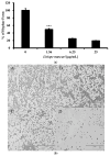
L. muscari inhibited the transition of C. albicans from yeast to hyphae. (a) L. muscari inhibited C. albicans filamentation induced in RPMI 1640. C. albicans (1 × 10⁶ cells/mL) and L. muscari (1.56, 6.25, and 25 μg/mL) were incubated at 37 °C for 4 h. (b) Images of C. albicans cells grown in RPMI 1640. L. muscari was added at concentrations of 1.56, 6.25, and 25 μg/mL. A: RPMI 1640 control, B: L. muscari 1.56 μg/mL, C: L. muscari 6.25 μg/mL, D: L. muscari 25 μg/mL. (c) L. muscari inhibited C. albicans filamentation induced in 10% FBS YPD medium. C. albicans (1 × 10⁶ cells/mL) and L. muscari (1.56, 6.25, and 25 μg/mL) were incubated at 37 °C for 4 h. (d) Images of C. albicans cells incubated in 10% FBS YPD medium. L. muscari was added at concentrations of 1.56, 6.25, and 25 μg/mL. A: 10% FBS YPD control, B: L. muscari 1.56 μg/mL, C: L. muscari 6.25 μg/mL, D: L. muscari 25 μg/mL. Scale bar: 200 μm. (***: p-value < 0.001).
Figure 3
Figure 3
L. muscari inhibited the transition of C. albicans from yeast to hyphae. (a) L. muscari inhibited C. albicans filamentation induced in RPMI 1640. C. albicans (1 × 10⁶ cells/mL) and L. muscari (1.56, 6.25, and 25 μg/mL) were incubated at 37 °C for 4 h. (b) Images of C. albicans cells grown in RPMI 1640. L. muscari was added at concentrations of 1.56, 6.25, and 25 μg/mL. A: RPMI 1640 control, B: L. muscari 1.56 μg/mL, C: L. muscari 6.25 μg/mL, D: L. muscari 25 μg/mL. (c) L. muscari inhibited C. albicans filamentation induced in 10% FBS YPD medium. C. albicans (1 × 10⁶ cells/mL) and L. muscari (1.56, 6.25, and 25 μg/mL) were incubated at 37 °C for 4 h. (d) Images of C. albicans cells incubated in 10% FBS YPD medium. L. muscari was added at concentrations of 1.56, 6.25, and 25 μg/mL. A: 10% FBS YPD control, B: L. muscari 1.56 μg/mL, C: L. muscari 6.25 μg/mL, D: L. muscari 25 μg/mL. Scale bar: 200 μm. (***: p-value < 0.001).
Figure 4
Figure 4
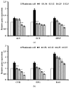
L. muscari decreased the expression of genes for hyphae-specific regulation, extracellular matrix, Ras1-cAMP-Efg1, MAP kinase, and Cph2-Tec1 in C. albicans. (a) Relative expression of genes regulating hyphae-specific genes. (b) Relative expression of genes regulated by the Ras1-cAMP-Efg1 pathway. (c) Relative expression of genes regulating extracellular matrix genes. (d) Relative expression of genes related to the Cph2-Tec1 pathway and the MAP kinase pathway. Total RNA was extracted from C. albicans, including control and biofilms treated with the indicated concentrations of L. muscari, synthesized cDNA, and examined by qPCR using the corresponding primers. (*: p-value <0.05, ***: p-value < 0.001).
Figure 4
Figure 4
L. muscari decreased the expression of genes for hyphae-specific regulation, extracellular matrix, Ras1-cAMP-Efg1, MAP kinase, and Cph2-Tec1 in C. albicans. (a) Relative expression of genes regulating hyphae-specific genes. (b) Relative expression of genes regulated by the Ras1-cAMP-Efg1 pathway. (c) Relative expression of genes regulating extracellular matrix genes. (d) Relative expression of genes related to the Cph2-Tec1 pathway and the MAP kinase pathway. Total RNA was extracted from C. albicans, including control and biofilms treated with the indicated concentrations of L. muscari, synthesized cDNA, and examined by qPCR using the corresponding primers. (*: p-value <0.05, ***: p-value < 0.001).
Figure 5
Figure 5
L. muscari lacks the ability to inhibit the growth of C. albicans.

Similar articles

References

    1. Atriwal T., Chawla M., Hussain A., Alajmi M.F., Abid M. Reactive oxygen mediated apoptosis as a therapeutic approach against opportunistic Candida albicans. Adv. Protein Chem. Struct. Biol. 2021;155:25–49. doi: 10.1016/bs.apcsb.2021.07.002. - DOI - PubMed
    1. Pfaller M.A., Diekema D.J. Epidemiology of invasive candidiasis: A persistent public health problem. Clin. Microbiol. Rev. 2007;20:133–163. doi: 10.1128/CMR.00029-06. - DOI - PMC - PubMed
    1. Chandra J., Kuhn D.M., Mukherjee P.K., Hoyer L.L., McCormick T., Ghannoum M.A. Biofilm formation by the fungal pathogen Candida albicans: Development, architecture, and drug resistance. J. Bacteriol. 2001;183:5385–5394. doi: 10.1128/JB.183.18.5385-5394.2001. - DOI - PMC - PubMed
    1. Benedict K., Jackson B.R., Chiller T., Beer K.D. Estimation of direct healthcare costs of fungal diseases in the United States. Clin. Infect. Dis. 2019;68:1791–1797. doi: 10.1093/cid/ciy776. - DOI - PMC - PubMed
    1. Koo H., Allan R.N., Howlin R.P., Stoodley P., Hall-Stoodley L. Targeting microbial biofilms: Current and prospective therapeutic strategies. Nat. Rev. Microbiol. 2017;15:740–755. doi: 10.1038/nrmicro.2017.99. - DOI - PMC - PubMed

LinkOut - more resources