

Evaluating the Margins of Breast Cancer Tumors by Using Digital Breast Tomosynthesis with Deep Learning: A Preliminary Assessment
- PMID: 38786329
- PMCID: PMC11119441
- DOI: 10.3390/diagnostics14101032
Evaluating the Margins of Breast Cancer Tumors by Using Digital Breast Tomosynthesis with Deep Learning: A Preliminary Assessment
Abstract
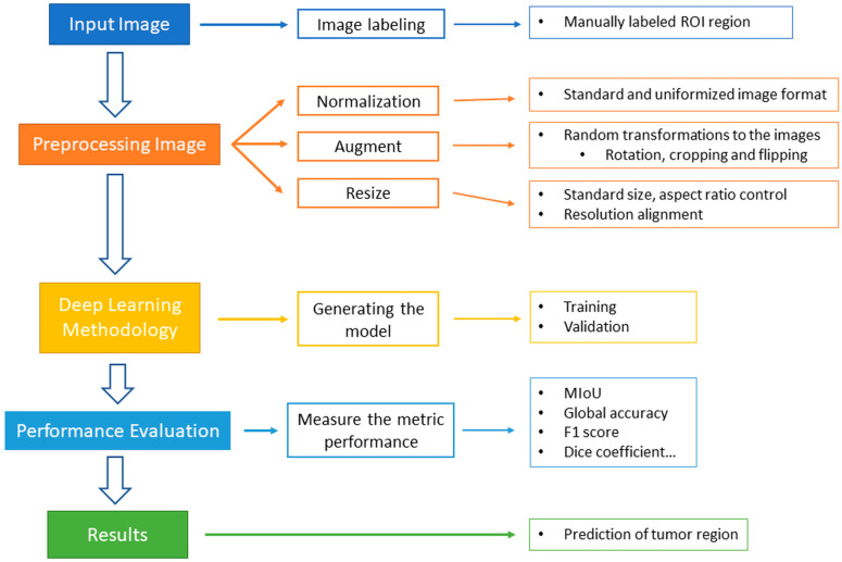
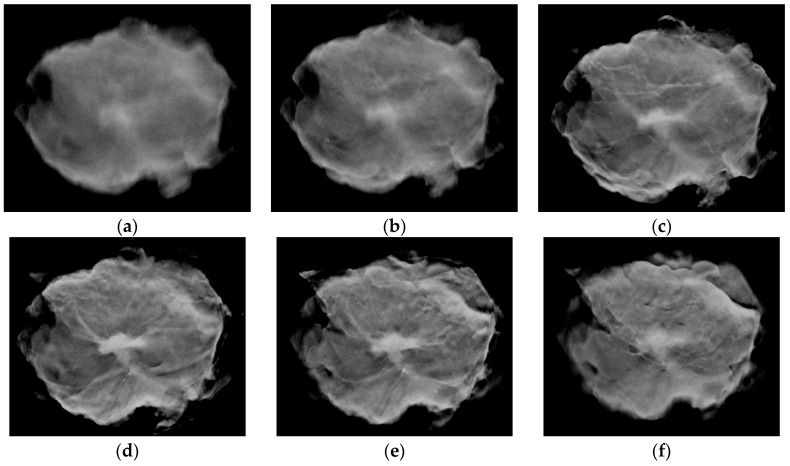
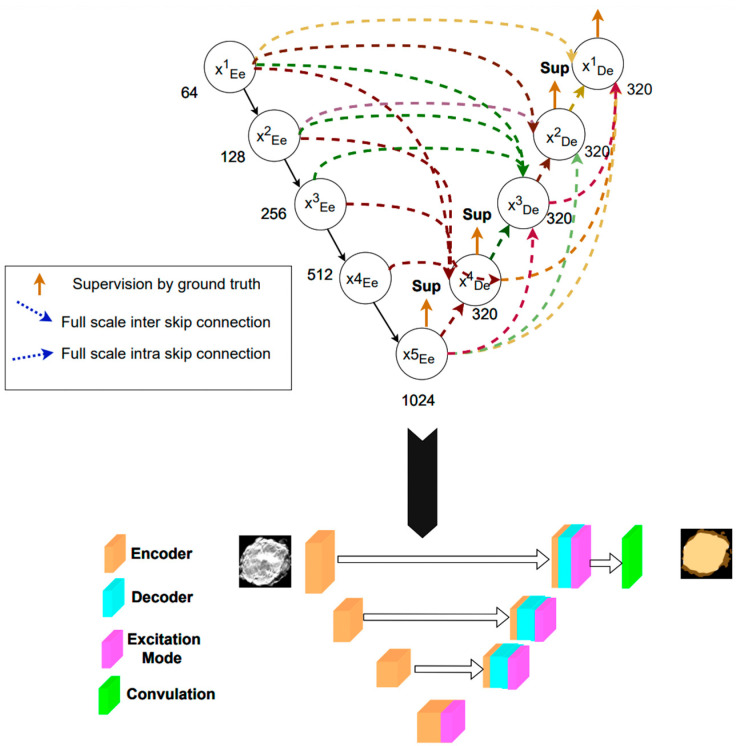
Background: The assessment information of tumor margins is extremely important for the success of the breast cancer surgery and whether the patient undergoes a second operation. However, conducting surgical margin assessments is a time-consuming task that requires pathology-related skills and equipment, and often cannot be provided in a timely manner. To address this challenge, digital breast tomosynthesis technology was utilized to generate detailed cross-sectional images of the breast tissue and integrate deep learning algorithms for image segmentation, achieving an assessment of tumor margins during surgery.
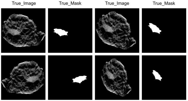
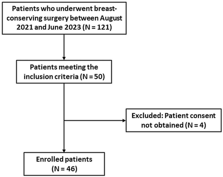
Methods: this study utilized post-operative tissue samples from 46 patients who underwent breast-conserving treatment, and generated image sets using digital breast tomosynthesis for the training and evaluation of deep learning models.
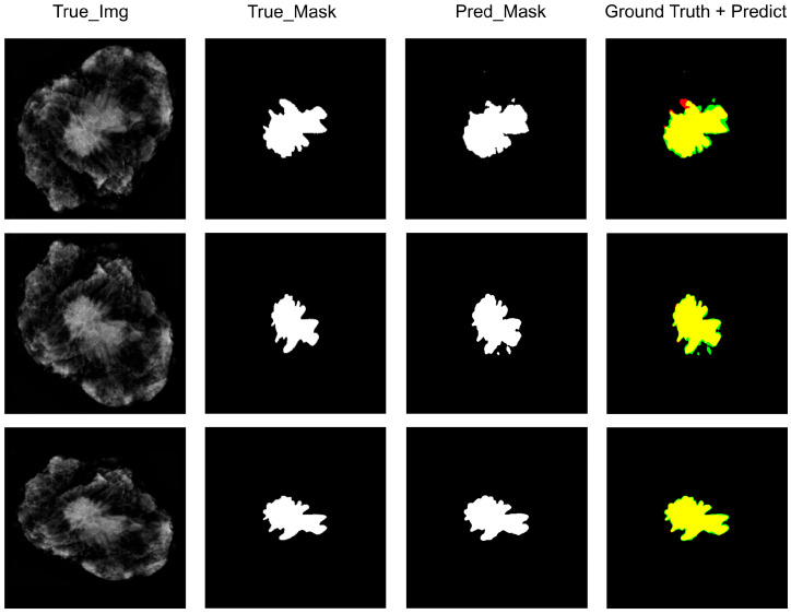
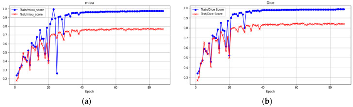
Results: Deep learning algorithms effectively identifying the tumor area. They achieved a Mean Intersection over Union (MIoU) of 0.91, global accuracy of 99%, weighted IoU of 44%, precision of 98%, recall of 83%, F1 score of 89%, and dice coefficient of 93% on the training dataset; for the testing dataset, MIoU was at 83%, global accuracy at 97%, weighted IoU at 38%, precision at 87%, recall rate at 69%, F1 score at 76%, dice coefficient at 86%.
Conclusions: The initial evaluation suggests that the deep learning-based image segmentation method is highly accurate in measuring breast tumor margins. This helps provide information related to tumor margins during surgery, and by using different datasets, this research method can also be applied to the surgical margin assessment of various types of tumors.
Keywords: breast cancer; cancer surgery; deep learning; surgical precision.
Conflict of interest statement
The authors declare no conflicts of interest.
Figures
Similar articles
-
Automated Tumor Segmentation in Breast-Conserving Surgery Using Deep Learning on Breast Tomosynthesis.J Imaging Inform Med. 2025 Mar 3. doi: 10.1007/s10278-025-01457-y. Online ahead of print. J Imaging Inform Med. 2025. PMID: 40032761
-
Diagnostic accuracy of resection margin in specimen radiography: digital breast tomosynthesis versus full-field digital mammography.Radiol Med. 2021 Jun;126(6):768-773. doi: 10.1007/s11547-021-01337-9. Epub 2021 Feb 24. Radiol Med. 2021. PMID: 33625658
-
A novel adaptive cubic quasi-Newton optimizer for deep learning based medical image analysis tasks, validated on detection of COVID-19 and segmentation for COVID-19 lung infection, liver tumor, and optic disc/cup.Med Phys. 2023 Mar;50(3):1528-1538. doi: 10.1002/mp.15969. Epub 2022 Oct 6. Med Phys. 2023. PMID: 36057788 Free PMC article.
-
Znet: Deep Learning Approach for 2D MRI Brain Tumor Segmentation.IEEE J Transl Eng Health Med. 2022 May 23;10:1800508. doi: 10.1109/JTEHM.2022.3176737. eCollection 2022. IEEE J Transl Eng Health Med. 2022. PMID: 35774412 Free PMC article.
-
Deep Learning Computer-Aided Diagnosis for Breast Lesion in Digital Mammogram.Adv Exp Med Biol. 2020;1213:59-72. doi: 10.1007/978-3-030-33128-3_4. Adv Exp Med Biol. 2020. PMID: 32030663 Review.
Cited by
-
Supporting intraoperative margin assessment using deep learning for automatic tumour segmentation in breast lumpectomy micro-PET-CT.NPJ Breast Cancer. 2025 Aug 9;11(1):88. doi: 10.1038/s41523-025-00797-w. NPJ Breast Cancer. 2025. PMID: 40783490 Free PMC article.
References
-
- Keating J.J., Fisher C., Batiste R., Singhal S. Advances in Intraoperative Margin Assessment for Breast Cancer. Curr. Surg. Rep. 2016;4:15. doi: 10.1007/s40137-016-0136-3. - DOI
Grants and funding
LinkOut - more resources
Full Text Sources
