Review
. 2024 May 20;9(5):306.
doi: 10.3390/biomimetics9050306.

Advancement in Cancer Vasculogenesis Modeling through 3D Bioprinting Technology

Affiliations
Review

Advancement in Cancer Vasculogenesis Modeling through 3D Bioprinting Technology

Arvind Kumar Shukla et al. Biomimetics (Basel). .

Abstract

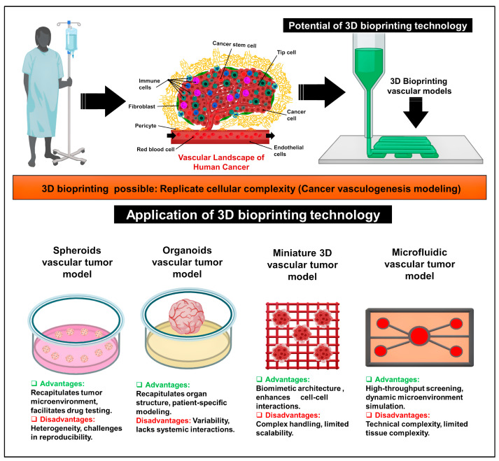
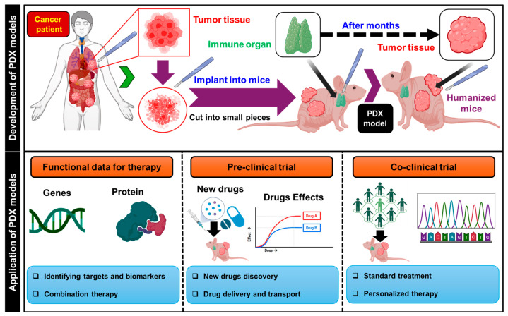
Cancer vasculogenesis is a pivotal focus of cancer research and treatment given its critical role in tumor development, metastasis, and the formation of vasculogenic microenvironments. Traditional approaches to investigating cancer vasculogenesis face significant challenges in accurately modeling intricate microenvironments. Recent advancements in three-dimensional (3D) bioprinting technology present promising solutions to these challenges. This review provides an overview of cancer vasculogenesis and underscores the importance of precise modeling. It juxtaposes traditional techniques with 3D bioprinting technologies, elucidating the advantages of the latter in developing cancer vasculogenesis models. Furthermore, it explores applications in pathological investigations, preclinical medication screening for personalized treatment and cancer diagnostics, and envisages future prospects for 3D bioprinted cancer vasculogenesis models. Despite notable advancements, current 3D bioprinting techniques for cancer vasculogenesis modeling have several limitations. Nonetheless, by overcoming these challenges and with technological advances, 3D bioprinting exhibits immense potential for revolutionizing the understanding of cancer vasculogenesis and augmenting treatment modalities.

Keywords: 3D bioprinting technology; cancer; cancer microenvironment; cancer modeling; vasculogenesis.


Conflict of interest statement

The authors declare no conflicts of interest.

Figures

Figure 1
Figure 1
Application of 3D bioprinting technology for cancer vascular models. These technologies include a spheroid vascular tumor model, organoid vascular tumor model, miniature 3D vascular tumor model, and microfluidic vascular tumor model.
Figure 2
Figure 2
Schematic showing the development (upper panel) and application (lower panel) of patient-derived xenograft (PDX) models. These models were established by transplanting tumor tissue derived from patients into immunodeficient or humanized immune-deficient mice. Subsequently, they were utilized to explore the functionality of cancer vasculogenesis and to identify optimal therapeutic strategies.
Figure 3
Figure 3
(A) The experimental setup involved establishing the SKOV3 cell-derived xenograft (CDX) model in nude mice. HER2-CAR-T cells were infused into the CDX model via the tail vein, alongside GFP-T cells. (B) (a) The results of the tumor growth curve in different experimental groups. (b) Kaplan–Meier survival analysis: the combination of HER-CAR-T cells with the CA4P group had a longer survival time compared to those in the other groups. ** p < 0.01, *** p < 0.001. ns indicates not significant. (c) The optical densitometric statistical analysis of the individual bands between the groups. (C) The effect of combination therapy on blood vessels and HER2 expression based on immunohistochemical analysis. Reproduced/adapted with permission [96].
Figure 4
Figure 4
Different types of 3D bioprinter according to methods depositing bioinks: (a) extrusion-based bioprinter (EBB), (b) droplet-based, and (c) laser-based bioprinter. Reproduced/adapted with permission [109].
Figure 5
Figure 5
(A) Three-dimensional bioprinted breast cancer–vessel–bone model for drug testing. (a) Schematic of one-step 3D bioprinting for modeling the metastasis process and drug testing. (b) Computer-aided design and photograph of the co-culture model. (B) (a) Live/dead staining of cell-laden scaffolds cultured for 1 d and 7 d, along with immunostaining of relevant markers after 14 d of culture. (b) Cell viability within the co-culture model post-bioprinting. (C) Vascularization within the co-culture model. (a) Vascular channel morphology, distribution of green fluorescent protein (GFP), human umbilical vein endothelial cells (HUVECs), and immunofluorescence images [136]. The red circle indicates the internal cavity and the white dashed box outlines the entire vessel region. (b) The growth of GFP-HUVECs over 14 d. White dashed lines indicate the boundaries of the vessels, and white arrows point to vessels infiltrating the interior of the tumor and bone chambers. (c) Immunofluorescence images and mean fluorescence intensity (MFI) of CD31 after 14 d of co-culture. * p < 0.05. Reproduced/adapted with permission [136].
Figure 6
Figure 6
(A) Advances in 3D bioprinting for personalized pathological studies. (a) Schematic of a 3D bioprinted tissue cultured on transwells. (b) H&E and Masson’s trichrome images of bioprinted tumor tissues with MCF-7 and HUVECs in the cancer compartment and HMFs and HUVECs in the stromal compartment, cultured for 7 or 10 d. Each histological image on the right is a magnified view of the dashed box in the corresponding image. (c) Immunostaining results showing endothelial network in the engineered tissue. The white dashed boxes were magnified and shown on the right side of the respective results. KRT8/18: keratin 8/18, VIM: vimentin. Reproduced/adapted with permission [144]. (B) The development of glioblastoma (GBM)-on-a-chip through 3D bioprinting for personalized pathological studies. (a) Photographs showing a GBM-on-a-chip model with GBM cells (blue) or HUVECs (magenta) printed on gas-permeable silicone on a glass slide covered by a glass slip. (b) Compartmental subdivision into core, intermediate, and peripheral areas with GBM cells and an external layer with HUVECs. (c) Oxygen level simulation performed within the hydrogel. (d) Immunostaining images of the core, intermediate, and peripheral zones using pimonidazole for hypoxic cells, Ki67 for oxygenated proliferating cells, and DAPI for cell nuclei. Scale bar: 200 μm. PM: pimonidazole. Reproduced/adapted with permission [43].
Figure 7
Figure 7
Schematic showing the design of a preclinical drug screening platform for personalized medicine by 3D bioprinting technology.
Figure 8
Figure 8
(A) Fabrication of liver lobule-like constructs. (a) Schematic depicting liver lobule-like structures produced via GelMA hydrogel beads by the dot extrusion printing system. (b) Images display layered lobule-like structures. Scale: 1 mm. (c) Live/dead analysis of C3A cells pre- and post-printing. Scale: 1 mm. (B) Endothelialized liver lobule-like constructs: (a) Schematic outlining four-step printing of endothelialized liver lobule-like constructs. (b) Bright-field and F-actin fluorescent images exhibit constructs on days 1, 7, and 14. Scale: 2 mm. Live/dead analysis of constructs with varied GelMA concentrations over 14 d. Scale: 200 µm. (C) In vitro evaluation: (a) F-actin staining reveals cell morphology at days 1 and 14. The white dashed boxes were magnified and shown on the right side of the respective results. Scale: 200 µm. (b) Cell viability at days 1, 7, and 14. (c) Cell diameter distributions after the 14-day cultivation with different GelMA concentrations. (D) Drug evaluation: (a) Live/dead images illustrate 3D liver cancer models post-sorafenib incubation. Scale: 200 µm. (b) Statistical analysis of cell viability in both constructs at varied drug concentrations. * p < 0.05, ** p < 0.01. ns indicates not significant. Reproduced/adapted with permission [156].
Figure 9
Figure 9
Schematic of a microfluidic biosensor utilizing Cas13 for cancer diagnosis. Blood samples from non-small-cell lung carcinoma (NSCLC) patients are directly inputted into the device. Nucleic acids are isolated and directed into wells containing reagents for detecting DNA mutations or quantifying overexpressed NSCLC-associated RNAs. Cas13 is activated by targeting RNAs, leading to the cleavage of target RNAs and fluorescent reporter RNAs. Tumor DNAs are amplified using recombinase polymerase amplification, followed by transcription with T7. Cas13 binds to mutation-containing transcripts, cleaving fluorescent reporter RNAs for signal detection.
Figure 10
Figure 10
(A) (a) Portable heater module with a USB microscope and heat platform for a 3D-printed array facilitates loop-mediated isothermal amplification (LAMP). (b) Assembly schematic illustrating the multifunctional, leak-proof bonding of 3D printed reactor array for nucleic acid amplification tests (NAATs). (c) Representative photos showing a colorimetric LAMP assay detecting N. meningitides at different colony-forming units (CFU) per reaction. (B) (a) LAMP curves for P. falciparum exhibiting sensitivity from 0.1 to 1000 pg per reaction. (b) Calibration curve showing the relationship between log target concentration and amplification for P. falciparum. (c) LAMP curves for N. meningitidis ranging from 50 to 5000 CFU per reaction. (d) Calibration curve for N. meningitidis correlates log target concentration to amplification. WarmStart® LAMP master mix was utilized. (C) (a,b) LAMP curves and calibration show quantitative detection of P. falciparum gDNA in plasma samples at spiked concentrations. (c,d) LAMP curves and calibration for quantitative detection of N. meningitidis bacteria in cerebrospinal fluid samples at varying concentrations. WarmStart® LAMP master mix was employed. Reproduced/adapted with permission [164].

Similar articles

References

    1. Baiguera S., Ribatti D. Ribatti, Endothelialization approaches for viable engineered tissues. Angiogenesis. 2013;16:1–14. doi: 10.1007/s10456-012-9307-8. - DOI - PubMed
    1. Yin S., Zhang W., Zhang Z., Jiang X. Recent advances in scaffold design and material for vascularized tissue-engineered bone regeneration. Adv. Healthc. Mater. 2019;8:1801433. doi: 10.1002/adhm.201801433. - DOI - PubMed
    1. Hantusch B. Fundamentals of Vascular Biology. Springer; Berlin/Heidelberg, Germany: 2019. Morphological and functional characteristics of blood and lymphatic vessels; pp. 1–43.
    1. Fleischer S., Tavakol D.N., Vunjak-Novakovic G. From arteries to capillaries: Approaches to engineering human vasculature. Adv. Funct. Mater. 2020;30:1910811. doi: 10.1002/adfm.201910811. - DOI - PMC - PubMed
    1. Popel A.S. Theory of oxygen transport to tissue. Crit. Rev. Biomed. Eng. 1989;17:257. - PMC - PubMed

LinkOut - more resources