

Quantity and Size of Titanium Particles Released from Different Mechanical Decontamination Procedures on Titanium Discs: An In Vitro Study
- PMID: 38786521
- PMCID: PMC11119952
- DOI: 10.3390/dj12050123
Quantity and Size of Titanium Particles Released from Different Mechanical Decontamination Procedures on Titanium Discs: An In Vitro Study
Abstract
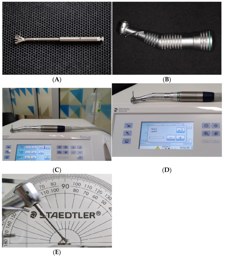
Complications such as peri-implantitis could ultimately affect the survival of a dental implant. The prevention and treatment of peri-implant diseases require managing bacterial biofilm and controlling environmental risks, including the presence of pro-inflammatory titanium (Ti) particles in the peri-implant niche. Objectives included the evaluation of the size and quantity of Ti particles released from moderately roughened Ti surfaces during common mechanical surface decontamination methods. One hundred and forty moderately roughened Ti discs were divided into seven groups (n = 20 per group); six groups received mechanical decontamination procedures (ultrasonic scaling (US) with a metal tip and poly-ether-ketone (PEEK) under low and medium power settings, air-polishing with erythritol powder, and Ti brush), and the control group underwent air-water spray using a dental triplex. The rinsing solution was collected for Ti mass analysis using inductively coupled plasma mass spectrometry (ICPMS), as well as for Ti particle size and count analysis under scanning electron microscopy (SEM) with energy-dispersive spectroscopy (EDS). US metal tip instrumentation generated 34.00 ± 12.54 μg and 34.44 ± 6.08 μg of Ti under low and medium power settings, respectively. This amount of Ti generation was significantly higher than other instrumentation methods. The mean Ti particle size of the US groups ranged from 0.89 ± 0.27 μm to 1.25 ± 0.24 μm. No statistically significant difference was found in the particle size among US groups and Ti brush group (1.05 ± 0.11 μm), except for US with the PEEK tip, where a significantly smaller mean particle diameter was found at the low power setting (0.89 ± 0.27 μm). Mechanical instrumentation can produce Ti particulates and modify the implant surfaces. US using a metal tip generated the highest amount of Ti with smaller Ti size particles compared to all other commonly used mechanical surface instrumentations. The EDS analysis confirmed Ti in PEEK US tips. It can be suggested that deterioration from the PEEK US tip and Ti brush, as observed under SEM, is an additional source of Ti release during Ti surface decontamination.
Keywords: dental implants; inductively coupled plasma mass spectrometry; instrumentation; peri-implantitis; scanning electron microscopy; titanium.
Conflict of interest statement
The authors declare no conflict of interest.
Figures


Similar articles
-
Effects of Mechanical Methods Used in Peri-implantitis Treatment on Implant Surface Decontamination and Roughness.J Vis Exp. 2025 Mar 14;(217). doi: 10.3791/67778. J Vis Exp. 2025. PMID: 40163409
-
The effect of piezoelectric ultrasonic instrumentation on titanium discs: a microscopy and trace elemental analysis in vitro study.Int J Dent Hyg. 2016 Aug;14(3):191-201. doi: 10.1111/idh.12142. Epub 2015 Jun 11. Int J Dent Hyg. 2016. PMID: 26094557
-
In Vitro Comparison of Titanium Disc Surface Roughness and Bacterial Colonization After Ultrasonic Instrumentation With Three Different Tips.J Oral Implantol. 2024 Oct 1;50(5):537-543. doi: 10.1563/aaid-joi-D-24-00049. J Oral Implantol. 2024. PMID: 39023858
-
Effect of implant cleaning on titanium particle dissolution and cytocompatibility.J Periodontol. 2021 Apr;92(4):580-591. doi: 10.1002/JPER.20-0186. Epub 2020 Sep 11. J Periodontol. 2021. PMID: 32846000
-
Decontamination of titanium implant surface and re-osseointegration to treat peri-implantitis: a literature review.Int J Oral Maxillofac Implants. 2012 Sep-Oct;27(5):1043-54. Int J Oral Maxillofac Implants. 2012. PMID: 23057016 Review.
References
-
- Berglundh T., Armitage G., Araujo M.G., Avila-Ortiz G., Blanco J., Camargo P.M., Chen S., Cochran D., Derks J., Figuero E., et al. Peri-implant diseases and conditions: Consensus report of workgroup 4 of the 2017 World Workshop on the Classification of Periodontal and Peri-Implant Diseases and Conditions. J. Periodontol. 2018;89:S313–S318. doi: 10.1002/jper.17-0739. - DOI - PubMed
Grants and funding
LinkOut - more resources
Full Text Sources
