. 2024 Apr 23;22(5):190.
doi: 10.3390/md22050190.

Inhibition Effects and Mechanisms of Marine Compound Mycophenolic Acid Methyl Ester against Influenza A Virus

Affiliations

Inhibition Effects and Mechanisms of Marine Compound Mycophenolic Acid Methyl Ester against Influenza A Virus

Zihan Wang et al. Mar Drugs. .

Abstract

Influenza A virus (IAV) can cause infection and illness in a wide range of animals, including humans, poultry, and swine, and cause annual epidemics, resulting in thousands of deaths and millions of hospitalizations all over the world. Thus, there is an urgent need to develop novel anti-IAV drugs with high efficiency and low toxicity. In this study, the anti-IAV activity of a marine-derived compound mycophenolic acid methyl ester (MAE) was intensively investigated both in vitro and in vivo. The results showed that MAE inhibited the replication of different influenza A virus strains in vitro with low cytotoxicity. MAE can mainly block some steps of IAV infection post adsorption. MAE may also inhibit viral replication through activating the cellular Akt-mTOR-S6K pathway. Importantly, oral treatment of MAE can significantly ameliorate pneumonia symptoms and reduce pulmonary viral titers, as well as improving the survival rate of mice, and this was superior to the effect of oseltamivir. In summary, the marine compound MAE possesses anti-IAV effects both in vitro and in vivo, which merits further studies for its development into a novel anti-IAV drug in the future.

Keywords: Akt-mTOR-S6K pathway; MAE; anti-viral effect; influenza A virus; viral pneumonia.


Conflict of interest statement

The authors declare no conflict of interest.

Figures

Figure 1
Figure 1
Inhibition effects of MAE against IAV infection in vitro. (a). Structure of MAE. (bd) The cytotoxicity of MAE in MDCK, Vero and A549 cells. Values are means ± S.D. (n = 3). (eg) Anti-IAV (PR8, Aichi, Virginia09; MOI = 0.1) activity of MAE (2.5–80 μM) was determined by CPE inhibition assay at 24 h p.i. Values are means ± S.D. (n = 3). (h) The inhibition of different concentrations of MAE (2.5–40 μM) on IAV multiplication was evaluated by HA assay. Values are means ± S.D. (n = 3).
Figure 2
Figure 2
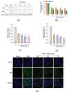
Inhibition of MAE on the protein and mRNA expression of IAV. (a,b) The inhibition of MAE on IAV multiplication was also evaluated by Western blot assay of virus NP and NS1 proteins in MDCK cells (a). Quantification of immunoblot for the ratio of NP or NS1 protein to β-actin was also shown (b). Values are means ± S.D. (n = 3). ** p < 0.01 vs. virus control group (PR8). (c,d) Quantitative RT-PCR assay of virus M1 (c) or NP (d) mRNA in MAE-treated cells was performed. Values are means ± S.D. (n = 3). ** p < 0.01 vs. virus control group (PR8). (e) PR8 (MOI = 1.0)-infected MDCK cells were treated with or without MAE after virus adsorption and then incubated at 37 °C for 4 h. After that, NP protein expression was determined by immunofluorescence assay. Scale bar represents 25 μm.
Figure 3
Figure 3
Influence of different treatment conditions of MAE on IAV infection. (a) The schematic diagram of different treatment conditions. (b) MDCK cells were infected with PR8 virus (MOI = 1.0) under four treatment conditions of MAE (20 μM) and the antiviral activity was determined by HA assay at 24 h p.i. Values are means ± S.D. (n = 3). * p < 0.05, ** p < 0.01 vs. PR8 group. (c,d) Effects of the MAE on HA protein expression under different treatment conditions. Quantification of immunoblot for the ratio of HA to β-actin was also shown (d). Values are means ± S.D. (n = 3). * p < 0.05, ** p < 0.01 vs. PR8 group. (e,f) PR8 (MOI = 1.0)-infected MDCK cells were treated with MAE (20 μM) for different time intervals, after which (at 8 h p.i.) the virus yields were determined via Western blot assay of virus HA protein (e). Quantification of immunoblot for the ratio of HA to β-actin was also shown (f). Values are means ± S.D. (n = 3). ** p < 0.01 vs. virus control group (PR8).
Figure 4
Figure 4
Enhancement of Akt-mTOR-S6K pathway by MAE in IAV-infected cells. (a) The inhibition effects of MAE and anti-HA antibody on PR8 virus-induced aggregation of chicken erythrocytes were evaluated by hemagglutination inhibition (HI) assay. (b) Inactivated PR8 virus was incubated with indicated concentrations of MAE or Zanamivir (20 μM), and the NA activity was determined by a fluorescent assay. Values are means ± S.D. (n = 3). ** p < 0.01 vs. Control group. (c) PR8 (MOI = 1.0) infected A549 cells were treated with or without MAE at indicated concentrations after removal of virus inoculums. At 5 h p.i., PB1 and p-mTOR protein were determined by Western blotting. (d,e) Quantification of immunoblot for the ratio of PB1 or p-mTOR to β-actin. Values are means ± S.D. (n = 3). ** p < 0.01 vs. PR8 group. (f) PR8 (MOI = 1.0) infected A549 cells were treated with or without MAE at indicated concentrations after removal of virus inoculums. At 5 h p.i., p-Akt and p-S6K protein were determined by Western blotting. (g,h) Quantification of immunoblot for the ratio of p-Akt or p-S6K to β-actin. Values are means ± S.D. (n = 3). * p < 0.05, ** p < 0.01 vs. PR8 group. (ik) PR8 (MOI = 1.0) infected A549 cells were treated with or without MAE after removal of virus inoculums. At 5 h p.i., p-ERK1/2 and p-NF-κB protein were determined by Western blotting (i). Quantification of immunoblot for the ratio of p-ERK1/2 and p-NF-κB to β-actin (j,k). Values are means ± S.D. (n = 3).
Figure 5
Figure 5
The anti-IAV activities of MAE in vivo. (a) Schematic diagram of IAV mouse pneumonia model. (b) Survival rate. IAV-infected mice received oral therapy with MAE or other drugs for five days. Results are expressed as the percentage of survival, evaluated daily for 14 days. (c) The average body weights were monitored daily for 14 days and are expressed as a percentage of the initial value. Values are means ± S.D. (n = 10). (d) The pulmonary viral titers were evaluated by NA titer assay. Values are means ± S.D. (n = 4). * p < 0.05, ** p < 0.01 vs. PR8 group. (e) Histopathologic analyses of lung tissues on day 3 p.i. by hematoxylin-eosin (H&E) staining (×100).

Similar articles

Cited by

References

    1. Cheng J., Tao J., Li B., Shi Y., Liu H. The lncRNA HCG4 regulates the RIG-I-mediated IFN production to suppress H1N1 swine influenza virus replication. Front. Microbiol. 2024;14:1324218. doi: 10.3389/fmicb.2023.1324218. - DOI - PMC - PubMed
    1. Hussain M., Galvin H.D., Haw T.Y., Nutsford A.N., Husain M. Drug resistance in influenza A virus: The epidemiology and management. Infect. Drug Resist. 2017;20:121–134. doi: 10.2147/IDR.S105473. - DOI - PMC - PubMed
    1. Pleschka S. Overview of influenza viruses. Curr. Top. Microbiol. Immunol. 2013;370:1–20. - PubMed
    1. Hay A.J., Wolstenholme A.J., Skehel J.J., Smith M.H. The molecular basis of the specific anti-influenza action of amantadine. EMBO J. 1985;4:3021–3024. doi: 10.1002/j.1460-2075.1985.tb04038.x. - DOI - PMC - PubMed
    1. Hurt A.C., Ho H.T., Barr I. Resistance to anti-influenza drugs: Adamantanes and neuraminidase inhibitors. Expert. Rev. Anti Infect. Ther. 2006;4:795–805. doi: 10.1586/14787210.4.5.795. - DOI - PubMed

LinkOut - more resources