. 2024 May 15;22(5):221.
doi: 10.3390/md22050221.

Antiviral Shrimp lncRNA06 Possesses Anti-Tumor Activity by Inducing Apoptosis of Human Gastric Cancer Stem Cells in a Cross-Species Manner

Affiliations

Antiviral Shrimp lncRNA06 Possesses Anti-Tumor Activity by Inducing Apoptosis of Human Gastric Cancer Stem Cells in a Cross-Species Manner

Ahmad Khan et al. Mar Drugs. .

Abstract

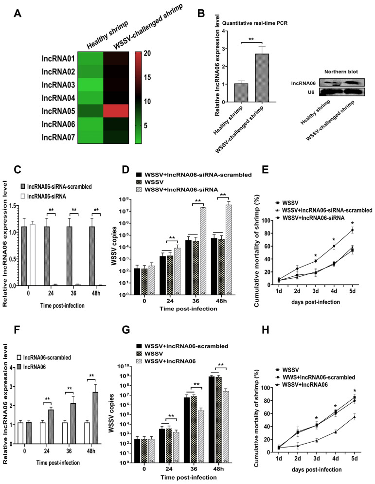
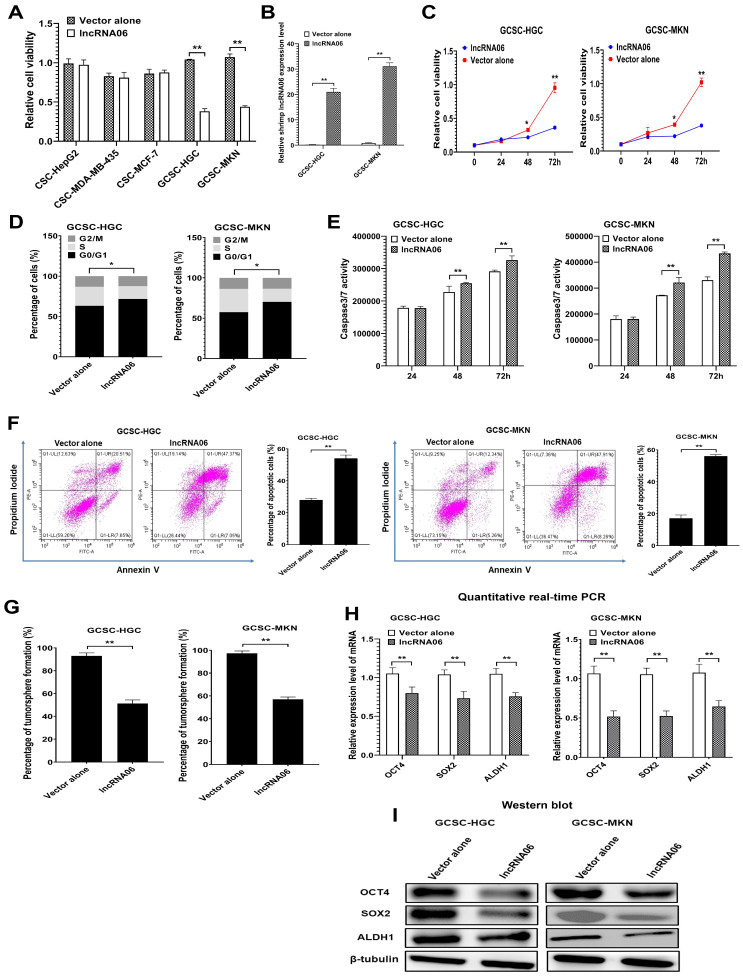
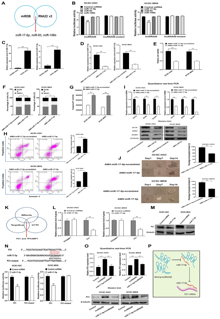
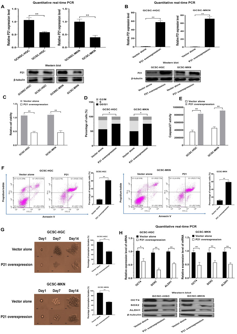
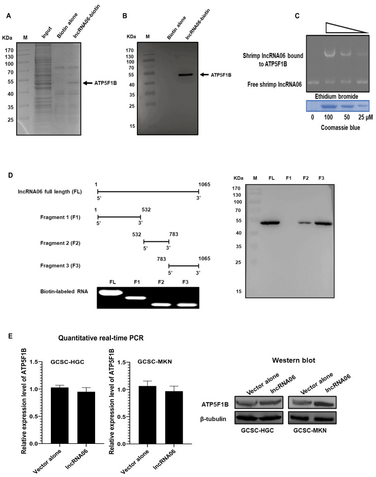
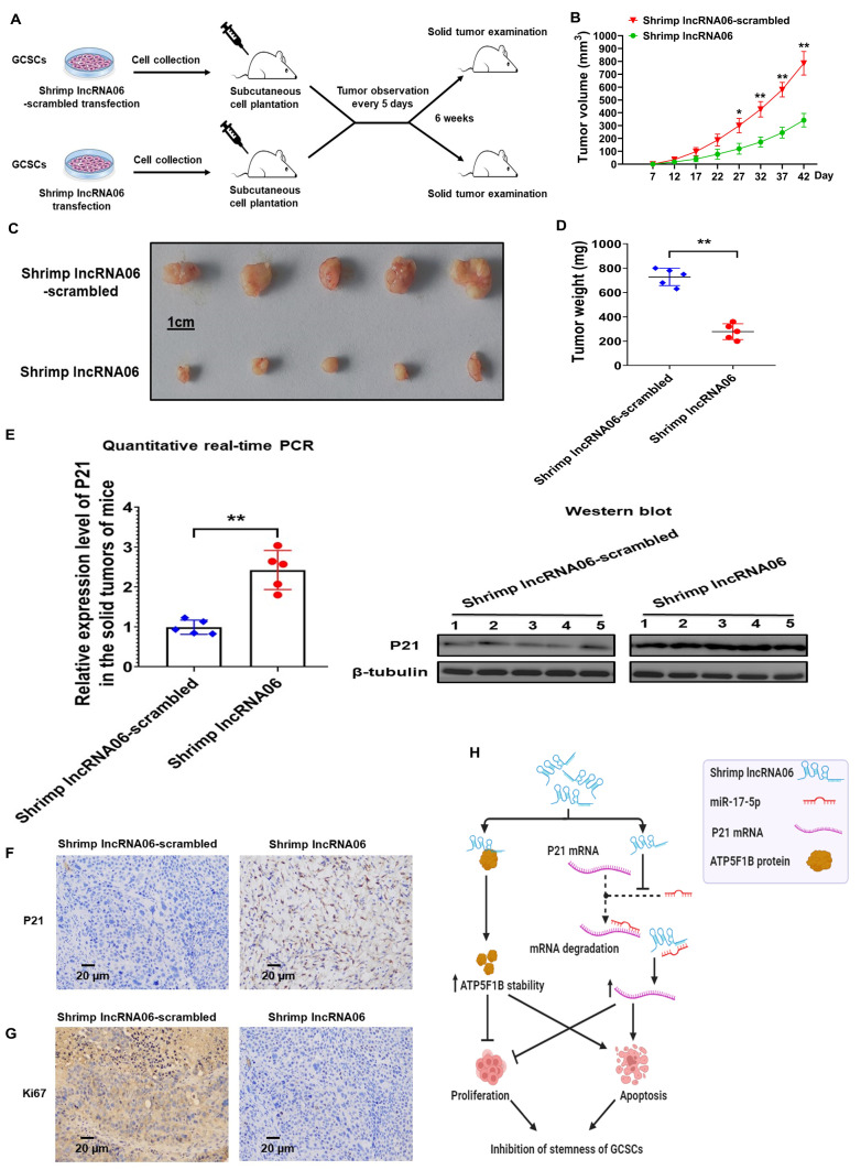
Virus infection causes the metabolic disorder of host cells, whereas the metabolic disorder of cells is one of the major causes of tumorigenesis, suggesting that antiviral molecules might possess anti-tumor activities by regulating cell metabolism. As the key regulators of gene expression, long non-coding RNAs (lncRNAs) play vital roles in the regulation of cell metabolism. However, the influence of antiviral lncRNAs on tumorigenesis has not been explored. To address this issue, the antiviral and anti-tumor capacities of shrimp lncRNAs were characterized in this study. The results revealed that shrimp lncRNA06, having antiviral activity in shrimp, could suppress the tumorigenesis of human gastric cancer stem cells (GCSCs) via triggering apoptosis of GCSCs in a cross-species manner. Shrimp lncRNA06 could sponge human miR-17-5p to suppress the stemness of GCSCs via the miR-17-5p-p21 axis. At the same time, shrimp lncRNA06 could bind to ATP synthase subunit beta (ATP5F1B) to enhance the stability of the ATP5F1B protein in GCSCs, thus suppressing the tumorigenesis of GCSCs. The in vivo data demonstrated that shrimp lncRNA06 promoted apoptosis and inhibited the stemness of GCSCs through interactions with ATP5F1B and miR-17-5p, leading to the suppression of the tumorigenesis of GCSCs. Therefore, our findings highlighted that antiviral lncRNAs possessed anti-tumor capacities and that antiviral lncRNAs could be the anti-tumor reservoir for the treatment of human cancers.

Keywords: antiviral lncRNA; cross-species regulation; human gastric cancer stem cells; shrimp; tumorigenesis.


Conflict of interest statement

The authors declare no competing interests.

Figures

Figure 1
Figure 1
Antiviral activity of shrimp lncRNA06. (A) The upregulated lncRNAs in the WSSV-challenged shrimp. Shrimp were infected with WSSV. At 48 h post-infection, shrimp hemocytes were collected and subjected to lncRNA sequencing. The heatmap showed the upregulated lncRNAs in the WSSV-challenged shrimp. (B) Upregulation of lncRNA06 in virus-infected shrimp. At 48 h post-infection, the expression level of lncRNA06 in shrimp hemocytes was determined using quantitative real-time PCR (**, p < 0.01) and Northern blot. U6 was used as a control. (C) Silencing of lncRNA06 in shrimp. Shrimp were injected with lncRNA06-siRNA or lncRNA06-siRNA-scrambled. At different times after injection, shrimp hemocytes were collected to examine the lncRNA06 expression using quantitative real-time PCR (**, p < 0.01). U6 was used as a control. (D) Effects of lncRNA06 silencing on virus infection. The synthesized lncRNA06-siRNA and WSSV were co-injected into shrimp. At different time points post-infection, WSSV copies in hemocytes were detected (**, p < 0.01). (E) Influence of lncRNA06 silencing on shrimp mortality. The cumulative mortality of shrimp was monitored at different times (*, p < 0.05). (F) Overexpression of lncRNA06 in shrimp. The synthesized lncRNA06 was injected into shrimp. At different times after injection, the expression level of lncRNA06 was examined with quantitative real-time PCR. U6 was used as a control (**, p < 0.01). (G) Impact of lncRNA06 overexpression on WSSV infection. The WSSV copies of the virus-challenged shrimp overexpressing lncRNA06 were determined (**, p < 0.01). (H) Influence of lncRNA06 overexpression on shrimp mortality. The cumulative mortality of shrimp was examined every day (*, p < 0.05).
Figure 2
Figure 2
Effects of shrimp lncRNA06 on human gastric cancer stem cells. (A) Influence of shrimp lncRNA06 on the viability of cancer stem cells. Cancer stem cells were transfected with shrimp lncRNA06. The vector alone was used as a control (control). Forty-eight hours later, the cell viability was assessed (**, p < 0.01). (B) Expression of shrimp lncRNA06 in GCSCs. At 48 h after transfection, the expression level of lncRNA06 in GCSCs was analyzed by quantitative real-time PCR (**, p < 0.01). (C) Impact of shrimp lncRNA06 on the viability of GCSCs. At different times after transfection, the cell viability of GCSCs was examined (*, p < 0.05; **, p < 0.01). (D) Role of shrimp lncRNA06 in the cell cycle. At 48 h after transfection, the cell cycle was examined using flow cytometry (*, p < 0.05). (E) Detection of caspase 3/7 activity. The caspase 3/7 activity of GCSCs transfected with shrimp lncRNA06 or vector alone was determined (**, p < 0.01). (F) Detection of apoptosis using the annexin V assay. GCSCs were examined using flow cytometry at 48 h after transfection (**, p < 0.01). (G) Influence of shrimp lncRNA06 on the tumorsphere formation capacity of GCSCs. The lncRNA06-transfected GCSCs were subjected to tumorsphere formation assays. Seven days later, the percentage of tumorsphere formation was evaluated (**, p < 0.01). (H) Effects of shrimp lncRNA06 on the expression of stemness genes in GCSCs. The expression profiles of stemness genes in GCSCs were examined at 48 h after transfection (**, p < 0.01). (I) Western blot analysis of stemness genes in GCSCs. β-tubulin was used as a control.
Figure 3
Figure 3
The underlying mechanism of shrimp lncRNA06 in GCSCs. (A) Prediction of miRNAs interacting with shrimp lncRNA06. The prediction was performed, and the overlapped miRNAs were the potential targets of lncRNA06. (B) Direct interaction between miRNAs and shrimp lncRNA06. GCSCs were co-transfected with shrimp lncRNA06 and a miRNA. Forty-eight hours later, the firefly and renilla luciferase activities of the cells were examined (**, p < 0.01). (C) Expression of miR-17-5p in GCSCs and GCNSCs. The expression level of miR-17-5p was examined (**, p < 0.01). (D) Knockdown of miR-17-5p in GCSCs. GCSCs were transfected with AMO-miR-17-5p or AMO-miR-17-5p-scrambled. At 48 h after transfection, the expression of miR-17-5p was determined (**, p < 0.01). (E) Impact of miR-17-5p silencing on cell viability. At 48 h after transfection, the cell viability of AMO-transfected GCSCs was determined (**, p < 0.01). (F) Effects of miR-17-5p silencing on the cell cycle. At 48 h after transfection, the cell cycle of GCSCs transfected with AMO was examined using flow cytometry (*, p < 0.05). (G) Effects of miR-17-5p downregulation on the apoptosis of GCSCs. GCSCs transfected with AMO were subjected to caspase 3/7 activity detection at 48 h after transfection (**, p < 0.01). (H) Detection of apoptosis using annexin V assays. At 48 h after transfection, apoptosis of GCSCs was examined using annexin V assay (**, p < 0.01). (I) Influence of miR-17-5p silencing on the expression of stemness genes in GCSCs. At 48 h after transfection of AMO, the cells were subjected to quantitative real-time PCR (**, p < 0.01) and Western blot to examine the expression of stemness genes. (J) Influence of miR-17-5p silencing on the ability of tumorsphere formation in GCSCs. Tumorsphere formation of a single cell was shown on the left. The percentage of tumorsphere formation was examined at day 14 after transfection (**, p < 0.01). Scale bar: 10 μm. (K) The potential genes targeted by miR-17-5p. The overlapped genes were the potential targets of miR-17-5p. (L) Impact of miR-17-5p overexpression on the expression of target genes. At 48 h after transfection of miRNA, the gene expression was examined using quantitative real-time PCR (**, p < 0.01). (M) Western blot analysis of p21 in the miR-17-5p-overexpressed GCSCs. β-tubulin was used as a control. (N) Direct interaction between miR-17-5p and p21. GCSCs were co-transfected with miR-17-5p or control miRNA and p21 or p21-mutant. Forty-eight hours later, the firefly and renilla luciferase activities were examined (**, p < 0.01). (O) Influence of shrimp lncRNA06 on the expression of p21 in GCSCs. At 48 h after transfection, the expression level of p21 was determined using quantitative real-time PCR (**, p < 0.01) and Western blot. The cells without any treatment were used as controls. (P) Model for the underlying mechanism of shrimp lncRNA06 in GCSCs.
Figure 4
Figure 4
Role of p21 in GCSCs. (A) Differential expression of p21 in GCSCs and GCNSCs. The expression level of p21 was examined using quantitative real-time PCR (**, p < 0.01) and Western blot. β-tubulin was used as a control. (B) Overexpression of p21 in GCSCs. GCSCs were transfected with the recombinant plasmid expressing p21. At 48 h after transfection, the cells were subjected to quantitative real-time PCR (**, p < 0.01) and Western blot. (C) Influence of p21 overexpression on cell viability. GCSCs overexpressing p21 were subjected to cell viability assays at 48 h after transfection (**, p < 0.01). (D) Effects of p21 overexpression on the cell cycle. The cell cycle of p21-overexpressing GCSCs was examined at 48 h after transfection (*, p < 0.05). (E) Impact of p21 overexpression on apoptosis of GCSCs. At 48 h after transfection, the caspase 3/7 activity of GCSCs overexpressing p21 was determined (**, p < 0.01). (F) Detection of apoptosis using the annexin V assay. GCSCs overexpressing p21 were subjected to annexin V assays at 48 h after treatment (**, p < 0.01). (G) Influence of p21 overexpression on the ability of tumorsphere formation in GCSCs. GCSCs overexpressing p21 were subjected to tumorsphere-forming assays. Fourteen days later, the percentage of tumorsphere formation was evaluated. Scale bar: 10 μm (**, p < 0.01). (H) Expression of stemness genes in p21-overexpressed GCSCs. The expression of stemness genes in GCSCs overexpressing p21 were examined at 48 h after transfection using quantitative real-time PCR (*, p < 0.05; **, p < 0.01) and Western blot.
Figure 5
Figure 5
Influence of lncRNA06-protein interaction on GCSCs. (A) The protein interacted with shrimp lncRNA06. The lysate of GCSCs was incubated with lncRNA06-coupled beads, followed by elution of the proteins. The proteins were identified using mass spectrometry. The protein identified was indicated with an arrow. M, protein marker. (B) Western blot analysis of the protein that interacted with shrimp lncRNA06. The eluted proteins of the lncRNA06 pull-down assays were examined using Western blot. M, protein marker. (C) Direct interaction between shrimp lncRNA06 and human ATP5F1B protein. Shrimp lncRNA06 was incubated with the recombinant ATP5F1B protein at different concentrations. The mixture was separated by agarose gel electrophoresis to detect RNAs (up). The protein used was separated by SDS-PAGE, followed by Coomassie brilliant blue staining (down). (D) The sites of shrimp lncRN06 engaging in interaction with the ATP5F1B protein. Shrimp lncRN06 was truncated (left) and then the biotin-labeled fragments were subjected to RNA pull-down assays using the lysate of GCSCs. The pulled-down proteins were analyzed by Western blot (right). (E) Impact of shrimp lncRNA06 on the stability of the ATP5F1B protein in GCSCs. At 48 h after transfection, the mRNA and protein levels of ATP5F1B in GCSCs were examined. β-tubulin was used as a control.
Figure 6
Figure 6
Role of shrimp lncRNA06 in the tumorigenesis of GCSCs in vivo. (A) Schematic diagram of tumorigenesis of GCSCs in NOD/SCID mice in vivo. GCSCs transfected with shrimp lncRNA06 were injected into NOD/SCID mice. The tumor volume was examined every 5 days, and the mice were sacrificed at week 6. (B) Evaluation of the solid tumor sizes resulting from mice receiving GCSCs transfected with shrimp lncRNA06. The horizontal axis indicated the days after cell inoculation into mice (*, p < 0.05; **, p < 0.01). (C) Effects of shrimp lncRNA06 on the tumor growth in mice. GCSCs expressing lncRNA06 were inoculated into mice. Six weeks later, the solid tumors of mice were examined. (D) Weight of the solid tumors of the mice inoculated with the lncRNA06-transfected GCSCs. The statistical significance of the difference between treatments was indicated with asterisks (**, p < 0.01). (E) The expression level of p21 in xenografts receiving GCSCs transfected with shrimp lncRNA06. The p21 expression was examined using quantitative real-time PCR (**, p < 0.01) or Western blot. β-tubulin was used as a control. (F) Immunohistochemical analysis of the expression of p21 in the solid tumor of mice. Brown represented the p21 protein, and blue represented the nuclei stained with hematoxylin. Scale bar: 20 μm. (G) Immunohistochemical analysis of the expression of Ki67 in the solid tumors of mice. Brown represented the Ki67 protein, and blue represented the nuclei. Scale bar: 20 μm. (H) Model for the underlying mechanism of shrimp lncRNA06 in tumorigenesis of GCSCs.

Similar articles

References

    1. Neu U., Mainou B.A. Virus interactions with bacteria: Partners in the infectious dance. PLoS Pathog. 2020;16:e1008234. doi: 10.1371/journal.ppat.1008234. - DOI - PMC - PubMed
    1. Lalbiaktluangi C., Yadav M.K., Singh P.K., Singh A., Iyer M., Vellingiri B., Zomuansangi R., Zothanpuia, Ram H. A cooperativity between virus and bacteria during respiratory infections. Front. Microbiol. 2023;14:1279159. doi: 10.3389/fmicb.2023.1279159. - DOI - PMC - PubMed
    1. Almand E.A., Moore M.D., Jaykus L.-A. Virus-Bacteria Interactions: An Emerging Topic in Human Infection. Viruses. 2017;9:58. doi: 10.3390/v9030058. - DOI - PMC - PubMed
    1. Galván-Alvarez D., Mendoza-Cano F., Hernández-López J., Sánchez-Paz A. Experimental evidence of metabolic disturbance in the white shrimp Penaeus vannamei induced by the Infectious Hypodermal and Hematopoietic Necrosis Virus (IHHNV) J. Invertebr. Pathol. 2012;111:60–67. doi: 10.1016/j.jip.2012.06.005. - DOI - PubMed
    1. Cui Y., Wan H., Zhang X. miRNA in food simultaneously controls animal viral disease and human tumorigenesis. Mol. Ther.-Nucleic Acids. 2021;23:995–1006. doi: 10.1016/j.omtn.2021.01.011. - DOI - PMC - PubMed