. 2024 May 10;16(5):219.
doi: 10.3390/toxins16050219.

Mutational Analysis of RIP Type I Dianthin-30 Suggests a Role for Arg24 in Endocytosis

Affiliations

Mutational Analysis of RIP Type I Dianthin-30 Suggests a Role for Arg24 in Endocytosis

Louisa Schlaak et al. Toxins (Basel). .

Abstract

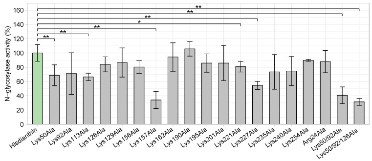
Saponin-mediated endosomal escape is a mechanism that increases the cytotoxicity of type I ribosome-inactivating proteins (type I RIPs). In order to actualize their cytotoxicity, type I RIPs must be released into the cytosol after endocytosis. Without release from the endosomes, type I RIPs are largely degraded and cannot exert their cytotoxic effects. Certain triterpene saponins are able to induce the endosomal escape of these type I RIPs, thus increasing their cytotoxicity. However, the molecular mechanism underlying the endosomal escape enhancement of type I RIPs by triterpene saponins has not been fully elucidated. In this report, we investigate the involvement of the basic amino acid residues of dianthin-30, a type I RIP isolated from the plant Dianthus caryophyllus L., in endosomal escape enhancement using alanine scanning. Therefore, we designed 19 alanine mutants of dianthin-30. Each mutant was combined with SO1861, a triterpene saponin isolated from the roots of Saponaria officinalis L., and subjected to a cytotoxicity screening in Neuro-2A cells. Cytotoxic screening revealed that dianthin-30 mutants with lysine substitutions did not impair the endosomal escape enhancement. There was one particular mutant dianthin, Arg24Ala, that exhibited significantly reduced synergistic cytotoxicity in three mammalian cell lines. However, this reduction was not based on an altered interaction with SO1861. It was, rather, due to the impaired endocytosis of dianthin Arg24Ala into the cells.

Keywords: N-glycosylase; basic amino acid residues; endocytosis; endosomal escape enhancer; ribosome-inactivating proteins; triterpene saponins; type I RIP.


Conflict of interest statement

The authors declare no conflicts of interest.

Figures

Figure 9
Figure 9
Protein structure of Hisdianthin and its mutant Hisdianthin Arg24Ala and structure of SO1861. (AD) Homology model of Hisdianthin and Hisdianthin Arg24Ala using Phyre2 and Mol* Viewer [63,64]. The high-resolution structure of dianthin-30 served as template (1.4 Å; PDB ID 1RL0) [65]. The homology models indicate confidence and coverage levels of 100%. (A,B) Tertiary structure of Hisdianthin. The residue Arg24 (highlighted in light green) is surface-accessible and interacts via hydrogen bonds with the residues Arg28, Ala179 and Ala178. (C,D) Tertiary structure of Hisdianthin Arg24Ala. After inserting an alanine at position 24, the hydrogen bonds with Ala178 and Ala179 no longer exist. Ala24 now interacts only with Arg28. (E) Structure of the bisdesmosidic triterpene saponin SO1861 [36].
Figure 1
Figure 1
SDS-PAGE (12.5%, Coomassie Brillant Blue stain) analysis of Hisdianthin and Hisdianthin mutants. The purified protein fractions obtained after rapid centrifugation-based Ni-NTA purification are visualized. In total, 0.6 µg of protein was applied to each pocket. Protein marker (M, in kDa) and the reference protein Hisdianthin have the same position in each gel.
Figure 2
Figure 2
Native MS analysis to study the binding of SO1861 to Hisdianthin. Samples were dissolved in 50 mM of ammonium acetate buffer (pH 6.24), and spectra were recorded with a constant protein concentration (Hisdianthin, 10 µM) and an increasing ligand concentration of SO1861 (0 to 100 µM) from top to bottom. Raw spectra are shown on the left, and corresponding deconvoluted spectra showing the experimental average protein masses are shown on the right. With the addition of SO1861, weak additional signals (highlighted by red arrows) indicate the binding of SO1861 to Hisdianthin in a 1:1 stoichiometry.
Figure 3
Figure 3
N-glycosylase activity of Hisdianthin mutants in comparison with native Hisdianthin. The adenine-releasing assay was performed with a protein concentration of 169 nM and 21.4 µM of A30 substrate overnight at 37 °C. The activities of Hisdianthin mutants were calculated as percentages related to the activity of Hisdianthin. Lysine substitution at positions 50, 113, 157, 221 and 227 and combined alanine substitutions at positions 50/92 and 50/92/126 significantly reduced the N-glycosylase activity. Values represent the means ± standard deviation of three independent measurements; n = 3 (significance: * p ≤ 0.05; ** p ≤ 0.01; Mann–Whitney U test).
Figure 4
Figure 4
Cytotoxic screening of Hisdianthin mutants in Neuro-2A cells. (A) Hisdianthin mutants Lys50Ala to Lys129Ala. (B) Hisdianthin mutants Lys156Ala to Lys190Ala. (C) Hisdianthin mutants Lys195Ala to Lys235Ala. (D) Hisdianthin mutants Lys240Ala to Arg24Ala. Cytotoxicity is represented using cell growth data. The cell confluence is shown as normalized cell index, which was calculated by dividing the cell confluence at each time point by the cell confluence at the reference time point of 25 h (time point after sample addition). In total, cell confluence was recorded over 72 h using the live-cell imaging CytoSMART Omni system. Then, 24 h after seeding the cells, 1 nM of Hisdianthin mutant either with the addition of 1 µg/mL of SO1861 or without the addition of SO1861 (1 nM Hisdianthin mutant ± 1 µg/mL SO1861) was added and incubated for 48 h. Control cells were treated the same way. As a positive control, 1 nM of native Hisdianthin ± 1 µg/mL SO1861 was used, and as a negative control, PBS ± 1 µg/mL SO1861 was used. Samples without the addition of SO1861 should have no effect on cell growth. The addition of SO1861 should reveal the cytotoxic effect of the Hisdianthin mutants and the Hisdianthin control. The samples without the addition of SO1861 are shown as solid lines, while the samples with the addition of SO1861 are shown as dashed lines. Data are shown as means of three independent measurements, each in triplicate (n = 3). For clarity, the standard deviation is not shown.
Figure 5
Figure 5
Cytotoxicity of Hisdianthin Arg24Ala in three cell lines. A2058 cells (A), HCT116 cells (B), and Neuro-2A cells (C) were incubated with 0.1 nM and 1 nM of Hisdianthin and Hisdianthin Arg24Ala ± 1 µg/mL of SO1861 for 48 h, respectively. Control cells were equivalently treated with PBS ± 1 µg/mL of SO1861. After 48 h of incubation, an MTT assay was performed. The presented cell viability was calculated relative to the control cells. The Arg24Ala mutation caused significantly reduced synergistic cytotoxicity (+SO1861) in all cell lines compared with native Hisdianthin. Data are shown as means ± standard deviation of three independent measurements, each in triplicate; n = 3 (Significance: **** p ≤ 0.0001, Student’s t-test).
Figure 6
Figure 6
Cytotoxicity of high concentrations of Hisdianthin Arg24Ala. Neuro-2A cells were incubated with either 10 nM, 100 nM or 1000 nM Hisdianthin and Hisdianthin Arg24Ala without the addition of SO1861 (A) and with the addition of 1 µg/mL of SO1861 (B) for 48 h. Control cells were treated equivalently with PBS ± 1 µg/mL of SO1861. At the end of incubation, an MTT assay was performed. (A) Without the addition of SO1861, Hisdianthin Arg24Ala was not cytotoxic even at high concentrations, whereas native Hisdianthin showed cytotoxicity above a concentration of 100 nM, even without the addition of SO1861. The cytotoxicity of the native and mutant forms differed significantly above a concentration of 100 nM. Data are shown as means ± standard deviation of three independent measurements, each in triplicate; n = 3 (significance: ** p ≤ 0.01; **** p ≤ 0.0001; Student’s t-test). (B) the addition of SO1861 caused an increase in cytotoxicity in both forms. The cytotoxicity differences in both forms were significant at 10 nM and at 1000 nM. Data are shown as means ± standard deviation of three independent measurements, each in triplicate; n = 3 (significance: * p ≤ 0.05; ** p ≤ 0.01; Mann–Whitney U test).
Figure 7
Figure 7
Characterization of Hisdianthin-CF568 and Hisdianthin Arg24Ala-CF568. (A) SDS-PAGE (12.5%, Coomassie Brillant Blue stain) of the labeling reaction of Hisdianthin und Hisdianthin Arg24Ala with CF®568 resulted in clean labeling products (each one a diffuse band at 30–33 kDa). Equivalent section: Left side is shown under daylight and right side at 366 nm. Lane I: Protein marker (in kDa). Lane II: Hisdianthin (2.0 µg). Lane III: Hisdianthin Arg24Ala (2.0 µg). Lane IV: Hisdianthin-CF568 (2.3 µg). Lane V: Hisdianthin Arg24Ala-CF568 (2.3 µg). (B) Fluorescence intensity of Hisdianthin-CF568 and Hisdianthin Arg24Ala-CF568 at 560/10 nm (excitation wavelength) and 595/35 nm (emission wavelength). No significant difference in intensity was measured at 100 nM or at 1000 nM of Hisdianthin-CF568 and Hisdianthin Arg24Ala-CF568. PBS was measured as a control. Data are shown as means ± standard deviation of three independent measurements, each in triplicate; n = 3 (significance: Mann–Whitney U test). (C) N-glycosylase activity of Hisdianthin-CF568 and Hisdianthin Arg24Ala-CF568. The adenine-releasing assay was performed with a protein concentration of 169 nM and 21.4 µM of A30 substrate at 37 °C overnight. Labeling with fluorescent dye CF®568 resulted in complete loss of activity in Hisdianthin-CF568 and Hisdianthin Arg24Ala-CF568. The means ± standard deviation of three independent measurements are shown; n = 3 (significance: ** p ≤ 0.01; Student’s t-test).
Figure 8
Figure 8
Endocytosis of Hisdianthin-CF568 and Hisdianthin Arg24Ala-CF568. Neuro-2A cells were incubated for 0, 2, 6, 16 and 24 h with 100 nM of Hisdianthin-CF568 and Hisdianthin Arg24Ala-CF568. After incubation with a labeled protein, the cell culture medium was removed, and cells were analyzed by flow cytometry using the peak height of the PE channel. Endocytosis was measured by means of fluorescence intensity increase. After 6 h, Hisdianthin-CF568 and Hisdianthin Arg24Ala-CF568 differed significantly in their endocytosis, where, first, Hisdianthin Arg24Ala-CF568 and, after 16 h, Hisdianthin-CF568 were more endocytosed. At least 10,000 cells per sample were measured. Only single cells were included in the analysis; doublets and debris were excluded. The means ± standard deviation of three independent measurements are shown; n = 3 (significance: * p ≤ 0.05; ** p ≤ 0.01; **** p ≤ 0.0001; Student’s t-test).

Similar articles

Cited by

References

    1. Endo Y., Tsurugi K. RNA N-glycosidase activity of ricin A-chain. Mechanism of action of the toxic lectin ricin on eukaryotic ribosomes. J. Biol. Chem. 1987;262:8128–8130. doi: 10.1016/S0021-9258(18)47538-2. - DOI - PubMed
    1. Endo Y., Mitsui K., Motizuki M., Tsurugi K. The mechanism of action of ricin and related toxic lectins on eukaryotic ribosomes. The site and the characteristics of the modification in 28 S ribosomal RNA caused by the toxins. J. Biol. Chem. 1987;262:5908–5912. doi: 10.1016/S0021-9258(18)45660-8. - DOI - PubMed
    1. Olsnes S., Pihl A. Different biological properties of the two constituent peptide chains of ricin, a toxic protein inhibiting protein synthesis. Biochemistry. 1973;12:3121–3126. doi: 10.1021/bi00740a028. - DOI - PubMed
    1. Guyette J.L., Serrano A., Huhn Iii G.R., Taylor M., Malkom P., Curtis D., Teter K. Reduction is sufficient for the disassembly of ricin and Shiga toxin 1 but not Escherichia coli heat-labile enterotoxin. Infect. Immun. 2023;91:e0033223. doi: 10.1128/iai.00332-23. - DOI - PMC - PubMed
    1. Timar J., McIntosh D.P., Henry R., Cumber A.J., Parnell G.D., Davies A.J. The effect of ricin B chain on the intracellular trafficking of an A chain immunotoxin. Br. J. Cancer. 1991;64:655–662. doi: 10.1038/bjc.1991.377. - DOI - PMC - PubMed

Publication types

LinkOut - more resources