. 2024 May 16;12(5):367.
doi: 10.3390/toxics12050367.

Titanium Dioxide Nanoparticles Induce Maternal Preeclampsia-like Syndrome and Adverse Birth Outcomes via Disrupting Placental Function in SD Rats

Affiliations

Titanium Dioxide Nanoparticles Induce Maternal Preeclampsia-like Syndrome and Adverse Birth Outcomes via Disrupting Placental Function in SD Rats

Haixin Li et al. Toxics. .

Abstract

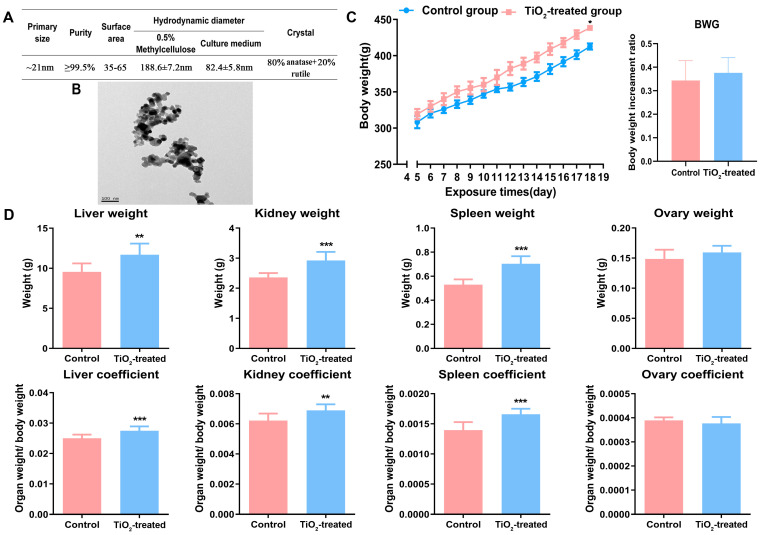
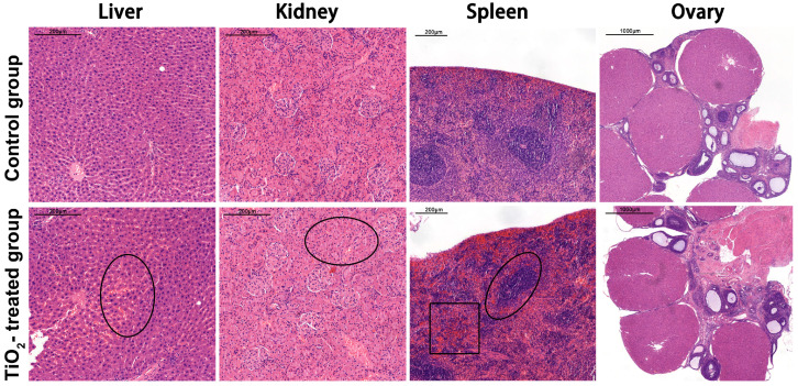
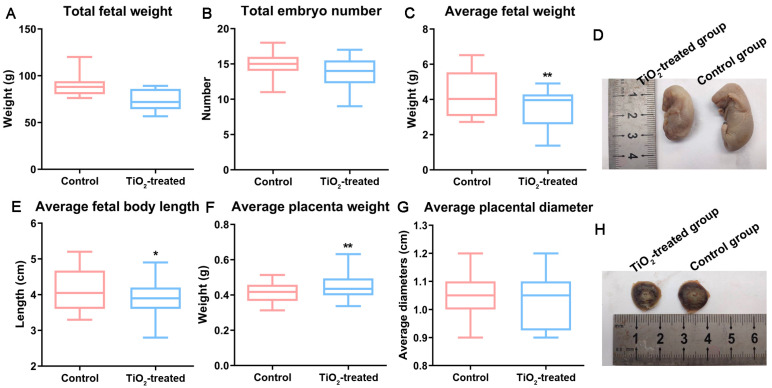
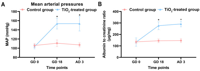
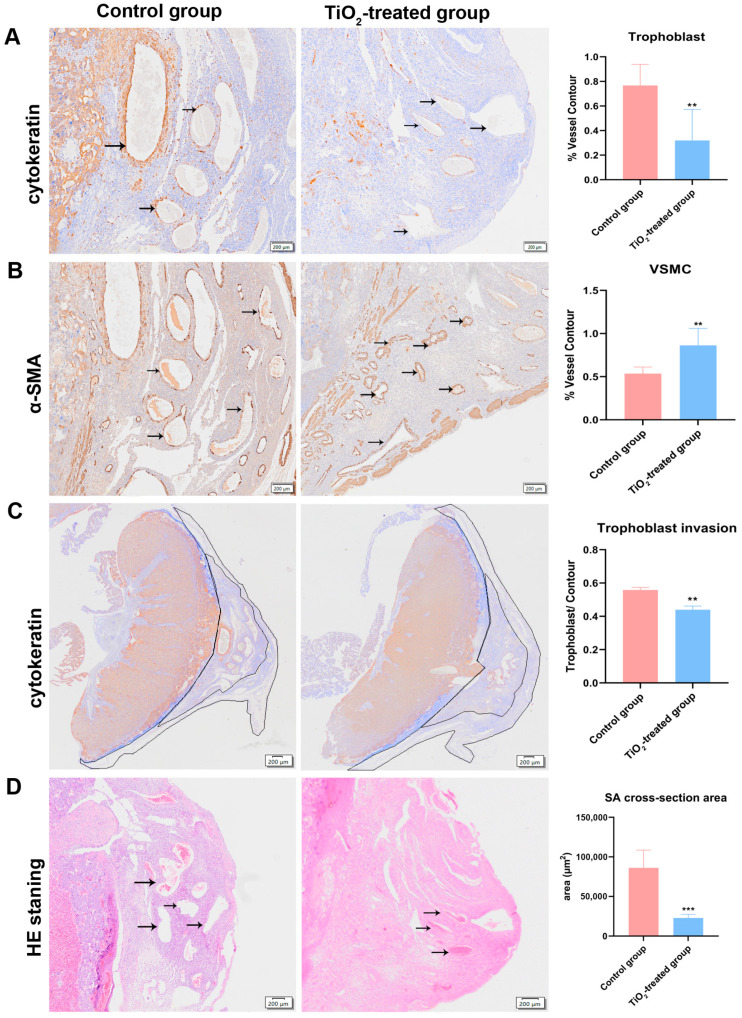
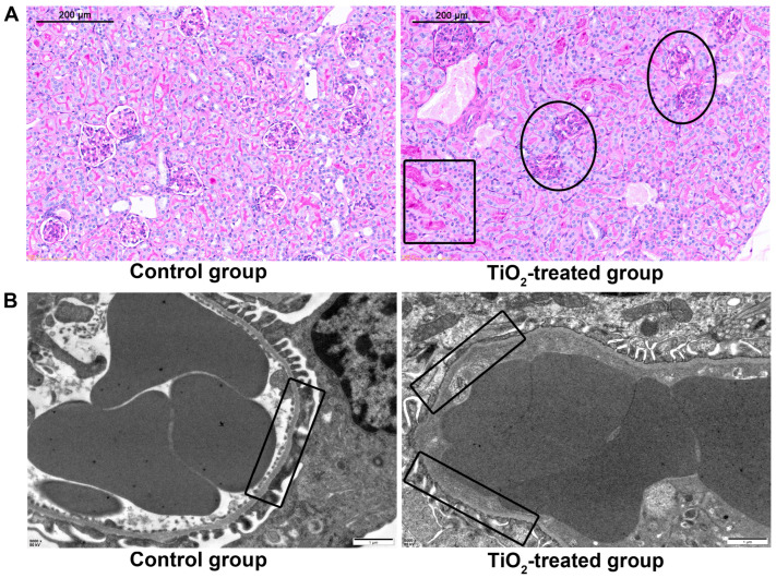
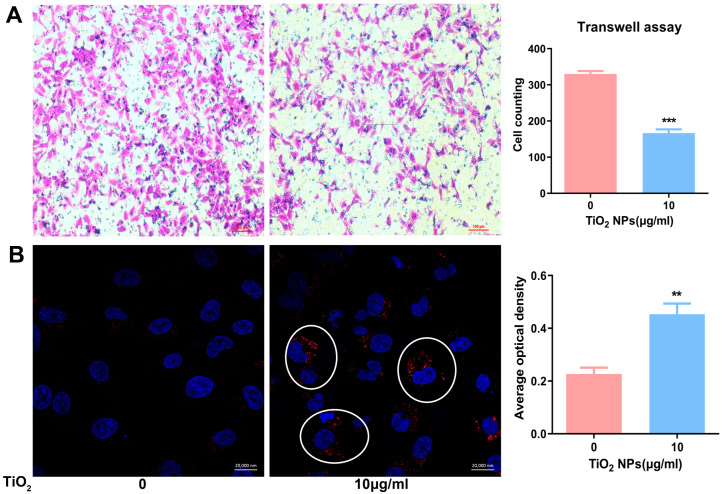
The escalating utilization of titanium dioxide nanoparticles (TiO2 NPs) in everyday products has sparked concerns regarding their potential hazards to pregnant females and their offspring. To address these concerns and shed light on their undetermined adverse effects and mechanisms, we established a pregnant rat model to investigate the impacts of TiO2 NPs on both maternal and offspring health and to explore the underlying mechanisms of those impacts. Pregnant rats were orally administered TiO2 NPs at a dose of 5 mg/kg body weight per day from GD5 to GD18 during pregnancy. Maternal body weight, organ weight, and birth outcomes were monitored and recorded. Maternal pathological changes were examined by HE staining and TEM observation. Maternal blood pressure was assessed using a non-invasive blood analyzer, and the urinary protein level was determined using spot urine samples. Our findings revealed that TiO2 NPs triggered various pathological alterations in maternal liver, kidney, and spleen, and induced maternal preeclampsia-like syndrome, as well as leading to growth restriction in the offspring. Further examination unveiled that TiO2 NPs hindered trophoblastic cell invasion into the endometrium via the promotion of autophagy. Consistent hypertension and proteinuria resulted from the destroyed the kidney GBM. In total, an exposure to TiO2 NPs during pregnancy might increase the risk of human preeclampsia through increased maternal arterial pressure and urinary albumin levels, as well as causing fetal growth restriction in the offspring.

Keywords: autophagy; placenta development; preeclampsia-like syndrome; pregnant model; titanium dioxide nanoparticles; trophoblastic cell function.


Conflict of interest statement

The authors declare no conflicts of interest.

Figures

Figure 1
Figure 1
(A,B) The characteristics of TiO2 NPs determined by a transmission electronic microscope (TEM), dynamic light scattering (DLS), and by the manufacturer’s report. (C) Maternal body weights were determined before the female rats were fed every morning. Body weight gain (BWG) = (mf − mi)/mi. “mf” represents the final body weight and “mi” represents the initial body weight. (D) The main organs (liver, kidney, spleen, and ovary) were weighed and the organ coefficients were calculated after the pregnant rats were executed on GD18. * p < 0.05, ** p < 0.01, *** p < 0.001. There were 7 rats in the control group and 8 rats in the exposure group.
Figure 2
Figure 2
The pathological changes in the maternal liver, kidney, spleen, and ovary after exposure were examined by light microscope after hematoxylin and eosin (HE) staining. Scale bar = 200 μm in the liver, kidney, and spleen. Scale bar = 1000 μm in the ovary. Pathological changes in the liver, kidney, and spleen were indicated with black circles, and hyperemia region was indicated with black square. There were 7 rats in the control group and 8 rats in the exposure group.
Figure 3
Figure 3
The main birth outcomes were recorded on GD18 after the mothers were executed. The total fetal weight (A), total embryo number (B), average fetal weight (C), average fetal body length (E), average placental weight (F), and placental diameter (G) were obtained from the control and TiO2-treated group. (D,H) show images of the fetuses and placenta. * p < 0.05, ** p < 0.01. There were 105 fetal rats in the control group and 95 fetal rats in the exposure group.
Figure 4
Figure 4
(A) The average maternal arterial pressures were measured using a non-invasive blood pressure analyzer before pregnancy (GD0), on the 18th day of gestation (GD18), and on the third day after delivery (AD3). (B) The maternal urinary protein levels were determined using spot urine samples, and the urinary albumin to creatinine ratio (ACR) was utilized to normalize the proteinuria. The data were indicated as mean ± SD. * p < 0.05. There were 7 rats in the control group and 8 rats in the exposure group.
Figure 5
Figure 5
(A) A placental invasion ability assessment was conducted using the percentage of interstitial trophoblast invasion into the mesometrial triangle (MT). The infiltrated trophoblast cells were identified using a cytokeratin-7 (CK-7) antibody. (B) Evidence of spiral artery (SA) remodeling was also identified through the α-actin-positive smooth muscle cells. (C) The ratio of the cytokeratin-7-positive trophoblast cell area to the MT area. (D) The cross-sectional areas and SA numbers were measured by HE staining. A quantitative analysis was carried out with the Olympus OlyVIA software. The data were indicated as mean ± SD. ** p < 0.01, *** p < 0.001. The obvious pathological changes were indicated with black arrows, and the trophoblast invasion areas were outlined. There were 7 rats in the control group and 8 rats in the exposure group.
Figure 6
Figure 6
(A) The glomerular basement membrane (GBM) and deposit of fibrin were revealed by periodic acid-Schiff (PAS) staining. (B) The ultra microstructure of the GBM was revealed by TEM after a series of sample preparations, bar = 1 μm. The glomerular lesions were indicated with black circles, and the fibrin deposition region was indicated with a black square. The normal and impaired GBM were highlighted with black squares in TEM images. There were 7 rats in the control group and 8 rats in the exposure group.
Figure 7
Figure 7
(A) The cellular migration and invasion ability of trophoblastic cell lines (HTR) were determined by a transwell assay; cell counting was obtained from five independent fields of the light microscope, and the data were presented as mean ± SD, scale bar = 100 μm. (B) The autophagy levels of HTR cells exposed to TiO2 NPs were examined by immunofluorescence; the nuclei were stained blued with DAPI and the autophagosomes were stained red with CY3. Their fluorescence density was measured with the Zeiss software (https://www.zeiss.com/microscopy/en/products/software/zeiss-zen.html accessed on 9 May 2024) within the laser confocal microscope software package. Scale bar = 20,000 nm. *** p < 0.001, ** p < 0.01. The autophagsomes are indicated by the white circles in the image.

Similar articles

References

    1. Baranowska-Wojcik E., Szwajgier D., Oleszczuk P., Winiarska-Mieczan A. Effects of Titanium Dioxide Nanoparticles Exposure on Human Health—A Review. Biol. Trace Elem. Res. 2019;193:118–129. doi: 10.1007/s12011-019-01706-6. - DOI - PMC - PubMed
    1. Warheit D.B., Donner E.M. Risk assessment strategies for nanoscale and fine-sized titanium dioxide particles: Recognizing hazard and exposure issues. Food Chem. Toxicol. 2015;85:138–147. doi: 10.1016/j.fct.2015.07.001. - DOI - PubMed
    1. Gulson B., McCall M.J., Bowman D.M., Pinheiro T. A review of critical factors for assessing the dermal absorption of metal oxide nanoparticles from sunscreens applied to humans, and a research strategy to address current deficiencies. Arch. Toxicol. 2015;89:1909–1930. doi: 10.1007/s00204-015-1564-z. - DOI - PubMed
    1. Shi H., Magaye R., Castranova V., Zhao J. Titanium dioxide nanoparticles: A review of current toxicological data. Part. Fibre Toxicol. 2013;10:15. doi: 10.1186/1743-8977-10-15. - DOI - PMC - PubMed
    1. Takeda K., Shinkai Y., Suzuki K., Yanagita S., Umezawa M., Yokota S., Tainaka H., Oshio S., Ihara T., Sugamata M. Health effects of nanomaterials on next generation. Yakugaku Zasshi. 2011;131:229–236. doi: 10.1248/yakushi.131.229. - DOI - PubMed

LinkOut - more resources